Tissue marker and method and apparatus for deploying the marker

a tissue marker and tissue technology, applied in the field of tissue marker systems, can solve the problems that bioactive materials can affect the therapy provided to patients, and achieve the effect of improving visibility

Inactive Publication Date: 2005-06-30
WINKEL AXEL
View PDF13 Cites 64 Cited by
  • Summary
  • Abstract
  • Description
  • Claims
  • Application Information

AI Technical Summary

Benefits of technology

[0016] To mark the tissue, the subject clip can be positioned next to a lesion like a tag. A marker having a bigger diameter than the lesion can be used and can surround the lesion in one or more dimensions like a cage. This cage can describe the position and size of the lesion and can, if desired, include a safety-margin. Surrounding the lesion makes the surgical or minimal invasive procedure to take out or destroy the lesion much more effective, as the size, position, and shape of the lesion can be discerned by the size, position, and shape of one or more markers used to appropriately mark the lesion.
[0018] In a specific embodiment, the subject marker can be modified to enhance visibility under MRI. Such modification can include the incorporation of a microcoil with the marker that can be imaged under MRI such that the marker can be found much faster under MRI than without the microcoil. In an embodiment, the microcoil can be incorporated in an implantable plastic capsule that is implanted in the marker.

Problems solved by technology

Such chemicals and / or bioactive materials can impact the therapy provided to the patient.

Method used


Image

  • Tissue marker and method and apparatus for deploying the marker
  • Tissue marker and method and apparatus for deploying the marker
  • Tissue marker and method and apparatus for deploying the marker

Examples


Embodiment Construction

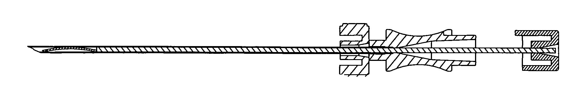
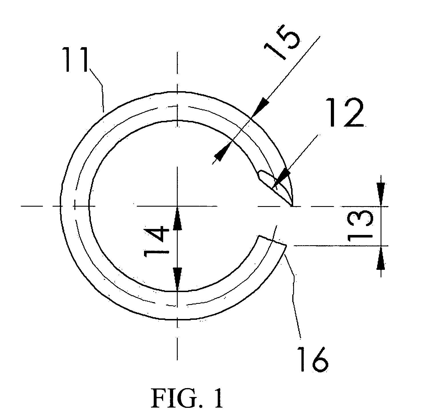
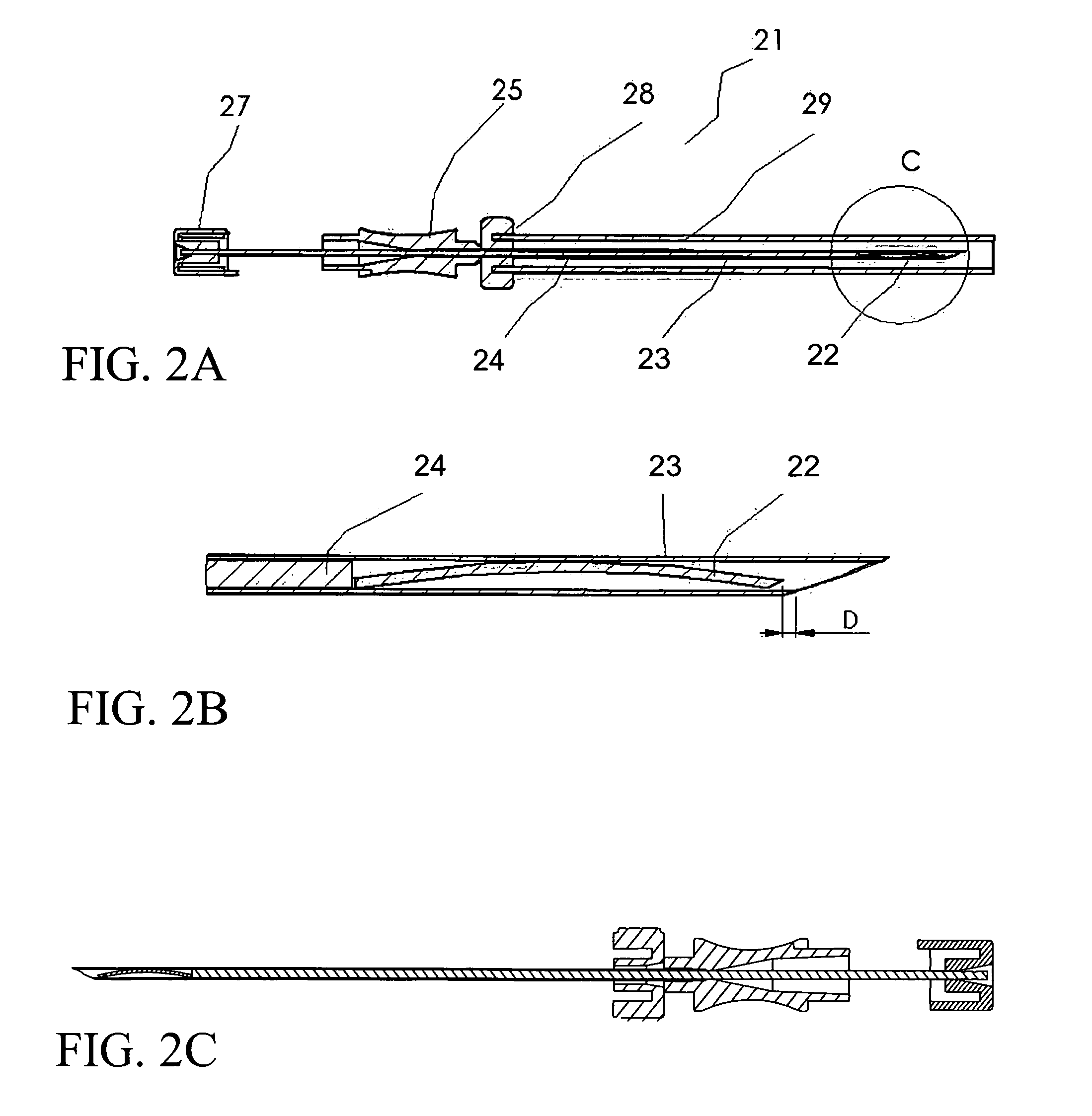
[0029] The subject invention relates to methods and apparatus for positioning a tissue marker in human or animal tissue. The subject invention also pertains to a tissue marker. Preferably, the subject tissue markers are visible under one or more imaging modalities such as, but not limited to, x-ray, ultrasound, and magnetic resonance imaging (MRI). In a specific embodiment, the subject marker is visible under magnetic resonance imaging (MRI).

[0030] In a specific embodiment, the subject invention incorporates a marker needle, an ejecting rod, and a tissue marker. The marker needle can penetrate the tissue to be marked such that the distal tip of the marker needle is positioned at the tissue location to be marked. The tissue marker can be inserted into the marker needle so as to be in an elongated position inside the marker needle. The marker can be inserted into the marker needle before or after positioning the marker needle. In a preferred embodiment, the marker is inserted into th...


PUM


Abstract

The subject invention pertains to a tissue marker. The subject invention also relates to methods and apparatus for deploying a tissue marker. In a specific embodiment, the subject marker is magnetic resonance imaging (MRI) compatible. In additional embodiment, various components of the subject marker deploying apparatus are MRI compatible. A specific marker in accordance with the subject invention is flexible such that the marker has an equilibrium shape, which the marker will have when deployed in tissue to be marked, and an elongated shape, which the marker can be bent into to be inserted into a marker needle.

Description

CROSS-REFERENCE TO RELATED APPLICATIONS [0001] This application claims the benefit of provisional patent application Ser. No. 60 / 525,205, filed Nov. 26, 2003, which is hereby incorporated by reference in its entirety.TECHNICAL FIELD [0002] The present invention relates in general to a tissue marker system, a method of deploying a tissue marker, and a tissue marker. A specific embodiment of the subject marker is compatible with magnetic resonance imaging (MRI) and is visible under MRI. In another embodiment, various components of the subject marker system are compatible with MRI such that the marker can be deployed under MRI. BACKGROUND [0003] Tissue markers are used in medicine to mark a suspicious lesion for surgical planning or for follow-up diagnostics. Markers are placed or deployed into tissue and remain in the tissue for a period of time. In order for markers not to move, markers are often clamped or stapled into tissue to hold them in place. Such markers which are clamped or ...

Claims


Application Information

Patent Timeline
Patent Type & Authority Applications(United States)
IPC IPC(8): A61B19/00
CPCA61B19/54A61B2019/5487A61B2019/5454A61B90/39A61B2090/3954A61B2090/3987
Inventor WINKEL, AXEL
Owner WINKEL AXEL
Why Eureka
Try Eureka