
Monoclonal antibodies against HMGB1

a monoclonal antibody and antibody technology, applied in the field of monoclonal antibodies against hmgb1, can solve the problems of elevated serum hmg1 levels that are toxi

Inactive Publication Date: 2005-07-14
THE FEINSTEIN INST FOR MEDICAL RES
View PDF16 Cites 53 Cited by
  • Summary
  • Abstract
  • Description
  • Claims
  • Application Information

AI Technical Summary

Benefits of technology

The present invention relates to antibodies or antigen-binding fragments that specifically bind to a vertebrate HMGB A box but do not specifically bind to non-A box epitopes of HMGB. These antibodies can inhibit the release of proinflammatory cytokines from a vertebrate cell treated with an HMGB protein. The invention also includes methods of detecting and identifying agents that bind to the HMGB protein. The technical effects of the invention include the development of tools for detecting and inhibiting the inflammatory response and treating inflammatory conditions.

Problems solved by technology

That work demonstrated that bacterial endotoxin (lipopolysaccharide (LPS)) activates monocytes / macrophages to release HMG1 as a late response to activation, resulting in elevated serum HMG1 levels that are toxic.

Method used


Image

  • Monoclonal antibodies against HMGB1
  • Monoclonal antibodies against HMGB1
  • Monoclonal antibodies against HMGB1

Examples


example 1

Materials and Methods

Generation of Monoclonal Antibodies to HMGB1

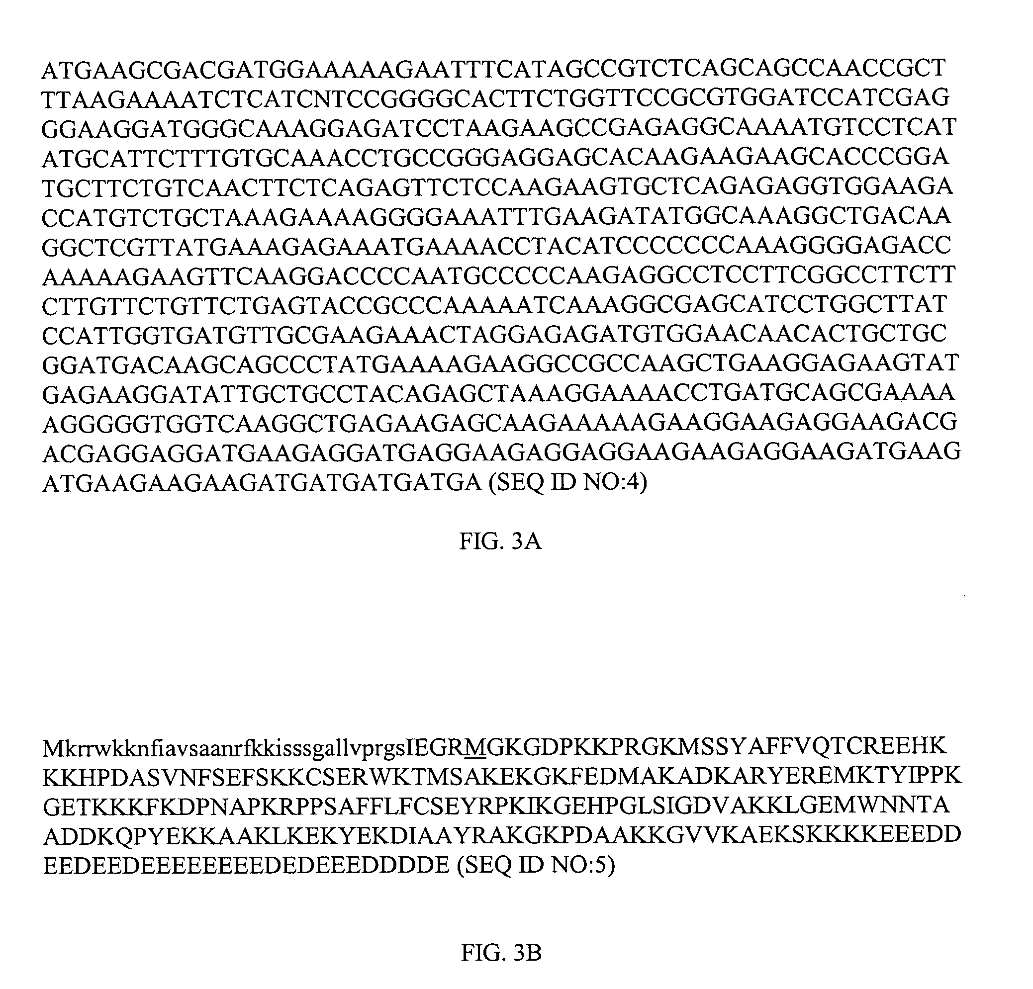
[0182] BALB / c mice were intraperitoneally immunized with 20 μg of recombinant CBP-Rat HMGB1 (CBP linked to amino acids 1-215 of rat HMGB1; nucleotide sequence of CBP-Rat HMGB1 is depicted as SEQ ID NO:4 and the amino acid sequence is depicted as SEQ ID NO:5 (see FIGS. 3A and 3B)) mixed with Freund's adjuvant at two-week intervals for 6 weeks. A final boost of 10 μg of the CBP-Rat HMGB1 in PBS was given intravenously after 8 weeks. Four days after the final boost, spleens from the mice were isolated and used for fusion. Fusion was carried out using standard hybridoma technique. The spleen was gently pushed through a cell strainer to obtain a single cell suspension. After extensive washing, spleen cells were mixed with SP2 / 0 myeloma cells. Polyethylene glycol (PEG) was added slowly, followed by media over a period of five minutes. The cells were washed and resuspended in DMEM containing 20% FCS and HAT, transferred to...

example 2

Identification and Characterization of Anti-HMGB1 Monoclonal Antibodies

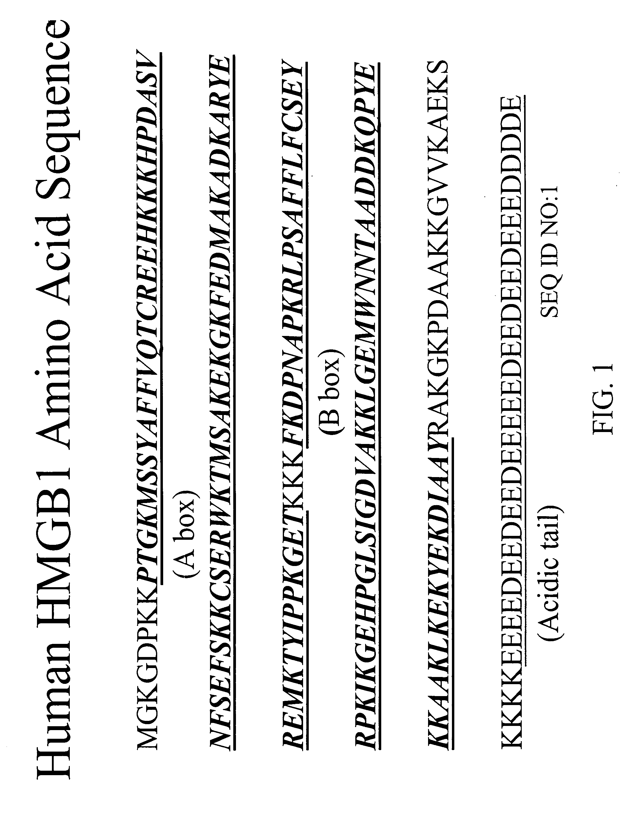
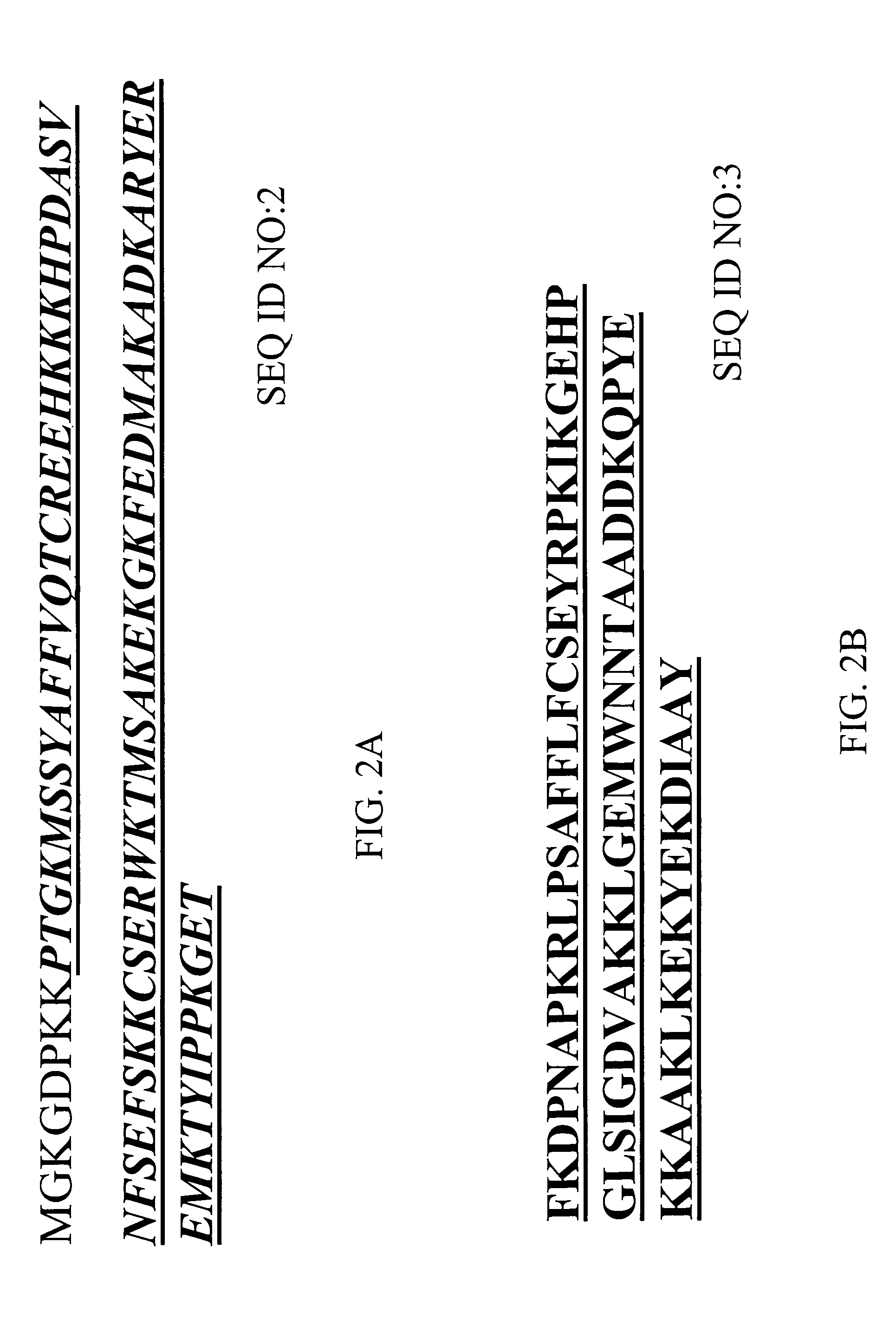
[0203] A number of novel anti-HMGB1 monoclonal antibodies have been isolated and purified from immunizations with either recombinant full-length rat HMGB1(SEQ ID NO:4; FIGS. 3A and 3B) or with a B-box polypeptide of human HMGB1 (SEQ ID NO:3; FIG. 2B). A table summarizing characteristics (clone name, immunogen, isotype, purified antibody binding domain, and results of in vivo CLP assays) of these antibodies is depicted in FIG. 7.

example 3

Determination of Selectivity of Anti-HMGB1 Monoclonal Antibodies

[0204] Experiments (e.g., ELISA, Western blot analysis) to determine the selectivity of the HMGB1 monoclonal antibodies revealed that particular anti-HMGB1 monoclonal antibodies are able to bind to the A box portion of HMGB1, the B box portion of HMGB1 and / or the whole HMGB1 protein. For example, as depicted in FIG. 7, anti-HMGB1 monoclonal antibodies were identified that can bind to the A box of HMGB1 (e.g., 6E6 HMGB1 mAb, 6H9 HMGB1 mAb, 10D4 HMGB1 mAb, 6H9 HMGB1 mAb, 2G7 HMGB1 mAb, 2G5 HMGB1 mAb, 4H11 HMGB1 mAb, 7H3 HMGB1 mAb, 9H3 HMGB1 mAb). Other monoclonal antibodies were identified that bind to the B box of HMGB1 (e.g., 2E11 HMGB1 mAb, 3G8 HMGB1 mAb, 3-5A6 HMGB1 mAb, 9G1 HMGB1 mAb, 4C9 HMGB1 mAb, 1C3 HMGB1 mAb, 5C12 HMGB1 mAb, 3E10 HMGB1 mAb, 7G8 HMGB1 mAb, 4A10 HMGB1 mAb).


PUM

PropertyMeasurementUnit
concentrationaaaaaaaaaa
volumeaaaaaaaaaa

Abstract

In various embodiments, the present invention is drawn to antibodies or antigen-binding fragments thereof that bind to a vertebrate high mobility group box (HMGB) polypeptide, methods of detecting and / or identifying an agent that binds to an HMGB polypeptide, methods of treating a condition in a subject characterized by activation of an inflammatory cytokine cascade and methods of detecting an HMGB polypeptide in a sample.

Description

RELATED APPLICATION [0001] This application claims the benefit of U.S. Provisional Application No. 60 / 502,568, filed Sep. 11, 2003. The entire teachings of the above application are incorporated herein by reference.BACKGROUND OF THE INVENTION [0002] Inflammation is often induced by proinflammatory cytokines, such as tumor necrosis factor (TNF), interleukin (IL)-1α, IL-1β, IL-6, macrophage migration inhibitory factor (MIF), and other compounds. These proinflammatory cytokines are produced by several different cell types, most importantly immune cells (for example, monocytes, macrophages and neutrophils), but also non-immune cells such as fibroblasts, osteoblasts, smooth muscle cells, epithelial cells, and neurons. These proinflammatory cytokines contribute to various disorders during the early stages of an inflammatory cytokine cascade. [0003] The early proinflammatory cytokines (e.g., TNF, IL-1, etc.) mediate inflammation, and induce the late release of high mobility group box 1 (HM...

Claims


Application Information

Patent Timeline
Patent Type & Authority Applications(United States)
IPC IPC(8): C07K14/47C07K16/18C12N5/20
CPCA61K2039/505C07K14/4718C07K16/24Y10S435/81C07K2317/565C07K2317/34C07K2316/96C07K2317/76A61P1/00A61P1/02A61P1/04A61P1/16A61P1/18A61P11/00A61P11/06A61P15/06A61P17/00A61P17/02A61P17/04A61P17/06A61P17/12A61P17/16A61P19/02A61P19/06A61P19/08A61P21/00A61P21/04A61P25/00A61P25/02A61P25/28A61P29/00A61P31/00A61P31/04A61P31/10A61P31/12A61P31/16A61P31/18A61P31/22A61P33/06A61P35/00A61P37/02A61P37/06A61P37/08A61P43/00A61P5/14A61P7/02A61P9/00A61P9/08A61P9/10A61P3/10
Inventor NEWMAN, WALTERQIN, SHIXINO'KEEFE, THERESAOBAR, ROBERT
Owner THE FEINSTEIN INST FOR MEDICAL RES
Learn More