Methods of diagnosing tissue fibrosis

a tissue fibrosis and tissue technology, applied in the field of fibrosis, can solve the problems of organ failure, liver disease or cancer end-stage, transplantation or death,

Active Publication Date: 2005-08-25
PROMETHEUS LAB
View PDF7 Cites 11 Cited by
  • Summary
  • Abstract
  • Description
  • Claims
  • Application Information

AI Technical Summary

Problems solved by technology

Progressive fibrosis of the liver, kidney, lungs and other organs frequently results in organ failure that leads to organ transplantation or death, affecting millions in the United States and worldwide.
Untreated, fibrosis of the liver leads to cirrhosis and eventually end-stage liver disease or cancer.
Diagnosis of the presence or severity of fibrotic liver disease is difficult, with liver biopsy currently the most reliable method available.
Unfortunately, liver biopsy has several limitations: pain in about 30% of patients; the risk of severe complications such as hemorrhage or infection; a death rate of 3 in 10,000; and the cost of hospitalization (Nord, Gastrointest. Endosc. 28:102-104 (1982); Cadranel et al., Hepatology 32:47-481 (2000); and Poynard et al., Can. J. Gastroenterol. 14:543-548 (2000)).
Furthermore, slowly progressive diseases such as hepatitis C require repeated biopsies for continual assessment of disease progression, thus compounding the risks and costs of the procedure.
Finally, biopsy can fail to detect disease because of the heterogeneous distribution of pathological changes in the liver; it is not surprising, then, that false negatives are seen in a significant percentage of cases biopsied (Nord, supra, 1982).
However, the performance of any single marker has not been good enough to substitute for the biopsy procedure in detecting or staging fibrosis.

Method used


Image

  • Methods of diagnosing tissue fibrosis
  • Methods of diagnosing tissue fibrosis
  • Methods of diagnosing tissue fibrosis

Examples


example i

Assays for β2-Macroglobulin, Hyaluronic Acid and Tissue Inhibitor of Metalloproteinases-1

A. Quantitation of Human α2-Macroglobulin (α2-MG)

[0158] Serum levels of human α2-macroglobulin were quantitated using the Beckman Array.RTM. 360 System as follows to determine α2-MG levels in the range of 0.75-270 mg / ml.

[0159] The Beckman Array® 360 system was used for determination of α2-MG concentrations. This system utilizes a nephelometer which measures the rate of light-scatter formation resulting from an immunoprecipitation reaction between α2-MG antigen in a sample with antibody to human α2-MG. After passing a beam of light through the solution in a flow cell, the intensity of light scattered by the formed macromolecular particles of insoluble complexes suspended in solution is detected and measured by the nephelometer. The increase in light scatter resulting from the antigen-antibody reaction is converted to a peak rate signal proportional to the α2-MG concentration in the sample. Th...

example ii

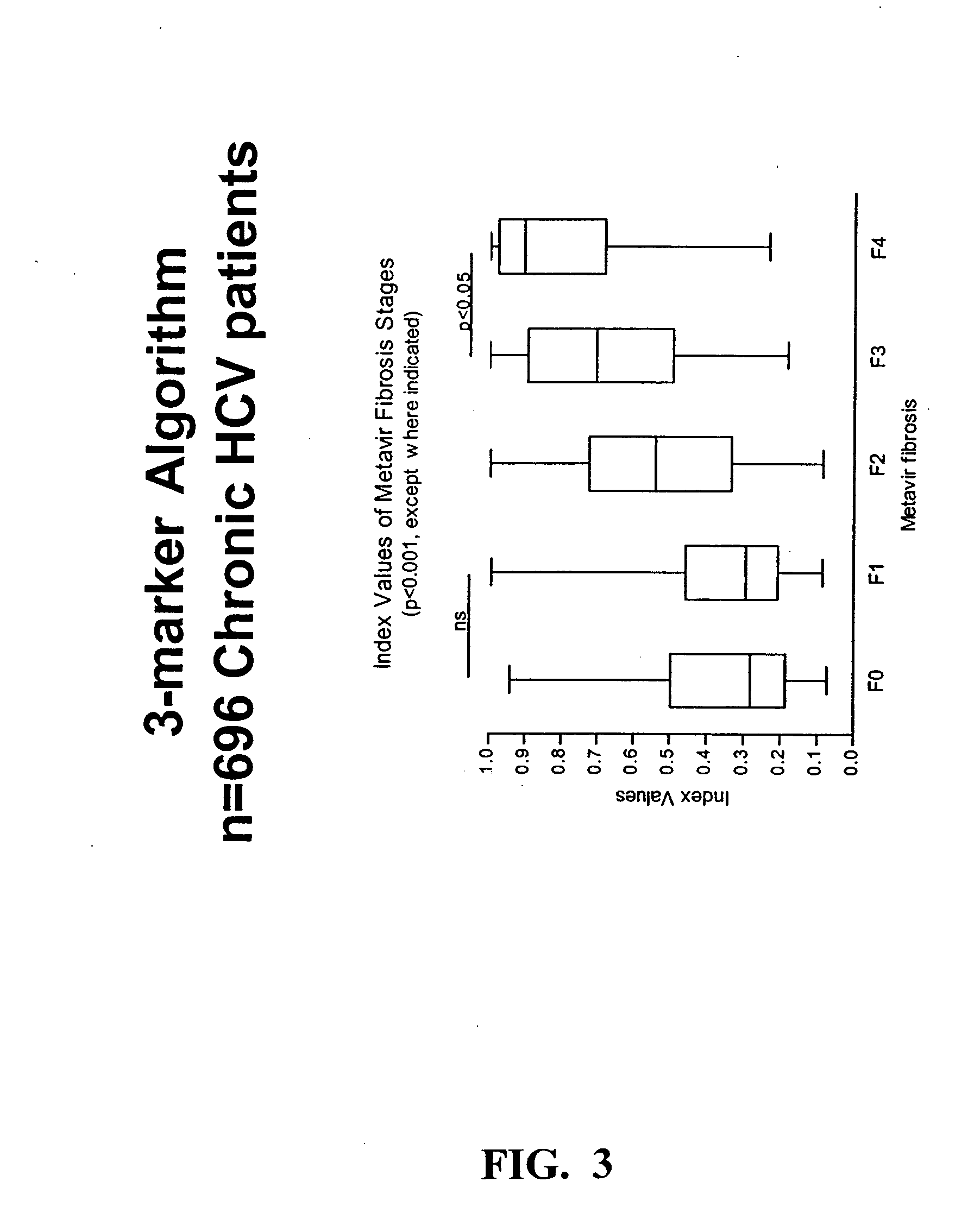
[0189] An algorithm was developed to differentiate no / mild from moderate / severe fibrosis in chronic HCV patients, as determined histologically from a liver biopsy, and staged using the Metavir system summarized in Table 2.

TABLE 2MetavirFibrosisstageDescriptionNo / mildF0No fibrosisF1Stellate enlargement of portaltract without septa formationModerate / severeF2Enlargement of portal tractwith rare septa formationF3Numerous septa without cirrhosisF4Cirrhosis

[0190] Serum samples from 696 chronic HCV patients were obtained, at or near the time of liver biopsy, from 4 study sites. Levels of the three markers of fibrosis, hyaluronic acid (HA), tissue inhibitor of metalloproteinase-1 (TIMP-1), and α2-macroglobulin (α2M, or A2M), were determined in each specimen by protein binding assay (HA), ELISA (TIMP-1) and nephelometry (A2M). Severity of fibrosis in patients was staged by histological staining of a biopsy specimen using the Metavir system. These variables were subjected to regression anal...

example 3

[0194] A second algorithm was determined using two markers: HA and TIMP1. The HA and TIMP1 levels from Example 2 were used in this algorithm. As for the three marker test above, sensitivity was maintained at 80%. Results are shown in Tables 5 and 6. With the two marker analysis and sensitivity of 80%, specificity of the test was 63.4%. In contrast, when A2M levels were analyzed with HA and TIMP1 levels as above, specificity of the test was 71.4%. Thus, addition of the A2M marker to the analysis improved the specificity of the test.

TABLE 5F0F1F2F3F4Test+35888490115412Test−5615755133284Total91245139103118696

[0195]

TABLE 6F2-F4F0-F1TotalTest+289123412Test−71213284Total360336696Prev51.7%Sens80.3%Spec63.4%PPV70.1%NPV75.0%Accuracy72.1%


PUM

PropertyMeasurementUnit
pHaaaaaaaaaa
pHaaaaaaaaaa
timeaaaaaaaaaa

Abstract

The present invention provides a method of diagnosing the presence or severity of tissue fibrosis in an individual by detecting α2-macroglobulin (α2-MG) in a sample from the individual; detecting hyaluronic acid (HA) in a sample from the individual; detecting tissue inhibitor of metalloproteinases-1 (TIMP-1) in a sample from the individual; and diagnosing the presence or severity of tissue fibrosis in the individual based on the presence or level of α2-MG, HA and TIMP-1.

Description

CROSS-REFERENCES TO RELATED APPLICATIONS [0001] This application claims the benefit of U.S. Provisional Application No. 60 / 514,034, filed Oct. 24, 2003, which is herein incorporated by reference for all purposes.FIELD OF THE INVENTION [0002] The present invention relates generally to the field fibrosis and, more specifically, to a panel of serological markers which together are diagnostic of tissue fibrosis. BACKGROUND OF THE INVENTION [0003] Progressive fibrosis of the liver, kidney, lungs and other organs frequently results in organ failure that leads to organ transplantation or death, affecting millions in the United States and worldwide. Hepatic fibrosis, for example, is the leading non-malignant gastrointestinal cause of death in the United States, and the progression of fibrosis is the single most important determinant of morbidity and mortality in patients with chronic liver disease. Furthermore, the process of fibrosis is common to liver diseases of many etiologies, includin...

Claims


Application Information

Patent Timeline
Patent Type & Authority Applications(United States)
IPC IPC(8): A61BC12Q1/68C12Q1/70G01N33/53G01N33/567G01N33/68
CPCC12Q1/6883G01N33/6893C12Q2600/158C12Q2600/112G01N2800/52
Inventor OH, ESTHERSMITH, CATHERINE
Owner PROMETHEUS LAB
Why Eureka
Try Eureka