Lung nodule detection and classification

a technology of computed tomography and lung cancer, applied in the field of automatic detection and classification of lung cancer, can solve the problems of low number of images that need to be interpreted in ct screening, no significant improvement in the survival rate of patients with lung cancer, and significant progress

Inactive Publication Date: 2005-09-22
RGT UNIV OF MICHIGAN
View PDF7 Cites 352 Cited by
  • Summary
  • Abstract
  • Description
  • Claims
  • Application Information

AI Technical Summary

Problems solved by technology

While breast, colon, and prostate cancer have seen improved survival rates within the 1974-1990 time period, there has been no significant improvement in the survival of patients with lung cancer.
One reason for the lack of significant progress in the fight against lung cancer may be due to the lack of a proven screening test.
Unfortunately, the number of images that needs to be interpreted in CT screening is high, particularly when a multi-detector helical CT detector and thin collimation are used to produce the CT images.
The analysis of CT images to detect lung nodules is a demanding task for radiologists due to the number of different images that need to be analyzed by the radiologist.
However, this methodology doubles the demand on the radiologists' time.
However, they also demonstrate large variations in performance, indicating that the computer vision techniques in this area have not been fully developed and are not at an acceptable level to use at a clinical setting.

Method used


Image

  • Lung nodule detection and classification
  • Lung nodule detection and classification
  • Lung nodule detection and classification

Examples


Embodiment Construction

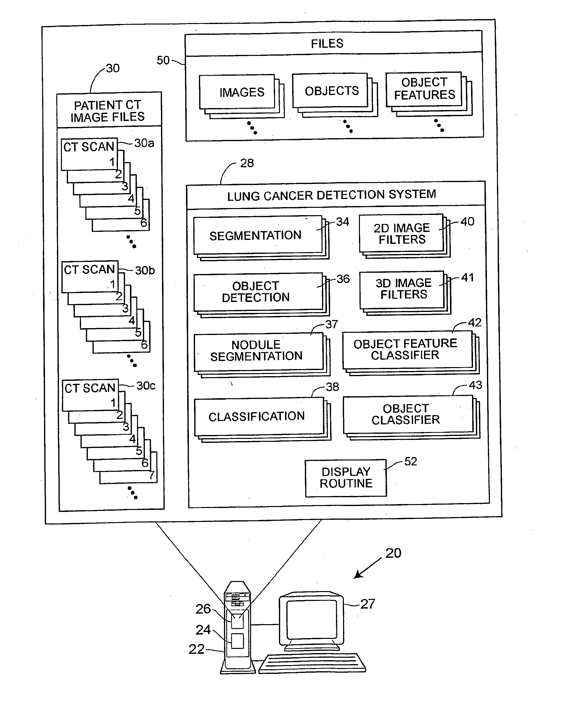
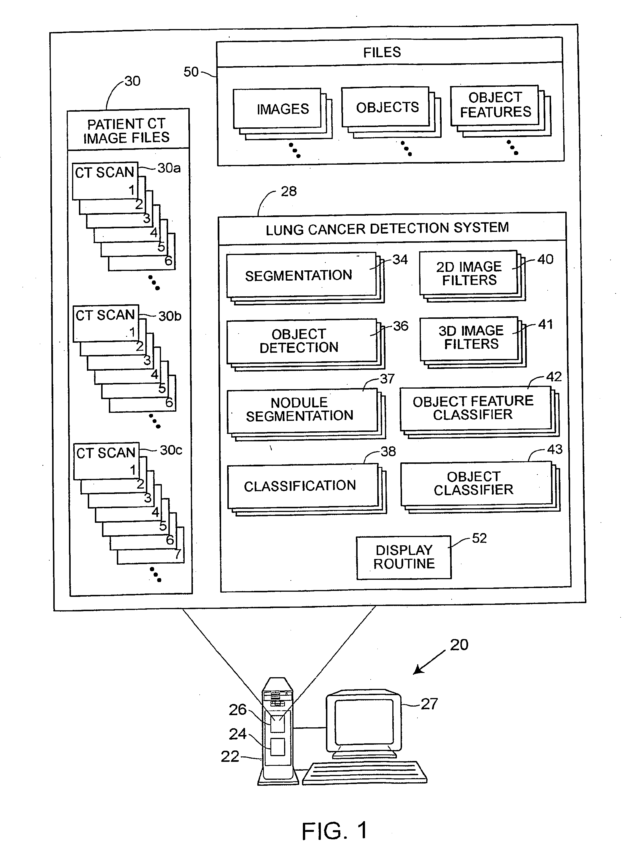
[0030] Referring to FIG. 1, a computer aided diagnosis (CAD) system 20 that may be used to detect and diagnose lung cancer or nodules includes a computer 22 having a processor 24 and a memory 26 therein and having a display screen 27 associated therewith, which may be, for example, a Barco MGD52I monitor with a P104 phosphor and 2K by 2.5K pixel resolution. As illustrated in an expanded view of the memory 26, a lung cancer detection and diagnostic system 28 in the form of, for example, a program written in computer implementable instructions or code, is stored in the memory 26 and is adapted to be executed on the processor 24 to perform processing on one or more sets of computed tomography (CT) images 30, which may also stored in the computer memory 26. The CT images 30 may include CT images for any number of patients and may be entered into or delivered to the system 20 using any desired importation technique. Generally speaking, any number of sets of images 30a, 30b, 30c, etc. (ca...


PUM


Abstract

A computer assisted method of detecting and classifying lung nodules within a set of CT images includes performing body contour, airway, lung and esophagus segmentation to identify the regions of the CT images in which to search for potential lung nodules. The lungs are processed to identify the left and right sides of the lungs and each side of the lung is divided into subregions including upper, middle and lower subregions and central, intermediate and peripheral subregions. The computer analyzes each of the lung regions to detect and identify a three-dimensional vessel tree representing the blood vessels at or near the mediastinum. The computer then detects objects that are attached to the lung wall or to the vessel tree to assure that these objects are not eliminated from consideration as potential nodules. Thereafter, the computer performs a pixel similarity analysis on the appropriate regions within the CT images to detect potential nodules and performs one or more expert analysis techniques using the features of the potential nodules to determine whether each of the potential nodules is or is not a lung nodule. Thereafter, the computer uses further features, such as speculation features, growth features, etc. in one or more expert analysis techniques to classify each detected nodule as being either benign or malignant. The computer then displays the detection and classification results to the radiologist to assist the radiologist in interpreting the CT exam for the patient.

Description

RELATED APPLICATIONS [0001] This claims the benefit under 35 U.S.C. §119(e) of U.S. Provisional Application Ser. No. 60 / 357,518, entitled “Computer-Aided Diagnosis (CAD) System for Detection of Lung Cancer on Thoracic Computed Tomographic (CT) Images” which was filed Feb. 15, 2002, the disclosure of which, in its entirety, is incorporated herein by reference and claims the benefit under U.S.C. §119(e) of U.S. Provisional Application Ser. No. 60 / 418,617, entitled “Lung Nodule Detection on Thoracic CT Images: Preliminary Evaluation of a Computer-Aided Diagnosis System” which was filed Oct. 15, 2002, the disclosure of which, in its entirety, is incorporated herein by reference.FIELD OF TECHNOLOGY [0002] This relates generally to computed tomography (CT) scan image processing and, more particularly, to a system and method for automatically detecting and classifying lung cancer based on the processing of one or more sets of CT images. DESCRIPTION OF THE RELATED ART [0003] Cancer is a ser...

Claims


Application Information

Patent Timeline
Patent Type & Authority Applications(United States)
IPC IPC(8): A61B6/03G06F19/00G06K9/00G06T7/00
CPCA61B6/03A61B6/466A61B6/583G06T2207/10081G06T2207/30061G06T7/0012
Inventor CHAN, HEANG-PINGSAHINER, BERKMANHADJIYSKI, LUBOMIR MZHOU, CHUANPETRICK, NICHOLAS
Owner RGT UNIV OF MICHIGAN
Why Eureka
Try Eureka