X-ray imaging system with automatic image resolution enhancement

a technology of x-ray imaging and image resolution, applied in the field of x-ray imaging systems, can solve the problems of patient discomfort, inability to read images, and inability to determine the appropriate areas of interest, etc., and achieve the effect of improving image resolution

Inactive Publication Date: 2005-10-27
VARIAN MEDICAL SYST TECH
View PDF21 Cites 10 Cited by
  • Summary
  • Abstract
  • Description
  • Claims
  • Application Information

AI Technical Summary

Benefits of technology

[0008] In accordance with the presently claimed invention, an automated X-ray imaging system and method are provided for producing a plurality of X-ray imaging signals having selectively enhanced image resolutions, e.g., for magnifying the field of view and providing a display image having features not otherwise visible to an unaided human eye. Successive doses of X-ray radiation are applied to a portion of the subject to produce corresponding image signals. Such doses of X-ray radiation are controlled by controlling X-ray radiation characteristics, such as intensity, focal spot size, focal spot location, focal spot shape, or collimation, to cause a subsequent image signal to differ from a prior image signal in one or more image characteristics, such as image resolution.

Problems solved by technology

While these problems can be minimized by the attending technician using collimators to isolate a region of interest, such technician is generally not qualified to read the images or determine the appropriate areas of interest.
Further, during many procedures, the patient is under some discomfort during the procedure and is, therefore, removed from the imaging system prior to any reading of the film or image.

Method used


Image

  • X-ray imaging system with automatic image resolution enhancement
  • X-ray imaging system with automatic image resolution enhancement
  • X-ray imaging system with automatic image resolution enhancement

Examples


Embodiment Construction

[0021] The following detailed description is of example embodiments of the presently claimed invention with references to the accompanying drawings. Such description is intended to be illustrative and not limiting with respect to the scope of the present invention. Such embodiments are described in sufficient detail to enable one of ordinary skill in the art to practice the subject invention, and it will be understood that other embodiments may be practiced with some variations without departing from the spirit or scope of the subject invention.

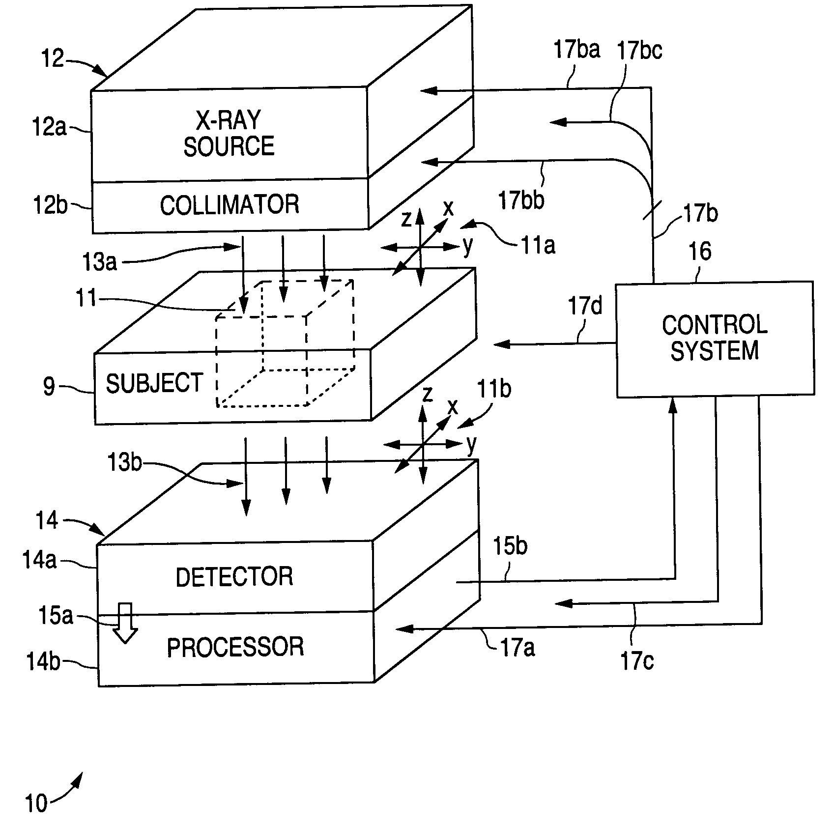
[0022] As discussed in more detail below, an x-ray imaging system in accordance with the presently claimed invention improves diagnostic x-ray image quality by using real-time computer analysis of an initial image, following which multiple images of regions of interest are taken using optimized imaging parameters. Optimization of the x-ray parameters includes collimation of the x-ray beam to the region of interest, as well as controlling foc...


PUM

PropertyMeasurementUnit
sizeaaaaaaaaaa
processing circuitryaaaaaaaaaa
voltageaaaaaaaaaa

Abstract

An automated X-ray imaging system and method for producing a plurality of X-ray imaging signals having selectively enhanced image resolutions, e.g., for magnifying the field of view and providing a display image having features not otherwise visible to an unaided human eye. Successive doses of X-ray radiation are applied to a portion of the subject to produce corresponding image signals. Such doses of X-ray radiation are controlled by controlling X-ray radiation characteristics, such as intensity, focal spot size, focal spot location, focal spot shape, or collimation, to cause a subsequent image signal to differ from a prior image signal in one or more image characteristics, such as image resolution.

Description

BACKGROUND OF THE INVENTION [0001] 1. Field of the Invention [0002] The present invention relates to x-ray imaging systems, and in particular, to automated x-ray imaging systems capable of providing selectively enhanced image resolutions, e.g., for magnifying the field of view and providing a display image having features not otherwise visible to an unaided human eye. [0003] 2. Description of the Related Art [0004] Medical x-ray imaging has been and continues to be a very important tool for medical diagnostics. Such systems typically use x-ray film or digital electronic image sensors to record the intensity of the photons that pass through the subject. Conventional automatic exposure systems are often used to control the x-ray exposure by controlling things such as the voltage or current driving the x-ray source or controlling the exposure time to achieve the best exposure to the entire volume of the subject being imaged. Such systems use a signal acquired at the exit side of the su...

Claims


Application Information

Patent Timeline
Patent Type & Authority Applications(United States)
IPC IPC(8): A61BA61B6/00G01N23/00G01N23/04G21K1/12H05G1/34H05G1/50H05G1/60
CPCA61B6/482A61B6/502G01N2223/206G01N23/04A61B6/542
Inventor HARDESTY, DAN
Owner VARIAN MEDICAL SYST TECH
Why Eureka
Try Eureka