Electrosurgical method and apparatus

a surgical method and electrosurgical technology, applied in the field of electrosurgical system, can solve the problems of tissue shrinkage, relatively slow process, charring and desiccation of tissue,

Active Publication Date: 2005-11-10
GYRUS ENT
View PDF14 Cites 509 Cited by
  • Summary
  • Abstract
  • Description
  • Claims
  • Application Information

AI Technical Summary

Benefits of technology

[0015] The control means is conveniently adapted to compare the inactive region temperature and the inactive region threshold temperature. In an alternative arrangement, the control means is adapted to calculate an extrapolated forecast of the inactive region temperature after a preset period of time, and compare the forecast with the inactive region threshold temperature. For example, the control means may calculate the rate of change of the temperature from a series of readings, and take action to adjust the power of the electromagnetic energy supplied by the generation means if it appears that the temperature is likely to exceed the threshold within a few seconds. In this way the apparatus is capable of responding in advance of the temperature threshold being breached, and can compensate for any delay between the power being altered and the change in power starting to take effect.

Problems solved by technology

The lesion is subsequently absorbed by the body with the result that tissue shrinkage is seen to occur.
It is well known that to produce a lesion the temperature of the tissue should be kept below 100° C. Temperatures above 100° C. are known to cause charring and desiccation of the tissue (which can be undesirable as the desiccated tissue is unable to absorb any further RF energy).
The problem with this requirement to maintain the probe temperature below 100° C. is that this can lead to a relatively slow process, requiring anything up to several minutes of treatment time in order to generate the lesion.
(see U.S. Pat. No. 4,411,266 for example), which can lead to even longer treatment times.
Another reason that many prior art lesion generation devices operate at relatively lower temperatures is the concern that the insulation covering the probe electrode will heat up to an unacceptable extent.
Even though the insulation is often set back from the tip of the electrode, any attempt to drive the probe at a level capable of causing relatively fast lesion generation runs the risk that the temperature of the insulation will rise also.
If the insulation rises to a temperature at which it, in addition to the electrode, is capable of causing tissue-altering effects, tissue which is not intended to be altered may become affected.
The problem with this type of cut-out arrangement is that, although it does guard against inadvertent tissue damage, the cut-out feature may be frequently triggered.

Method used


Image

  • Electrosurgical method and apparatus
  • Electrosurgical method and apparatus
  • Electrosurgical method and apparatus

Examples


Embodiment Construction

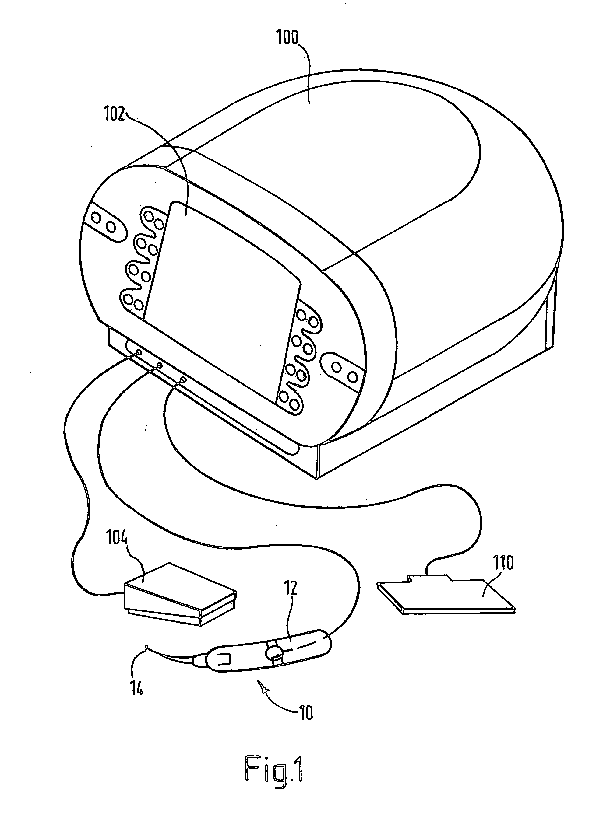
[0042]FIG. 1 shows the apparatus for a typical embodiment of an RF electrosurgical device for forming lesions in body tissue. The system comprises a controller 100 (including an RF power supply) with a user input and display panel 102. Also provided are a foot switch 104, an electrical grounding pad 110 and a probe 10 including a surgical handpiece 12 with a surgical electrode 14. The user input allows the user to input different parameters to affect lesion size, including treatment duration, and total energy delivery.

[0043] The controller 100 converts the low frequency electrical energy supplied by a wall connection (not shown) into the high frequency or RF energy necessary for surgery. The user input and display panel 102 displays relevant parameters and provides buttons and switches for user input to the control systems. The foot switch 104 connected to the controller provides means for switching the unit on and off. The surgical handpiece 12 is also connected to the controller ...


PUM


Abstract

Apparatus for forming a lesion in body tissue includes a probe having an active region with an electrode for contacting tissue to be treated, and an inactive region including an insulative sleeve around a portion of the electrode. The temperature of the inactive region is monitored using a temperature sensor. A controller supplies radio frequency energy to the electrode and samples signals from the temperature sensor. By performing a calculation using the sampled signals and a predetermined inactive region threshold temperature, and by adjusting the supplied radio frequency power, the inactive region of the probe can be maintained at or below an inactive region maximum temperature whilst the controller continues to supply radio frequency energy to the electrode. The probe has a second temperature sensor, mounted at a distal end of the electrode, the controller being configured to reduce the supplied radio frequency power when the electrode temperature reaches a predetermined maximum electrode temperature.

Description

FIELD OF THE INVENTION [0001] This invention relates to an electrosurgical system comprising an electrosurgical generator and a handpiece including electrosurgical electrodes. In particular, this invention is directed to electrosurgical systems capable of forming a lesion in body tissue, as is known for the treatment of various medical conditions including airway obstructions and sleep apnea. BACKGROUND OF THE INVENTION [0002] It is well known in the field of electrosurgery that there are two distinct tissue effects which can be achieved using radio frequency (RF) energy, depending on the temperature to which the tissue is raised. The first is the removal of tissue by vaporisation, in which the electrode or electrodes are subjected to relatively high temperatures (typically over 1000° C.). The second is the production of tissue necrosis without the removal of tissue in situ, and it is to this second type of system to which the present invention is directed. [0003] U.S. Pat. No. 5,84...

Claims


Application Information

Patent Timeline
Patent Type & Authority Applications(United States)
IPC IPC(8): A61B17/00A61B18/04A61B18/12A61B18/14
CPCA61B18/1206A61B18/1477A61B2017/00084A61B2018/00797A61B2018/00702A61B2018/00791A61B2018/00327
Inventor AMOAH, FRANCIS
Owner GYRUS ENT
Why Eureka
Try Eureka