
Jugular and subclavian access site dressing, anchoring system and method

Active Publication Date: 2005-12-15
TRI STATE HOSPITAL SUPPLY
View PDF10 Cites 7 Cited by
  • Summary
  • Abstract
  • Description
  • Claims
  • Application Information

AI Technical Summary

Benefits of technology

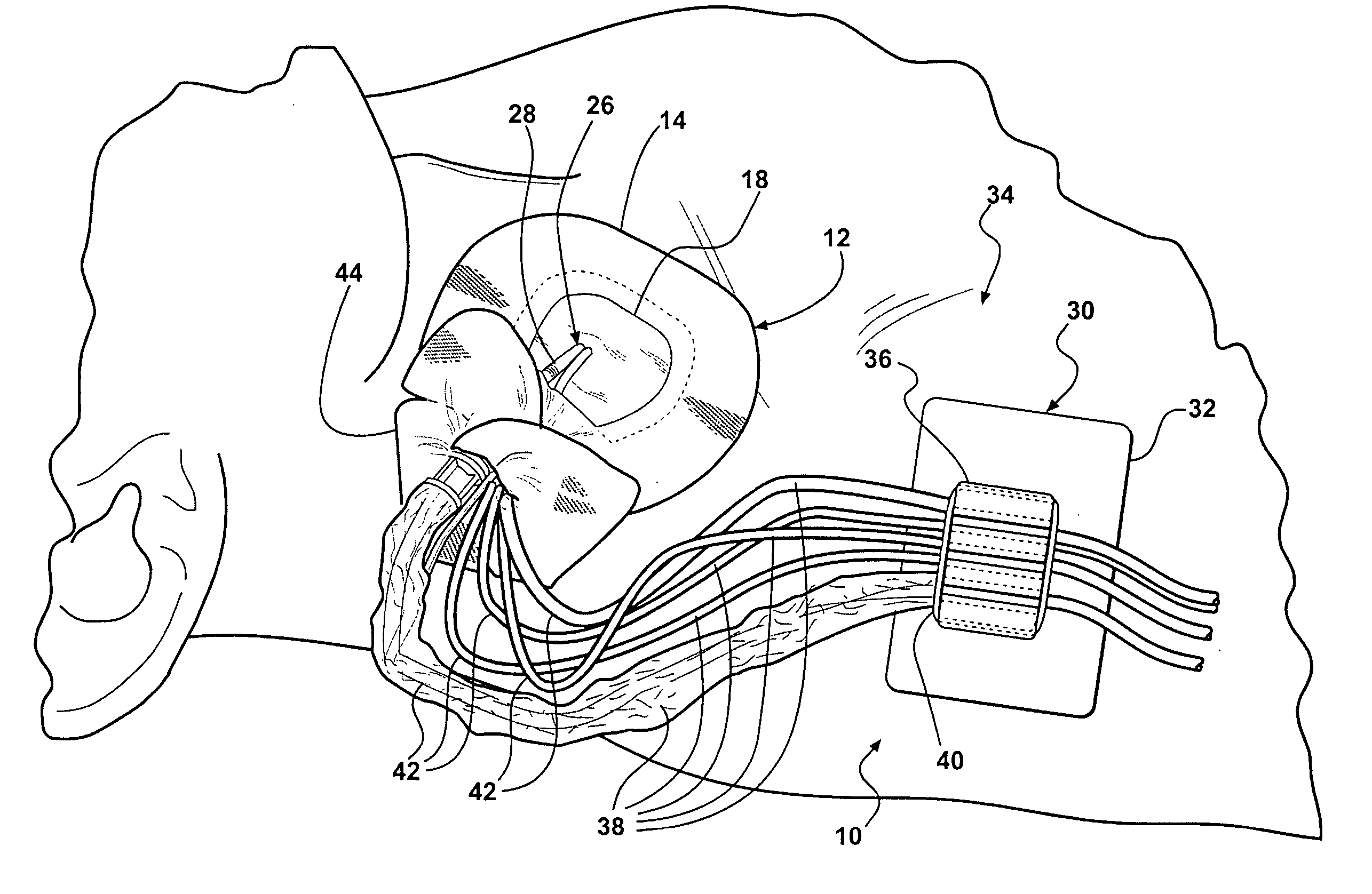
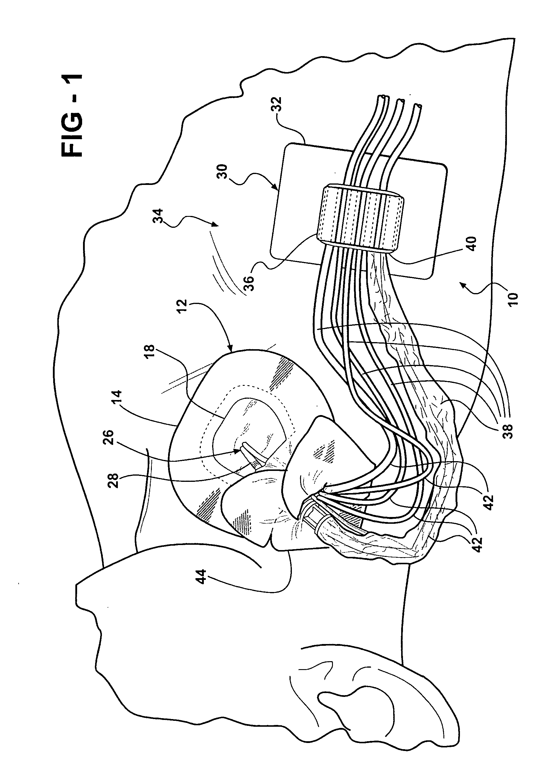
[0008] More particularly, the present invention provides an access site system that includes a dressing and a medical tubing anchor. The dressing may be a non-window or window dressing. Further, the connecting / medical tubes are arranged at and between the dressing and the medical tubing anchor in such a way as to eliminate the twisting and tearing forces exerted by the medical tubing. The dressing may wrap around the medical tubing, such as the side arm of an introducer sheath, to eliminate the tearing force caused by movement of the introducer sheath side arm. The medical tubing anchor eliminates the pulling force arising when dangling medical tubes catch on other objects or people's clothing by firmly anchoring the tubes. The present system may also provide for a strain relieving U-shaped bend in the medical tubing between the medical tubing anchor and the dressing. This stress relieving U-shaped bend flexes to and fro, up and down, left and right during any relative motion, in any direction between the anchor location and the catheter access site. It also eliminates the possibility of a tearing force arising at the dressing when a patient equipped with the present invention moves his or her head.
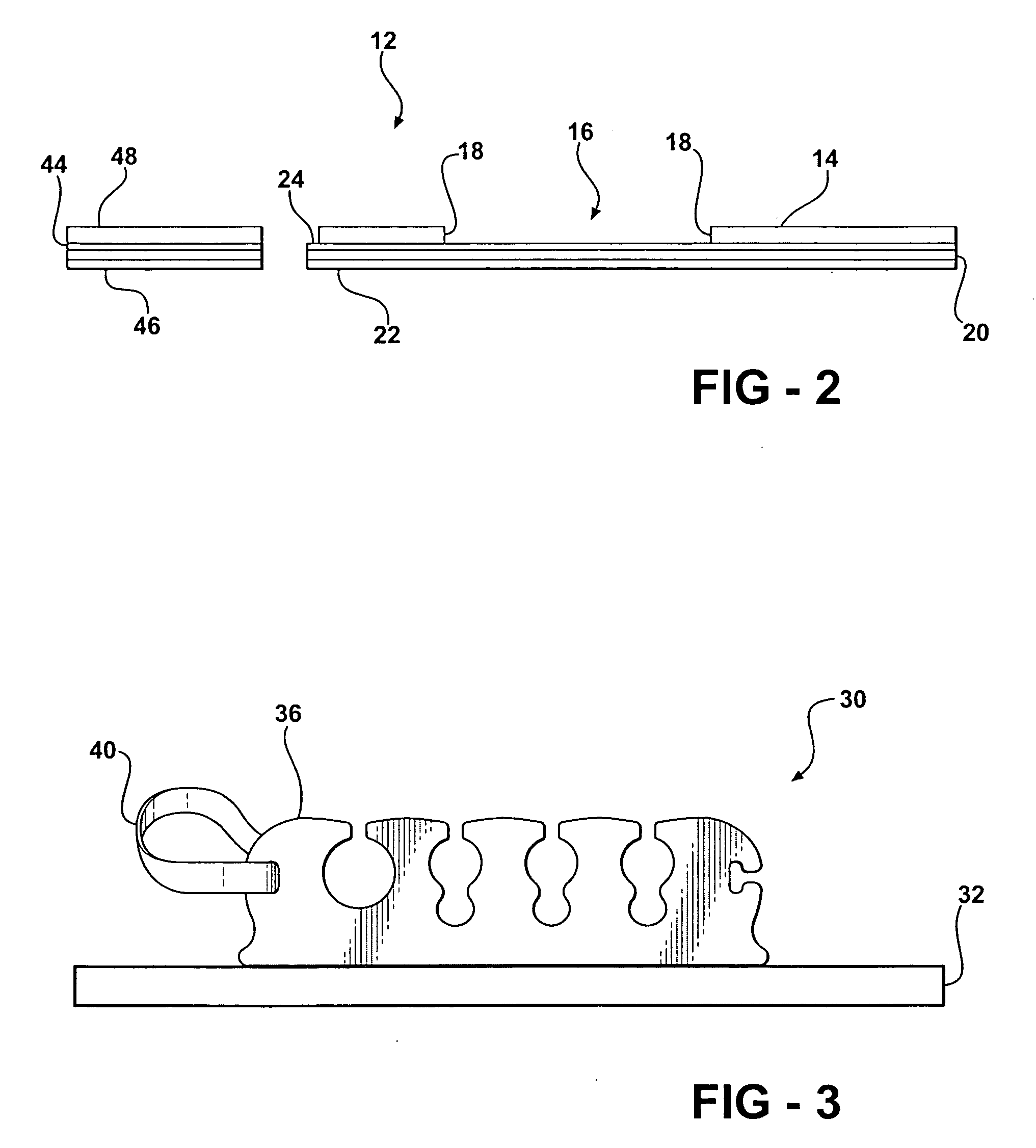
[0009] Specifically, a system adapted to tend a jugular or subclavian catheter and connecting tubing includes a dressing adapted to protect and cover a catheter access site and at least one catheter. The system further includes a medical tubing anchor including a base support adapted to mount to a patient's skin and a flexible anchor member on the base support adapted to receive and support the connecting tubing therein. The medical tubing anchor also includes a keeper on the anchor member for keeping tubing disposed in the anchor member from inadvertent release. The medical tubing anchor is spacedly disposed from the dressing. The connecting tubing connects with the catheter and the system, when rigged with adequate stress relieving tubing slack, allows for movement of the tubing without the tubing exerting a pulling force on the catheter.
[0012] In one embodiment, the method may further include the steps of: placing the adhesive side of the dressing onto an access site of a patient to secure medical tubing, to secure the catheter at the access site, and to protect the access site from contamination; inserting the medical tubing into the medical tubing anchor by squeezing and holding ends of the flexible anchor member and the base support to open tubing slots in the flexible anchor member; mounting the base support of the tubing anchor onto the skin of the patient on a side of the patient's body where the dressing is located such that the tubing anchor is spacedly disposed from the dressing; adjusting the medical tubing by squeezing and holding ends of the flexible anchor member and the base support of the medical tubing anchor to open the tubing slots so that the length of the medical tubing located between the dressing and the medical tubing anchor is greater than the distance between the access site and the medical tubing anchor, thereby causing the medical tubing to bend in a U shape between the dressing and the medical tubing anchor; and securing the medical tubing in the medical tubing anchor.
[0013] In an alternative embodiment, the method may further include the steps of: placing the adhesive side of the dressing onto an access site of a patient to secure medical tubing, to secure the catheter at the access site, and to protect the access site from contamination; mounting the base support of the tubing anchor onto the skin of the patient on a side of the patient's body where the dressing is located such that the tubing anchor is spacedly disposed from the dressing; after mounting the base support of the tubing anchor onto the skin, inserting the medical tubing into the medical tubing anchor by squeezing and holding ends of the flexible anchor member and the base support to open tubing slots in the flexible anchor member; after inserting the medical tubing into the medical tubing anchor, adjusting the medical tubing by squeezing and holding ends of the flexible anchor member and the base support of the medical tubing anchor to open the tubing slots so that the length of the medical tubing located between the dressing and the medical tubing anchor is greater than the distance between the access site and the medical tubing anchor, thereby causing the medical tubing to bend in a U shape between the dressing and the medical tubing anchor; and securing the medical tubing in the medical tubing anchor.
[0015] In yet another embodiment, the method may further include the steps of: placing the adhesive side of the dressing onto an access site of a patient to secure medical tubing, to secure the catheter at the access site, and to protect the access site from contamination; mounting the base support of the tubing anchor onto the skin of the patient on a side of the patient's body where the window dressing is located such that the tubing anchor is spacedly disposed from the dressing; inserting the medical tubing into the medical tubing anchor; adjusting the medical tubing so that the length of the medical tubing located between the dressing and the medical tubing anchor is greater than the distance between the access site and the medical tubing anchor, thereby causing the medical tubing to bend in a U shape between the dressing and the medical tubing anchor; and securing the medical tubing in the medical tubing anchor. The step of adjusting the medical tubing further may include adjusting the medical tubing such that the tubing is slack between the dressing and the tubing anchor.
[0016] Further, the step of providing a window dressing may further include providing a window dressing including a closure member having an adhesive side and an opposite non-adhesive side, wherein the closure member is adapted to overlie and close an end of the layer member. The method then may include the steps of: placing the adhesive side of the transparent film layer of the dressing onto an access site of a patient to secure medical tubing, to secure the catheter at the access site, and to protect the access site from contamination; placing the adhesive side of the closure member of the dressing onto an end of the layer member; mounting the base support of the tubing anchor onto the skin of the patient on a side of the patient's body where the window dressing is located such that the tubing anchor is spacedly disposed from the dressing; inserting the medical tubing into the medical tubing anchor; adjusting the medical tubing so that the length of the medical tubing located between the dressing and the medical tubing anchor is greater than the distance between the access site and the medical tubing anchor, thereby causing the medical tubing to bend in a U shape between the dressing and the medical tubing anchor; and securing the medical tubing in the medical tubing anchor.

Problems solved by technology

It is known in the art relating to jugular and subclavian access site dressings used during and shortly after medical procedures such as open heart or thoracic surgery that the dressings have a tendency to become dislodged from the patient's skin, thereby exposing the access site to harmful bacteria and other pathogens.
Further, in known prior art access site dressings, the side arm of an introducer sheath, which is at a 60 or 90 degree angle to the introducer sheath secured by the dressing, and other medical tubing and connectors, and stresses imparted thereto, can cause separation of the dressing from a patient's skin.
This is undesirable because a patient will often move his or her head and the dressing is ineffective if it releases from the patient's skin.
Also, the side arm tends not to stay flat against a patient's body but instead projects or lifts up off the body, which further loosens the dressing.
Even more, other medical tubing located at the access site, such as central venous catheters (CVC's), often dangle insecurely from the jugular access site.
When this tubing gets caught on other objects, such as medical instruments or medical personnel's clothing, the force of the tubing catching also can tear the dressing from the access site.
Further, known prior art access site dressings have not been able to accommodate all of the potential combinations of medical tubing that can exist at a jugular access site.
The prior art dressings have been able to accommodate some of these combinations but no prior art dressing has been suitable for use with all of these possible combinations.
Even those which do accommodate a few combinations often come loose from the skin within a day or less, and thus serve no useful clinical purpose.
Prior art access site dressings are capable of being used on either the right hand side or the left hand side, however, they do not secure and protect the catheters and access sites, and come loose prematurely on either the left or right side.
Furthermore, prior jugular and subclavian access site systems have not been able to suitably anchor the medical tubing that leads up to the access site.
As previously mentioned, this is problematic for the reason that this tubing tends to dangle from the patient and can be pulled if caught on other objects or people.
This not only puts a tearing pressure on the jugular access site dressing, but also can pull the medical tubing away from the access point on the patient's skin.
Another shortcoming of prior anchoring methods used in jugular access site systems is that the tubing anchors are not designed in a way to clear the gown of the patient worn by the patient during and after the operating procedure.
When a patient is wearing a hospital gown, the gown interferes with the tubing and / or the anchoring device used to anchor the tubing located at the jugular access site.
Finally, prior medical tubing anchors are not able to anchor more than one tube at a time.

Method used


Image

  • Jugular and subclavian access site dressing, anchoring system and method
  • Jugular and subclavian access site dressing, anchoring system and method
  • Jugular and subclavian access site dressing, anchoring system and method

Examples


Embodiment Construction

[0022] Referring now to the drawings in detail, numeral 10 generally indicates a system adapted to tend a jugular or subclavian catheter and connecting tubing that prevents inadvertent release of catheter(s) and dressing from a patient's body. The system can accommodate various sizes and combinations of connecting / medical tubing utilized at a jugular or subclavian access site, such as an introducer sheath with a side arm, an introducer sheath with a side arm in combination with a pulmonary artery catheter, and either a single, double, or triple lumen central venous catheter. Moreover, the system can be utilized on either a right-hand side or a left-hand side access site and the system eliminates the forces that cause the dressing to tear away from the skin around the access site.

[0023] As shown in FIGS. 1 through 3, a preferred system adapted to tend a jugular or subclavian catheter and connecting tubing 10 includes a window dressing 12 of the type including a layer member 14 havin...


PUM


Abstract

A system adapted to tend a jugular or subclavian catheter and connecting tubing includes a jugular and subclavian access site dressing and a medical tubing anchor. The dressing is adapted to protect and cover a catheter access site and the catheter. The medical tubing anchor is spacedly disposed from the dressing. The connecting tubing connects with the catheter and the system allows for movement of the tubing without the tubing exerting a pulling force on the catheter.

Description

TECHNICAL FIELD [0001] This invention relates to dermal dressings and medical tubing anchors for the protection of jugular or subclavian catheter access sites, and more particularly to a system for protecting, anchoring, and securing introducer sheaths and catheters at and around a jugular or subclavian access site. BACKGROUND OF THE INVENTION [0002] It is known in the art relating to jugular and subclavian access site dressings used during and shortly after medical procedures such as open heart or thoracic surgery that the dressings have a tendency to become dislodged from the patient's skin, thereby exposing the access site to harmful bacteria and other pathogens. Some common forms of traditional access site dressings include self-adherent protective bandage tape or clear film product alternatives that use non-sensitizing hypoallergenic adhesives to cover all or part of the indwelling catheter access site. Some dressings also combine non-woven tape and absorbent gauze-like materia...

Claims


Application Information

Patent Timeline
IPC IPC(8): A61M5/32A61M25/02
CPCA61M2025/028A61M25/02
Inventor PROPP, DONALD J.
Owner TRI STATE HOSPITAL SUPPLY
Learn More