Ophthalmic clip and associated surgical method

a surgical method and clip technology, applied in the field of ophthalmic clip and associated surgical method, can solve the problems of gradual and irreversible loss of vision, and achieve the effect of extending the healing process

Inactive Publication Date: 2006-01-05
CARO NICHOLAS C
View PDF29 Cites 18 Cited by
  • Summary
  • Abstract
  • Description
  • Claims
  • Application Information

AI Technical Summary

Benefits of technology

[0010] In a further aspect of the invention, a method for applying the clip is also provided. Pursuant to the method, the location of the ciliary muscles in the eye are determined, and an incision is made in the conjunctiva to gain access to the sclera overlying the ciliary muscles. The incision is opened to expose the sclera and opposed pockets are made in the surface of the sclera for receiving the feet of a clip, as described above. The clip is attached to the eye by introducing the feet of the clip into the pockets made in the sclera, with the downward curve of the body of the clip compressing the surface of the sclera inwardly. The conjunctiva is then closed over the clip. Optionally, a fibrin adhesive may be applied to the conjunctiva after it is closed over the clip in order to expedite the healing process.

Problems solved by technology

However, the inconveniences associated with eyeglasses and contact lenses have prompted investigation into, and the development of, surgical techniques aimed at correcting presbyopia.
Primary open angle glaucoma occurs when the eye's drainage canals become clogged over time, causing a gradual and irreversible loss of vision.

Method used


Image

  • Ophthalmic clip and associated surgical method
  • Ophthalmic clip and associated surgical method
  • Ophthalmic clip and associated surgical method

Examples


Embodiment Construction

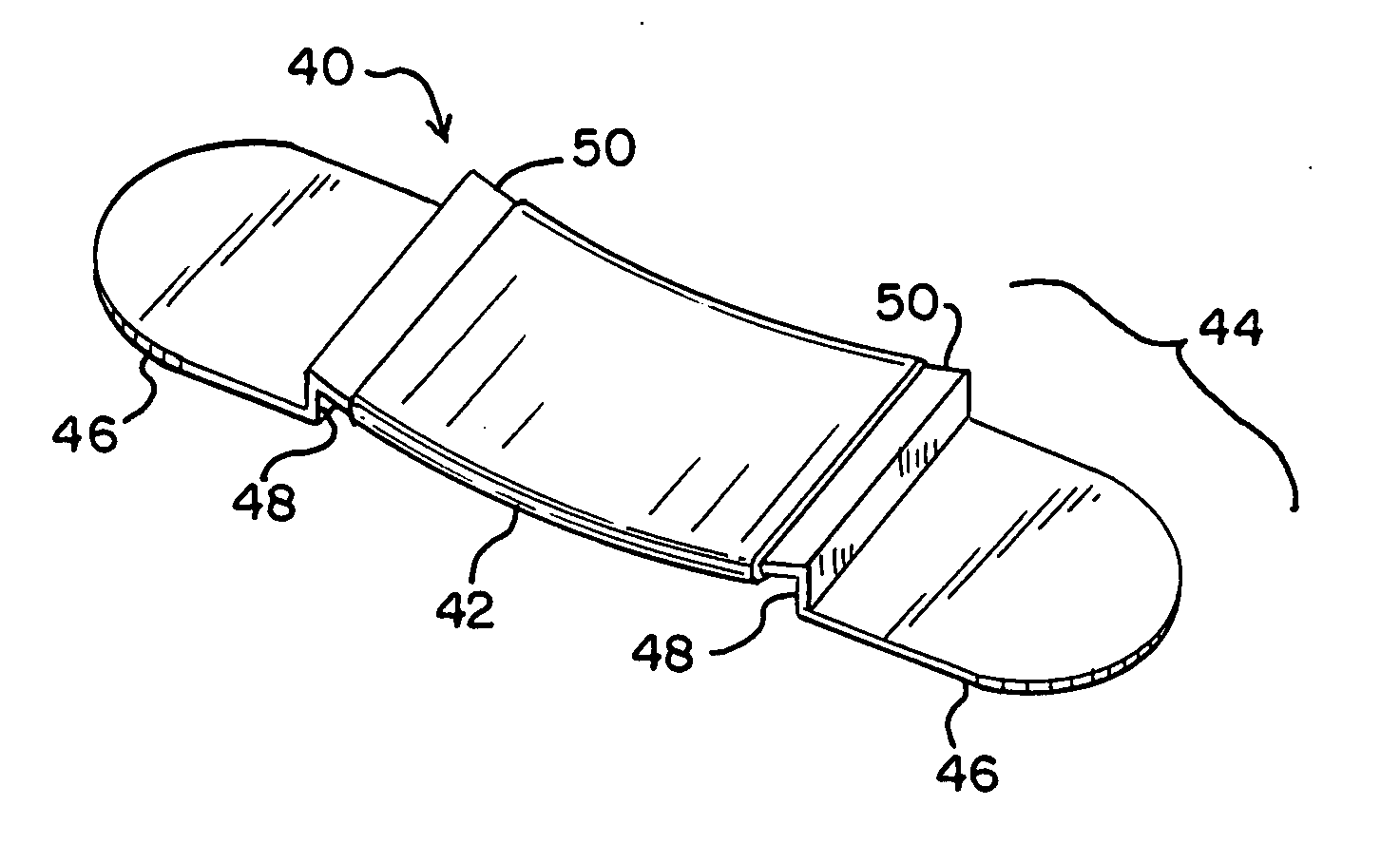
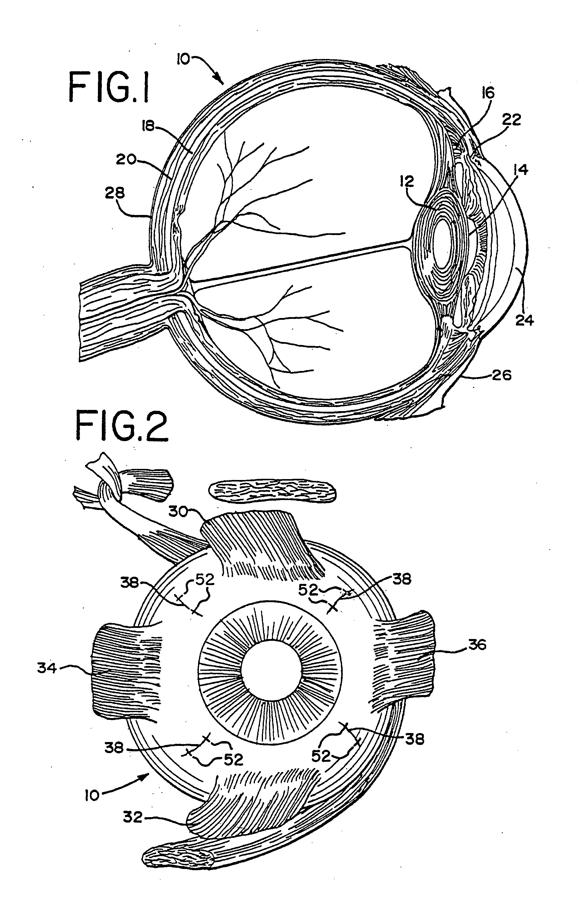
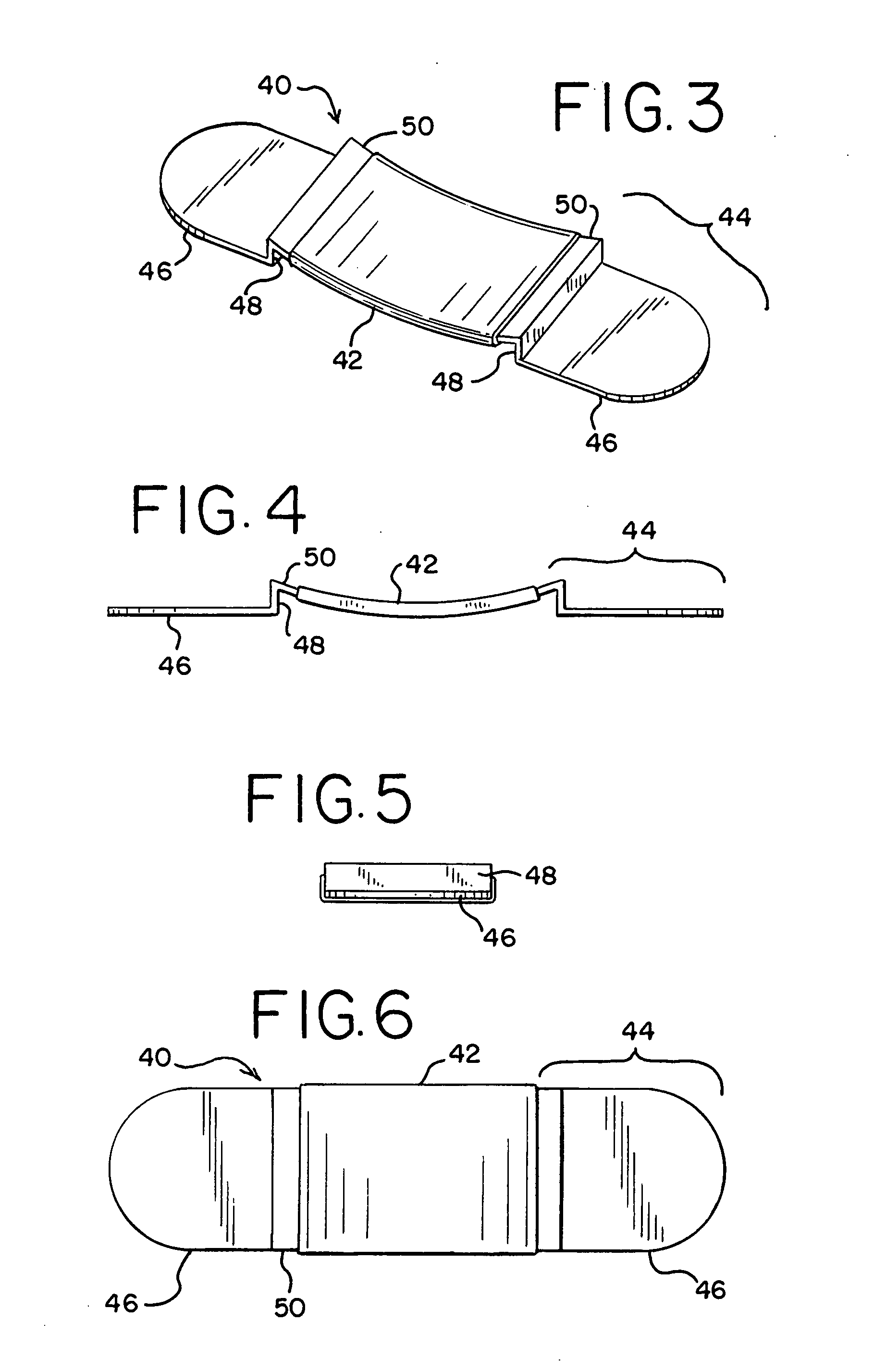
[0017] The method that utilizes the clip of the present invention is based upon the theory that the cause of presbyopia is the failure of the ciliary body to adjust the lens diameter in order to focus images onto the retina for close objects. The ciliary muscles change the lens diameter by using the sclera as support or fixation structure. As the sclera of the eye weakens due to age, the ciliary muscles lack the support needed in order to alter the lens diameter for focusing on close objects. Thus, in order to allow the ciliary muscle to alter the lens diameter to see close objects, the sclera must be supported or reinforced. Accordingly, an improved clip for reinforcing the sclera is provided, so as to form a stronger and more stable support for the ciliary muscles. The clip of the present invention accomplishes this by compressing or depressing the sclera. In effect, the sclera is strengthened, and the ciliary muscles are then able to again function properly to provide near vision...


PUM


Abstract

A method and clip for treating presbyopia and / or open angle glaucoma in which the sclera is supported or reinforced, while substantially maintaining the special relationship between the ciliary muscle and the lens. The method includes making an incision in the conjunctiva to gain access to the sclera overlying the ciliary muscle. The Tenon's capsules are moved laterally to expose the sclera, and opposed shallow pockets are made in the sclera. A clip, or series of clips, is provided having two opposed feet, which are received in the opposed pockets in the sclera. The Tenon's capsules are then slid over the clip and the conjunctiva is closed.

Description

[0001] The present invention is directed to an ophthalmic clip for treating vision disorders, such as presbyopia and / or glaucoma and an associated surgical method for application of the clip. BACKGROUND OF THE INVENTION [0002] Presbyopia is a vision disorder associated with aging resulting from the failure of the accommodation mechanism of the eye. The accommodative mechanism is driven principally by parasympathetic inervation of the ciliary smooth muscle. In the non-presbyopic eye, this causes the muscle to slide forward in a unified manner and produces an inward movement of the muscle. The result is a reduction in the diameter of the ciliary muscle collar that instigates a series of events leading to an ability to see near objects clearly. [0003] Presbyopia is most frequently treated by the use of reading glasses, bifocals, and progressive multi-focal contact lenses. However, the inconveniences associated with eyeglasses and contact lenses have prompted investigation into, and the...

Claims


Application Information

Patent Timeline
Patent Type & Authority Applications(United States)
IPC IPC(8): A61B17/08
CPCA61B17/122A61F9/00781A61F9/007
Inventor CARO, NICHOLAS C.
Owner CARO NICHOLAS C
Why Eureka
Try Eureka