Method for imaging in a medical interventional procedure by image subtraction

Inactive Publication Date: 2006-02-02
SIEMENS AG
View PDF5 Cites 54 Cited by
  • Summary
  • Abstract
  • Description
  • Claims
  • Application Information

AI Technical Summary

Benefits of technology

[0008] An object of the present invention is to provide a method for imaging in an interventional medical procedure, with which movement artifacts due to movements

Problems solved by technology

First, the background is no longer correctly subtracted such that image artifacts are created.
Additionally, it may occur that the position of the instrument obtained in the image is not correct relative to the shown blood vessel.
This error can lead, for example, to the catheter showing outside of the vessel in the image although it is actually located within the vessel.
In the extreme case, such fa

Method used


Image

  • Method for imaging in a medical interventional procedure by image subtraction
  • Method for imaging in a medical interventional procedure by image subtraction
  • Method for imaging in a medical interventional procedure by image subtraction

Examples


Example

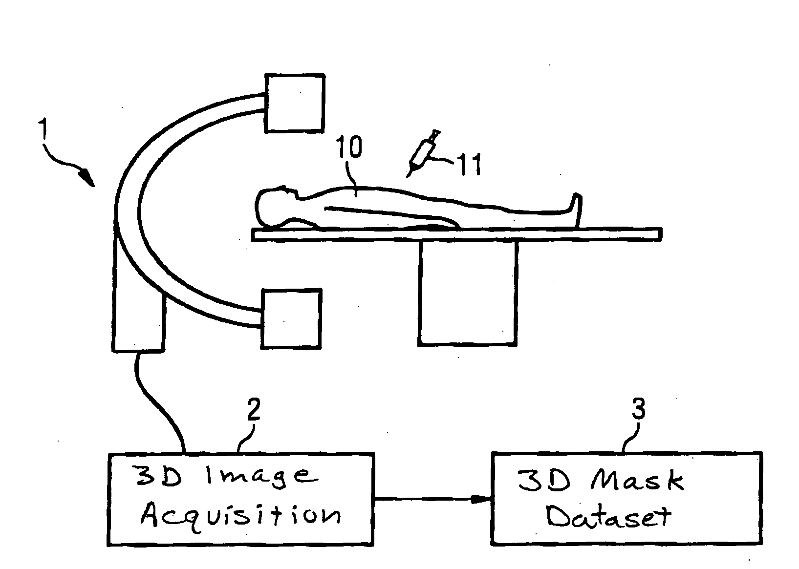
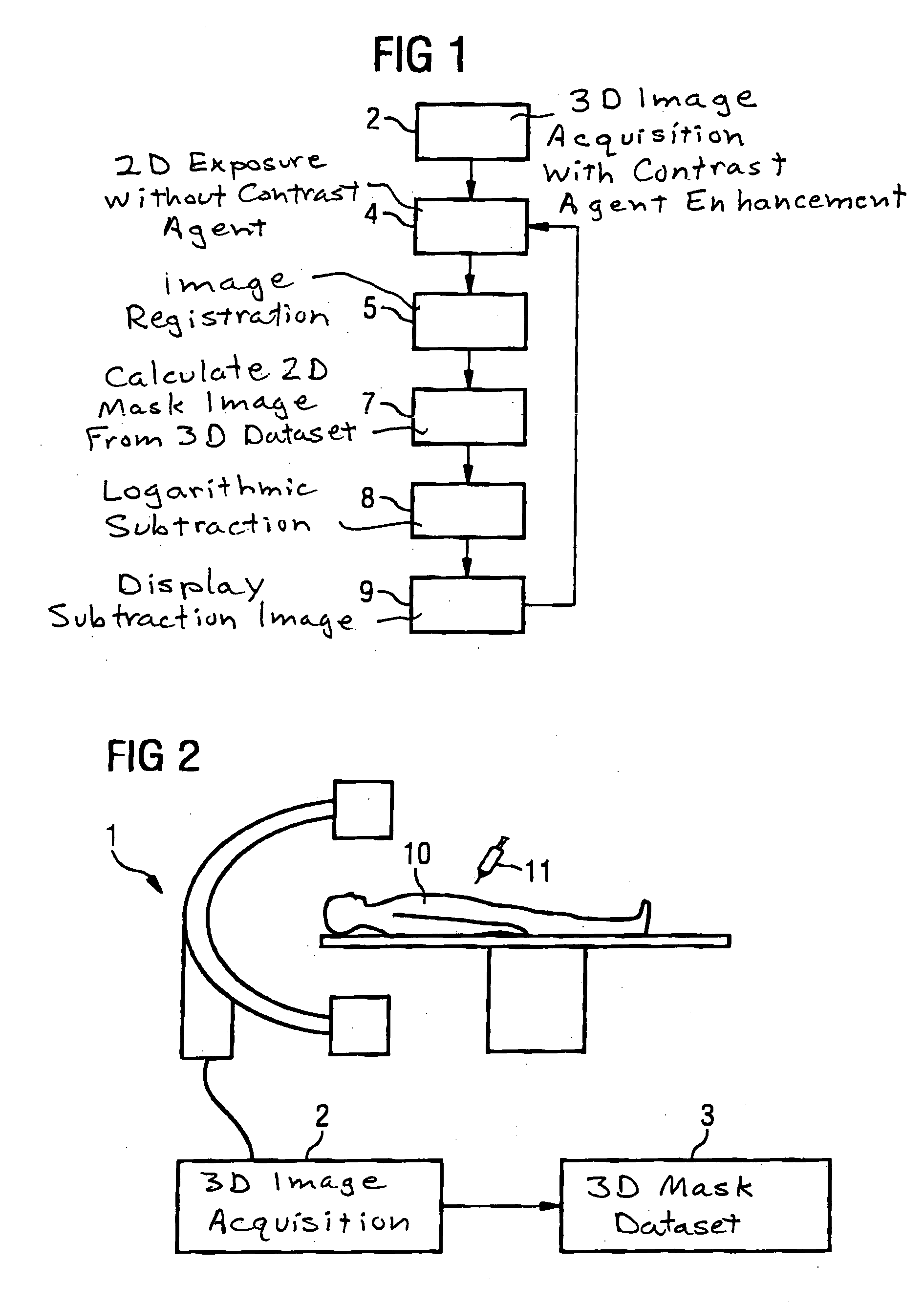
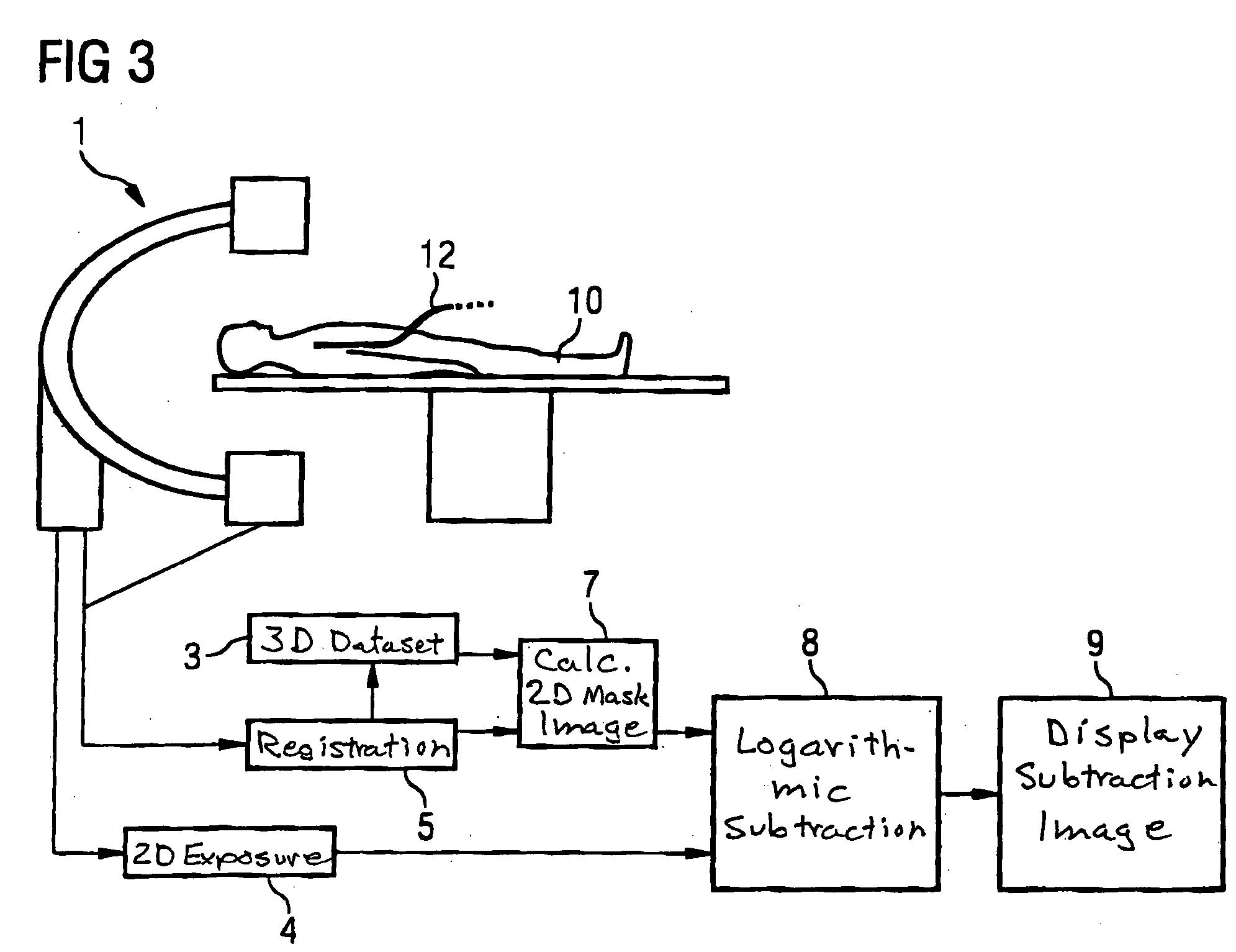
[0018]FIG. 1 shows an exemplary flowchart for implementation of the inventive method for imaging in an interventional procedure on a patient. A 3D volume data set of the patient, or at least of a body region of the patient, is initially generated with contrast agent injection. The acquisition of this data set can ensue either with an x-ray computed tomography system or with an angiography system with a C-arm.

[0019] The acquisition of the 3D volume data of a body region of interest of a patient 10 with a C-arm angiography system 1 is illustrated in FIG. 2. the 3D image acquisition 2 is implemented after a contrast agent injection 11. The image data acquired by image reconstruction from the measurement values of the 3D image acquisition 2 are stored as a 3D mask data set 3 and thus are available for later further processing. This first step of this acquisition of a 3D volume or mask data set can ensue with an x-ray dose that is reduced relative to that for diagnostic 3D rotation angi...


PUM


Abstract

In a method for imaging in a medical interventional procedure, in which an image of structures (in particular vessels) of a body region is generated during the procedure, image data of a first 2D x-ray image of the body region that is generated with a contrast agent enhancement of the structures, and image data of at least one second 2D x-ray image of the body region that is acquired without contrast agent enhancement of the structures, are subtracted from one another. The image data of the first 2D x-ray image are calculated from a 3D volume data set of a computed tomography exposure of the body region. Movement artifacts due to movements of the patient between the generation of a mask image and subsequent fluoroscopy images thus are prevented or at least reduced without time-consuming user interaction.

Description

BACKGROUND OF THE INVENTION [0001] 1. Field of the Invention [0002] The present invention concerns a method for imaging in a medical interventional procedure, in particular of the type using a pathfinder technique in which an image of structures (in particular vessels) of a body region is generated during the procedure, wherein image data of a first 2D x-ray image of the body region are generated with a contrast agent enhancement of the structures, and image data of at least one second 2D x-ray image of the body region are acquired without contrast agent enhancement of the structures, the images being subtracted from one another. [0003] 2. Description of the Prior Art [0004] The technique known as the pathfinder technique, also known by the term road mapping, is used in the selective catheterization of vessels in the framework of an interventional treatment. In these vessel interventions, the current position of an x-ray-absorbing catheter or guide wire is shown in a two-dimensional...

Claims


Application Information

Patent Timeline
IPC IPC(8): H05G1/64
CPCA61B6/12A61B6/463A61B6/5235A61B6/481A61B6/504A61B6/466
Inventor BOESE, JAN
Owner SIEMENS AG
Why Eureka
Try Eureka