Enhancement of sensitivity of fluorophore mediated biosensing and bioimaging

a fluorophore and biosensing technology, applied in the field of biosensing and bioimaging, can solve the problems of limiting the effectiveness of these fluorophores in biosensing/bioimaging applications, the level of biomarkers in a sample is usually extremely low, etc., and achieves enhanced fluorescence emission and fluorescence emission enhancement

Inactive Publication Date: 2006-02-02
UNIV OF LOUISVILLE RES FOUND INC
View PDF10 Cites 7 Cited by
  • Summary
  • Abstract
  • Description
  • Claims
  • Application Information

AI Technical Summary

Benefits of technology

The invention provides a device and method for improving the fluorescence emission in biosensors that use fluorophores. This is achieved by exposing the fluorescence complex to an enhancing agent, such as a nanometal particle or an organic solvent, which can increase the brightness and duration of the fluorescence signal. This improves the sensitivity and accuracy of the biosensors, making them more reliable and useful in various applications.

Problems solved by technology

The technical problem addressed in this patent is the need for highly sensitive and specific sensors to detect biomarkers in bio-samples, which are present in low levels and can be structurally homologous to other biomolecules. The use of fluorophores in biosensing and bioimaging applications is limited due to their low quantum yield caused by non-radiative decay, especially by self-quenching. The patent aims to enhance the sensitivity of fluorophore-mediated biosensing and bioimaging applications by improving the fluorescence of these fluorophores.

Method used


Image

  • Enhancement of sensitivity of fluorophore mediated biosensing and bioimaging
  • Enhancement of sensitivity of fluorophore mediated biosensing and bioimaging
  • Enhancement of sensitivity of fluorophore mediated biosensing and bioimaging

Examples


Embodiment Construction

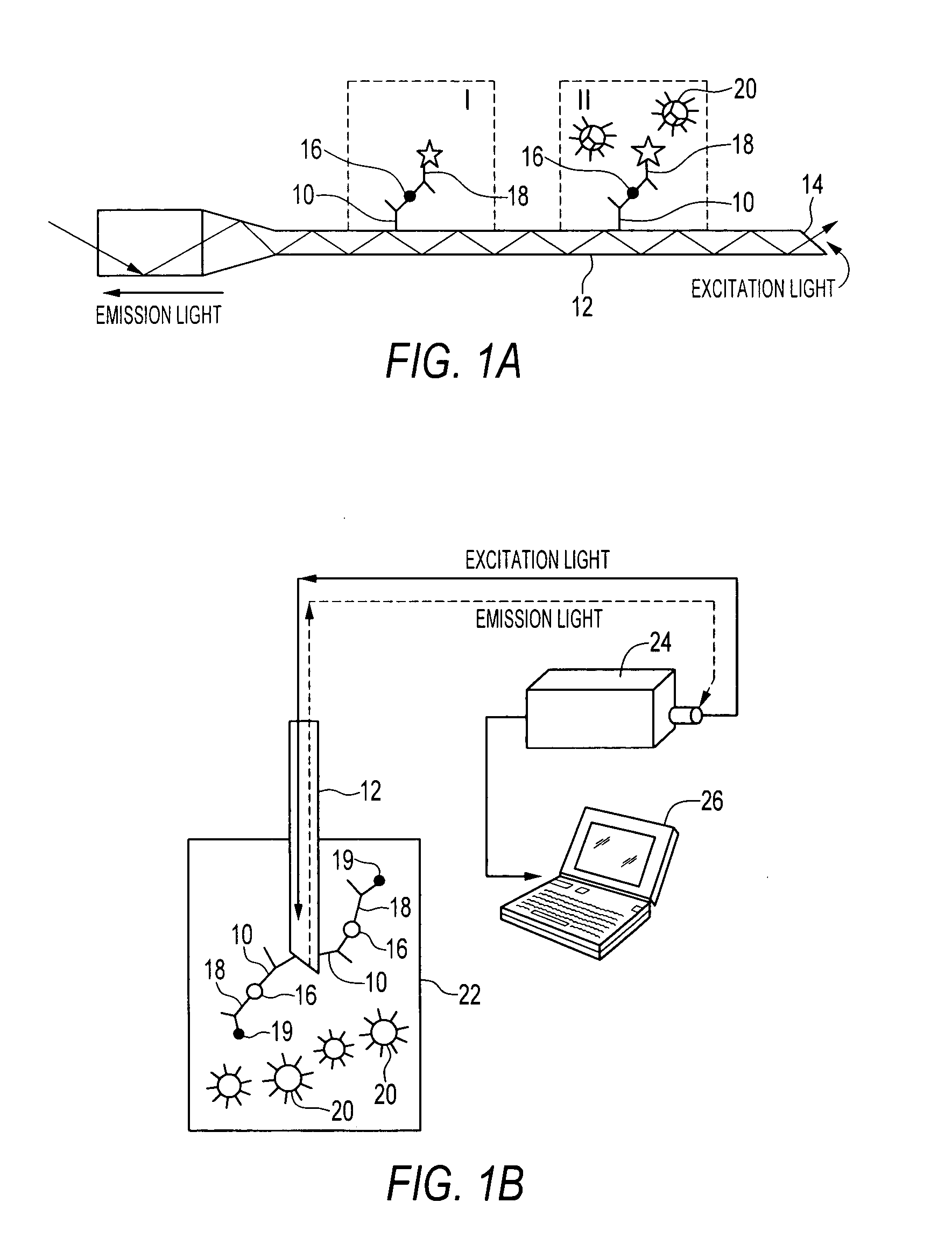
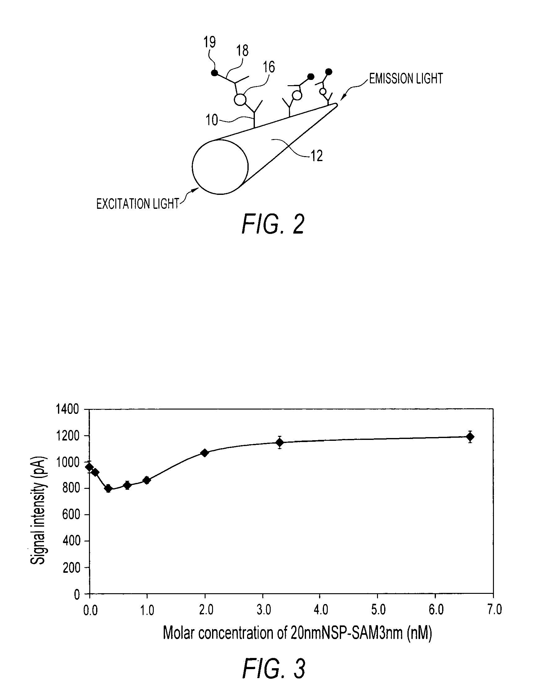
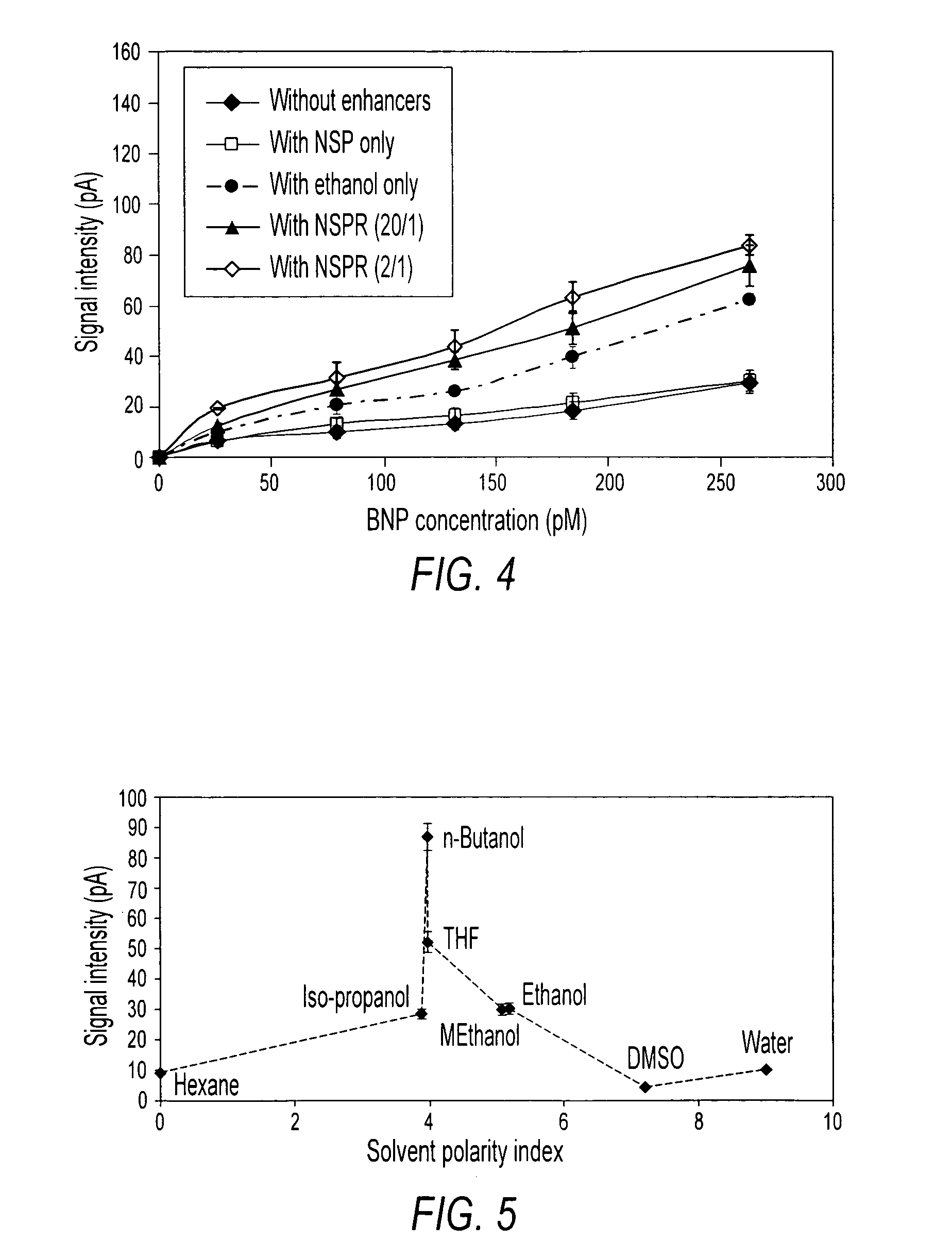
[0023] Fluorescence of a fluorophore is normally limited by self-quenching of the free electrons in the fluorophore, thereby limiting the sensitivity of biosensors with which they are used. Embodiments of the invention reduce or eliminate this self-quenching, which enhances fluorescence emission of the fluorophore, thereby enhancing the ability to detect the presence of and quantify a target analyte. Fluorescence of some fluorophores and of fluorophore mediated biosensors is also enhanced significantly by biocompatible organic solvents. Devices and methods of the invention include exposing a fluorophore-mediated sandwich immunoassay to an enhancing agent, where the enhancing agent may be a nanometal particle having a layer of predetermined thickness, an organic, biocompatible solvent, or a combination of both a nanometal particle having a layer of predetermined thickness and an organic, biocompatible solvent.

[0024] Electrons of fluorophores that are normally involved in self-quench...


PUM


Abstract

A method of enhancing fluorescence emission in a fluorophore-mediated sensing, biosensing, imaging, and bioimaging. An example of biosensing is a fluorophore-mediated sandwich immunoassay with a 1° monoclonal antibody against a target analyte and a fluorophore-linked 2° monoclonal antibody, exposing the immunoassay to an enhancing agent, applying excitation light to the immunoassay, and measuring an emission signal from the immunoassay.

Description


Claims


Application Information

Patent Timeline
Owner UNIV OF LOUISVILLE RES FOUND INC
Why Eureka
Try Eureka