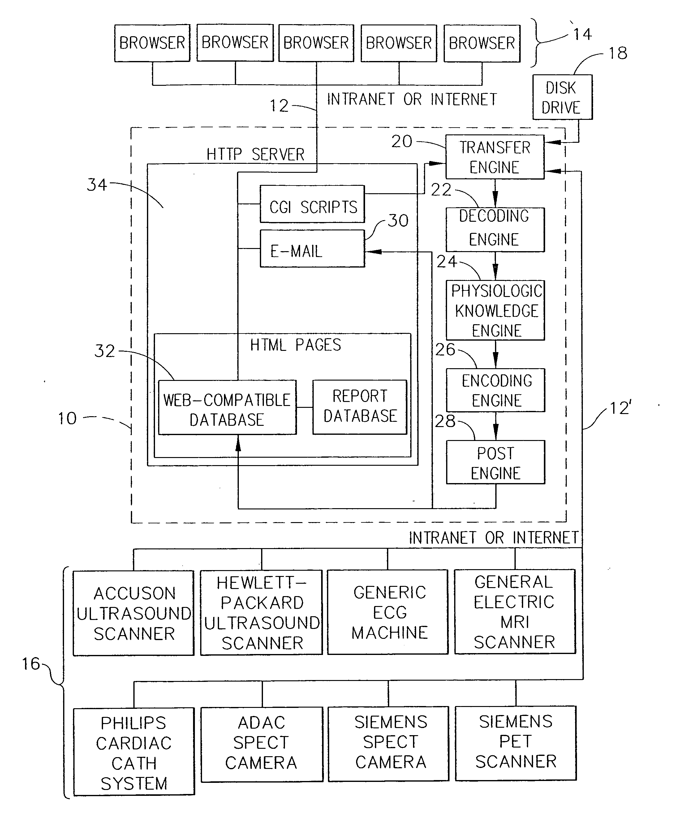
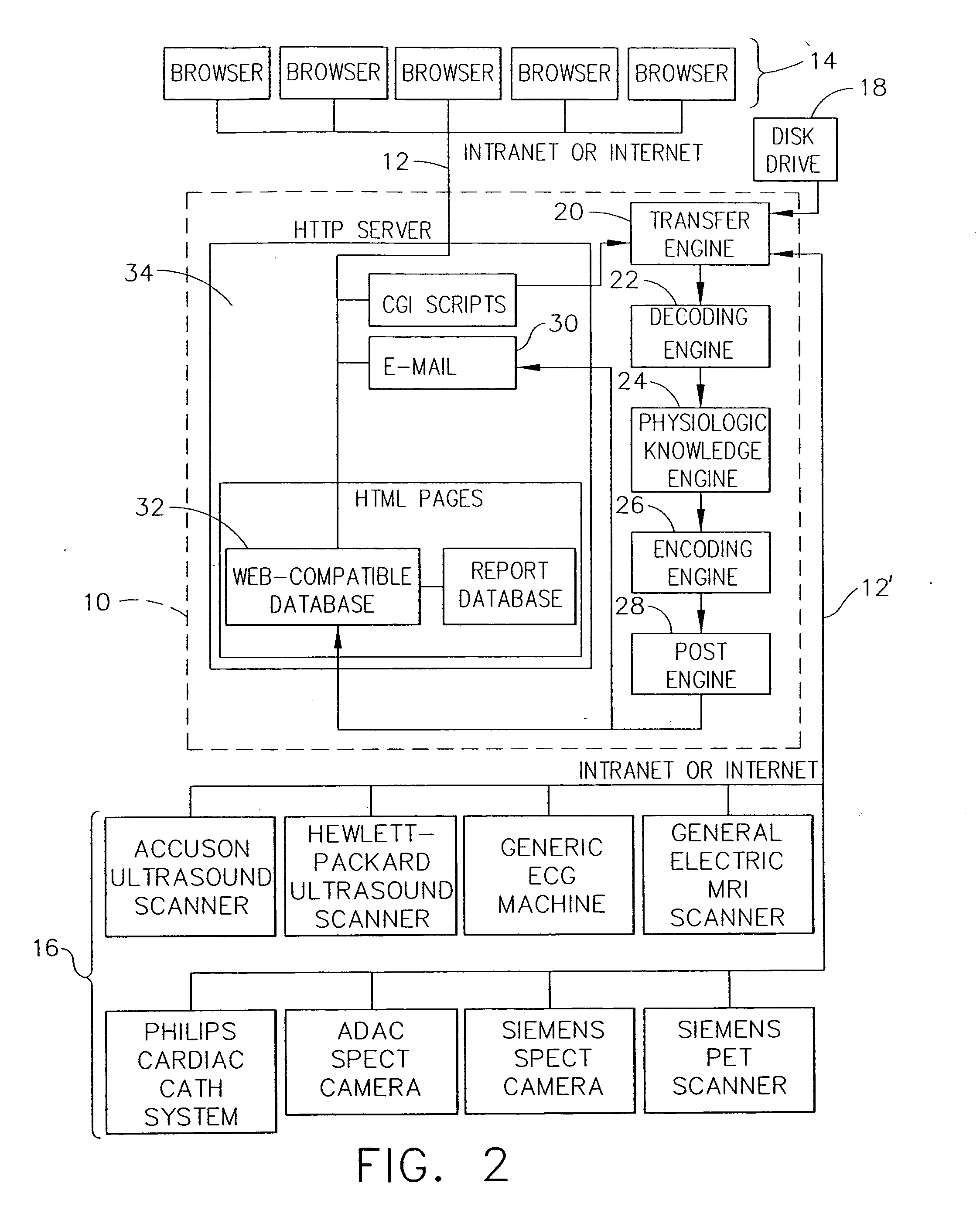
In certain situations, however, the particular manner in which the images are made available to physicians and their patients introduces obstacles to timely and accurate diagnoses of
disease.
These obstacles generally relate to the fact that each manufacturer of a medical imaging
system uses different and proprietary formats to store the images in digital form.
Although it is typically possible to “export” the images from a proprietary
workstation to an industry-standard format such as “
Digital Imaging Communications in
Medicine” (
DICOM), Version 3.0, several limitations remain as discussed subsequently.
The specialist physician, however, typically has not performed a clinical history and
physical examination of the patient and often is not aware of the patient's other test results.
Although this approach does allow for expert interpretation of the images by the specialist physician, several limitations are introduced for the
primary physician and for the patient, such as, for example: (1) The
primary physician does not see the images unless he travels to another department and makes a request; (2) It is often difficult to find the images for viewing because there typically is no formal procedure to accommodate requests to show the images to the
primary physician; (3) Until the written report is forwarded to the primary physician's office, it is often difficult to determine if the images have been interpreted and the report generated; (4) Each proprietary
workstation requires training in how to use the
software to view the images; (5) It is often difficult for the primary physician to find a
technician who has been trained to view the images on the proprietary
workstation; (6) The workstation
software is often “upgraded” requiring additional training; (7) The primary physician has to walk to different departments to view images from the same patient but different modalities; (8) Images from the same patient but different modalities cannot be viewed side-by-side, even using proprietary workstations; (9) The primary physician cannot show the patient his images in the physician's office while explaining the diagnosis; and (10) The patient cannot transport his images to another physician's office for a second opinion.
A similar barrier to accessing
medical information exists for physicians interested in providing real-time training to other physicians, and for physicians seeking real-time advice from other physicians based on the results of medical imaging.
Generally, such training and / or advice can only be obtained by having the second physician physically present in the same room or in an immediately adjacent room while the first physician performs the imaging procedure.
Teleradiology does not, however, allow for the examination of the images from any site other than the central location, precluding examination of the images by the primary physician and the patient.
One such limitation is the bandwidth of current Internet connections which, because of the
large size of medical images, result in transfer times which are unacceptably long.
The problem of bandwidth can be addressed by compressing the image data before transfer, but compression typically involves loss of
diagnostic information.
In addition, due to the size of the images the time required to process image data from an original format to a format which can be viewed by Internet browsers is considerable, meaning that systems designed to create Web Pages “
on the fly” introduce a
delay of seconds to minutes while the person requesting to view the images waits for the data to be processed.
Workstations allow images to be reordered or placed “side-by-side” for viewing, but again, an Internet
system would have to create new Web Pages “
on the fly” which would introduce further delays.
There are several disadvantages to using “
Java” to manipulate the image data.
First, the user must wait additional time while the “
Java” code is downloaded.
Third, the user must wait while the “
Java” code processes the image data, which is slow because the image files are large.
Fourth, “Java” code is relatively new and often causes browsers to “
crash.” Finally, due to the “crashing” problem “Java” programmers typically only test their code on certain browsers and computers, such as Microsoft Explorer on a PC, precluding widespread use by owners of other browsers and other computer platforms.
The approach of Wood, however, creates Web Pages “
on the fly,” meaning that the user must wait for the
image processing to complete.
In addition, the approach of Wood is limited to
ultrasound images generated by scanners manufactured by a single company, and does not enable viewing of images from modalities other than
ultrasound.
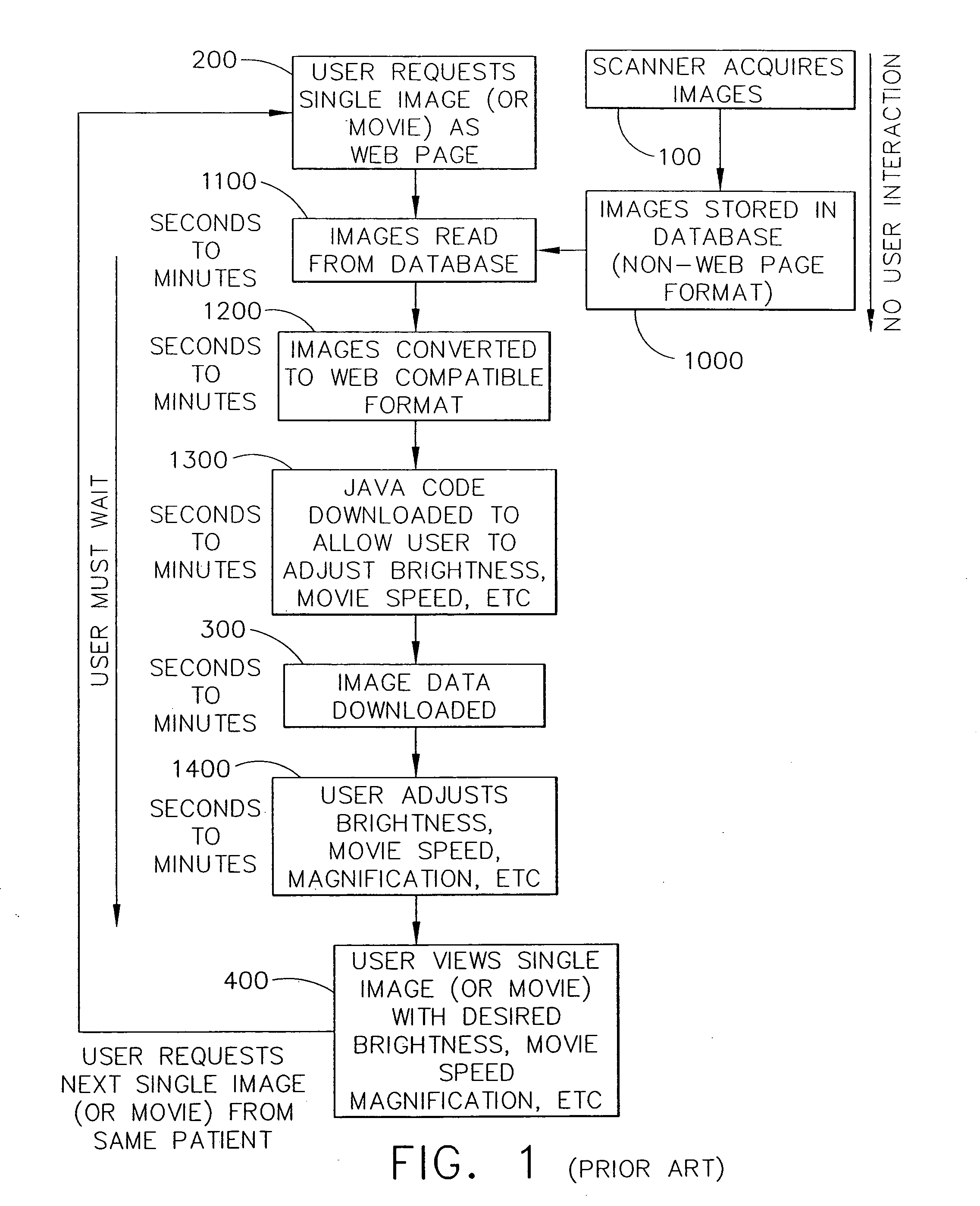
As can be seen in FIG. 1, serial processing of image data “on the fly” combined with extensive user interaction results in a slow, expensive, and unstable system.
Steps 1000-1400 result in extensive user interaction which results in the system being slow, expensive and unstable.


