Extraocular device

a technology of extraocular devices and ocular surfaces, which is applied in the direction of external electrodes, internal electrodes, therapy, etc., can solve the problems of permanent defect in the integrity of the globe, and achieve the effect of restoring, improving or preventing deterioration of visual perception

Inactive Publication Date: 2006-05-04
SYDNEY BIOTECH
View PDF28 Cites 64 Cited by
  • Summary
  • Abstract
  • Description
  • Claims
  • Application Information

AI Technical Summary

Benefits of technology

[0054] In another form, the invention relates to a method of restoring, improving or preventing deterioration of visual perception in a patient having a visual disorder, the method comprising applying an extraocular electrical impulse to a scleral surface and of an eye and thereby to retinal cells of the eye of the patient.

Problems solved by technology

Furthermore, there is no requirement for a cable passing through the sclera, which leaves a permanent defect in the integrity of the globe.

Method used


Image

  • Extraocular device
  • Extraocular device
  • Extraocular device

Examples


example 1

Development of a Retinal Prosthesis for Blind Patients Based on Extraocular Stimulation of the Eye with Disc Electrodes

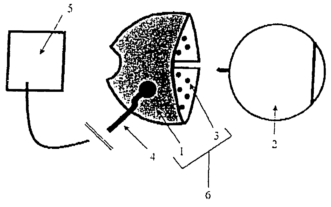
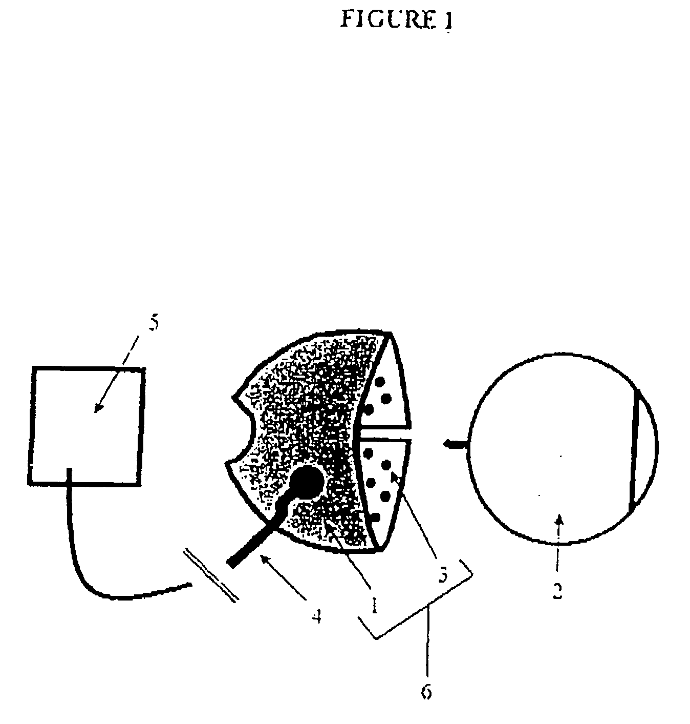
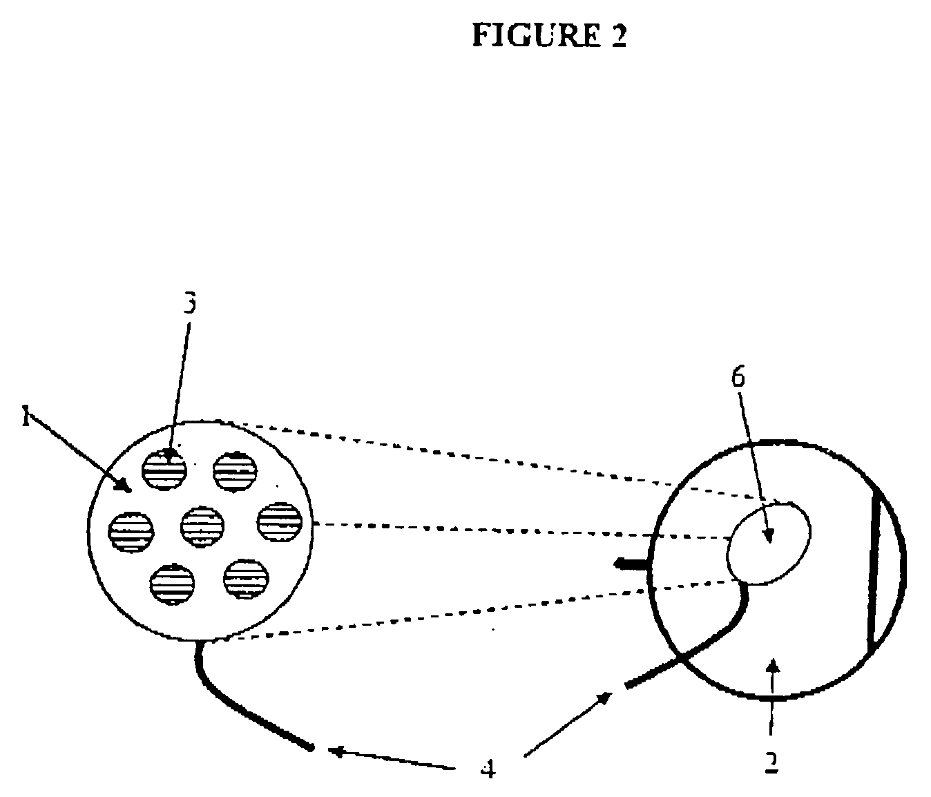
[0121] The present study was undertaken to investigate the feasibility of an extraocular approach to retinal stimulation. This is the first study of an extraocular retinal prosthesis, an approach to visual prosthesis development for which a device has hitherto not yet been developed (Maynard, 2001, above; Margalit et al., 2002, above; Humayan et al., 2003, above). The present inventors intend to adapt the neuroprosthetic technology that is available in the Nucleus 24 Auditory Brainstem Implant (Cochlear Ltd, Australia). This implant has an electrode array of 21 platinum disc electrodes, each of 700 μm diameters, with a 950 μm centre-to-centre inter-electrode spacing, arranged in 3 rows in a silicone carrier. A number of electrode types, configurations, and stimulus methods were investigated to optimise the development of an extraocular retinal implant for the resto...

example 2

Establishment of Suitable Threshold, Strength-Duration and Evoked Response Amplitude Data for Electrical Stimulation of the Retina with an Extraocular Device

Materials and Methods

[0146] Acute experiments were carried out in anaesthetised cats, with adherence to the ARVO statement for the use of animals in ophthalmic and vision research, the NIH principles of laboratory animal care and in accordance with the guidelines of the animal ethics committee of the University of New South Wales. In adult cats (n=3), anaesthesia was induced with an intramuscular injection of ketamine (20 mg / kg) and xylazine (1 mg / kg). The animals were given a preoperative dose of subcutaneous atropine (0.2 mg / kg) and dexamethasone (1.5 mg / kg). The pupils were dilated with atropine (1%) and phenylephrine (10%) eye drops. After intubation, the animals were ventilated and anaesthesia was maintained with a 70:30 mixture of nitrous oxide and oxygen, with 0.5-1% halothane. ECG, end-tidal carbon dioxide, and core b...


PUM


Abstract

A medical device for use on the human eye is described. The device is placed in an extrocular location in a patient and delivers an electrical current that stimulates the retina of patients who are blind or have vision disorders. It has at least one electrode that makes contact with the scleral surface of the eye, the electrode typically being activated by an electrical stimulator. The device produces electrical pulses which pass through the electrodes on the scleral surface of the eye, to activate the retina of the eye, which causes the patient to experience improved vision, visual sensations or the prevention of deterioration of vision. By this means, sight can be restored or improved where patients have disorders of their retina or other parts of their visual system.

Description

FIELD OF THE INVENTION [0001] The present invention relates generally to devices for enhancing visual perception, and more particularly to devices that generate electrical impulses in an extraocular location of an eye of a patient, to deliver electrical stimuli to a retinal region in the eye of the patient for restoration or enhancement of visual perception, or prevention of deterioration to visual perception. BACKGROUND OF THE INVENTION [0002] Loss or impairment of visual perception can result from a variety of causes. Examples include diseases that cause deterioration of the retina, such as Retinitis Pigmentosa, which affects over one million people worldwide, and can lead to profound blindness. Other such diseases include Usher syndrome, age-related macular degeneration, Stargardt macular dystrophy, Leber congenital amaurosis and Bardet-Biedl syndrome. [0003] There is currently no clinical means of restoring visual perception to patients who have developed blindness (no light per...

Claims


Application Information

Patent Timeline
Patent Type & Authority Applications(United States)
IPC IPC(8): A61N1/04
CPCA61F9/08A61N1/0543A61N1/36046
Inventor CHOWDHURY, VIVEKCORONEO, MINAS THEODOREMORLEY, JOHN WILLIAM
Owner SYDNEY BIOTECH
Why Eureka
Try Eureka