Method and device for radiographic imaging

a radiographic imaging and radiographic technology, applied in the field of radiographic imaging methods and devices, can solve the problems of limiting the subsequent interpretation possibilities of images, errors in diagnosis, etc., and achieve the effects of low irradiation level, useful clinical index, and low level of irradiation

Active Publication Date: 2006-09-14
EOS IMAGING
View PDF15 Cites 31 Cited by
  • Summary
  • Abstract
  • Description
  • Claims
  • Application Information

AI Technical Summary

Benefits of technology

[0008] By virtue of these provisions, it is possible to obtain, with a very limited number of images, that is to say with a relatively low level of irradiation, an index which takes account of the three-dimensional shape of the osseous body examined. It is thus possible to ascertain the distribution in volume of this index, which can be especially representative of the bone mineral density. The method according to an embodiment of the invention thus provides, with a relatively low level of irradiation, a useful clinical index f

Problems solved by technology

In addition, the methods of the prior art do not take into account the individual morphological characteristics of the patients, and this can lead to errors in diagnosis.
This l

Method used


Image

  • Method and device for radiographic imaging
  • Method and device for radiographic imaging
  • Method and device for radiographic imaging

Examples


Embodiment Construction

[0074] In the various figures, the same reference labels designate identical or similar elements.

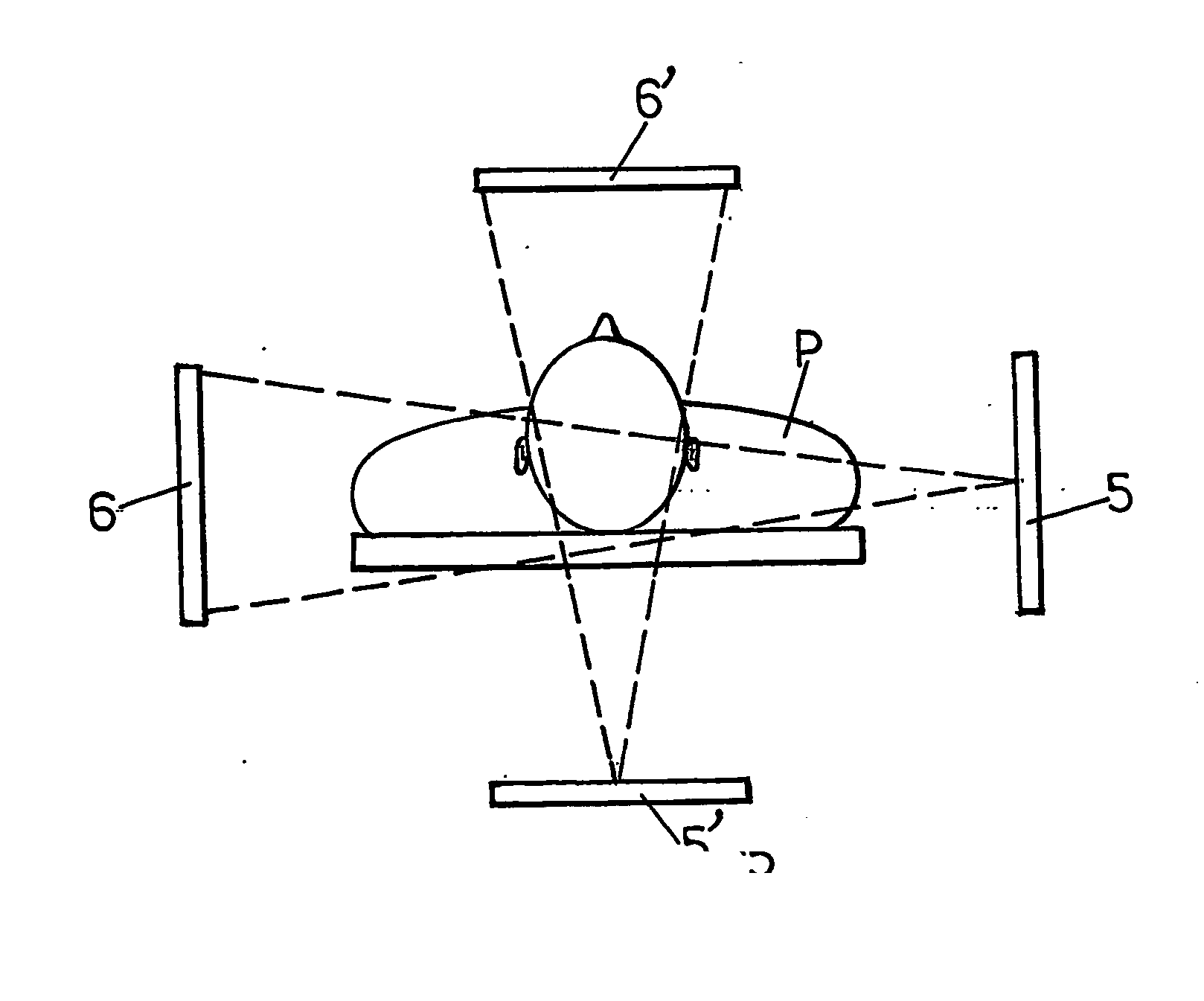
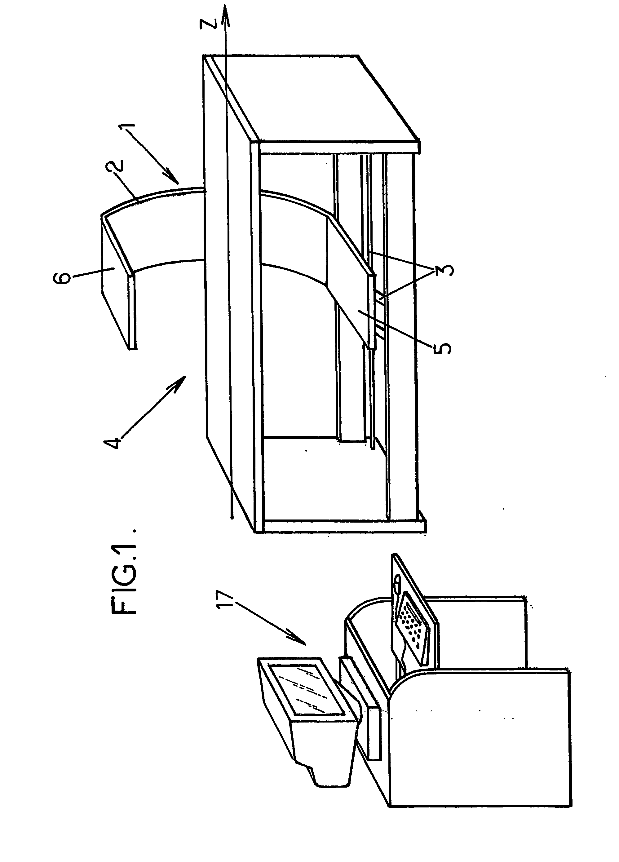
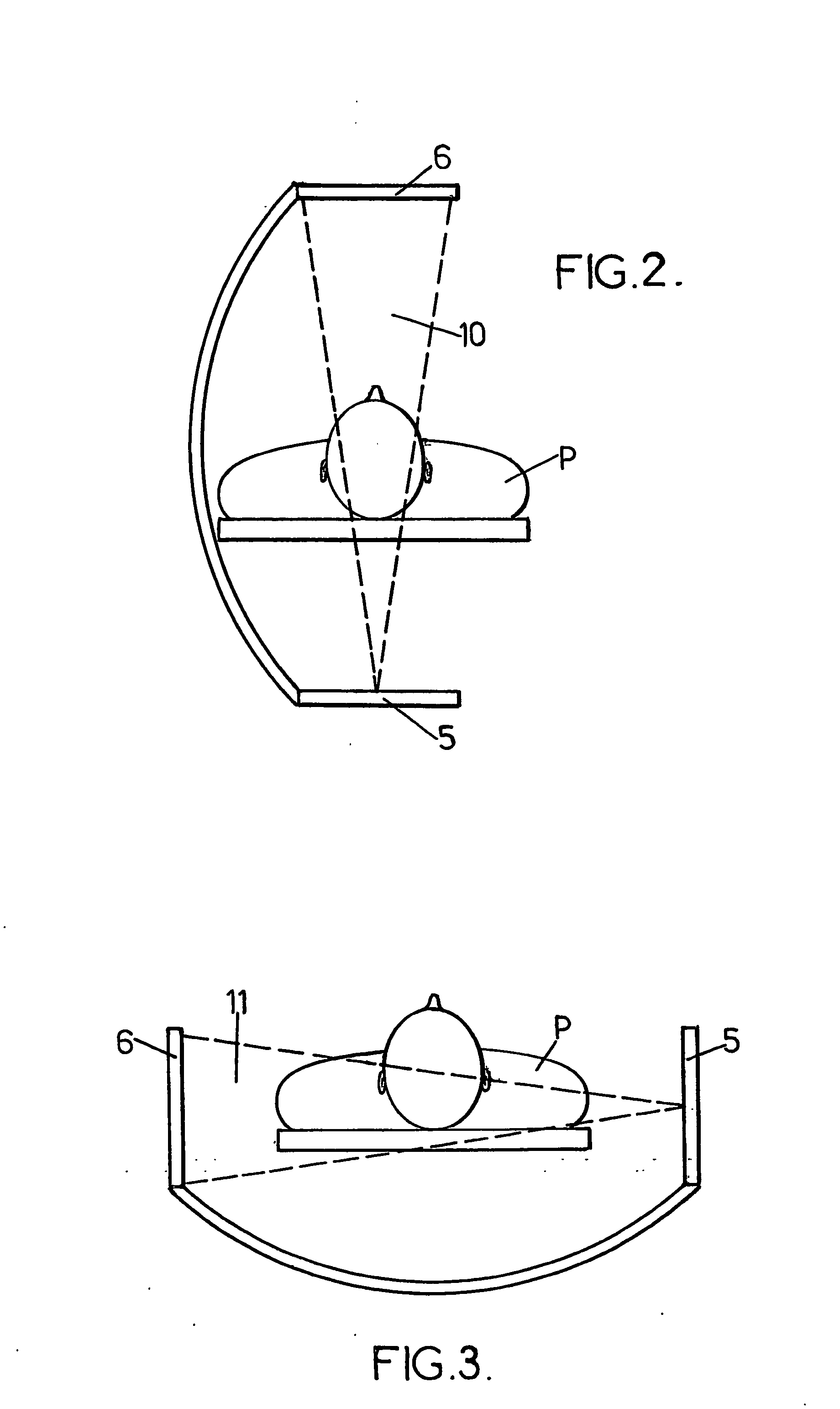
[0075]FIG. 1 shows an imaging device 1 for three-dimensional reconstruction of a composite index, such as the bone mineral density referred to a volume. This device comprises a mobile frame 2 which can be displaced by a motor on guides 3, in translation on a horizontal longitudinal axis Z and in rotation about this same horizontal axis Z.

[0076] This frame 2 encloses an observation field 4 in which a patient can be placed.

[0077] The mobile frame 2 comprises radiation-generating means and detection means.

[0078] These radiation-generating means and these detection means are of a type known to the skilled person (see, for example, document U.S. Pat. No. 5,778,045).

[0079] The radiation-generating means are formed by an X-ray source 5. They are designed to generate alternately an impulse corresponding to a high-energy spectrum and an impulse corresponding to a low-energy spectrum.

[0080] ...


PUM


Abstract

Method for radiographic imaging, in particular for measuring the bone mineral density of an osseous body, this method involving an operation which consists in determining the value of a composite index using, on the one hand, digitized radiological data, and, on the other hand, a three-dimensional generic model of said osseous body.

Description

FIELD OF THE INVENTION [0001] The present invention relates to methods and devices for radiographic imaging, in particular for measuring bone mineral density. DESCRIPTION OF THE RELATED ART [0002] More particularly, the invention concerns a method for radiographic imaging comprising a step (d) which consists in introducing, into calculation means, first digitized radiological data from signals delivered by means of detection of X-rays and corresponding to pixels of a first image of an anatomical part comprising an osseous body and scanned, in a first incidence, with a beam of X-rays having an energy spectrum distributed about at least two energies, these first data comprising, for each pixel, coordinates of the pixel in the first image and absorptiometry values designed to calculate the bone mineral density of the osseous body, referred to a surface area unit. [0003] This type of method is known, in particular from the document U.S. Pat. No. 5,778,045. It is used in particular for e...

Claims


Application Information

Patent Timeline
IPC IPC(8): G06K9/00G06T15/00A61B6/00G06T11/00
CPCA61B6/505
Inventor LE BRAS, ANTHONYKOLTA, SAMIMITTON, DAVIDSKALLI, WAFADE GUISE, JACQUES A.ROUX, CHRISTIANTEYSSEYRE, SEBASTIENFECHTENBAUM, JACQUES
Owner EOS IMAGING
Why Eureka
Try Eureka