Function image display method and device

a function image and display method technology, applied in tomography, instruments, applications, etc., can solve the problems of difficult to completely recognize whether the biological function abnormality is abnormal, symptom and danger, and the abnormality cannot be easily judged, so as to achieve efficient judgment of danger degree and easy to judge danger degree

Active Publication Date: 2006-09-28
FUJIFILM HEALTHCARE CORP
View PDF6 Cites 23 Cited by
  • Summary
  • Abstract
  • Description
  • Claims
  • Application Information

AI Technical Summary

Benefits of technology

[0005] It is an object of the present invention to provide an apparatus for diagnosing image, which is capable of easily judging the danger degree by summarizing the data obtained from a tomogram and the data obtained from a plurality of functional images into a piece of image by using a single apparatus for diagnosing image (modality). The object of the present invention is to provide an apparatus and a method for diagnosing image by displaying the data of a necessary portion only among the data in a plurality of required functional images, rendering the judgment free of confusion caused by complex data, and making it possible to efficiently judge the danger degree.

Problems solved by technology

However, since only one parameter is displayed, it is difficult to totally recognize whether the biological function abnormality, symptom and danger are of light degrees or serious degrees (hereinafter referred to as danger degree).
Besides, since color is displayed on the whole tomogram, the data are so complex that the abnormality cannot be easily judged.
However, it is difficult to bring the positions and the shapes into agreement.
Further, when the SPECT image and MR image are to be overlapped one upon the other, the SPECT image is deformed to meet the standard brain and is adjusted for its position with the MR image imposing a probability of losing the inherent shape of the patient's brain and of losing important data related to the shape.
When the patient's skull is deformed, in particular, the loss of data of the shape becomes a serious problem.

Method used


Image

  • Function image display method and device
  • Function image display method and device
  • Function image display method and device

Examples


embodiment 1

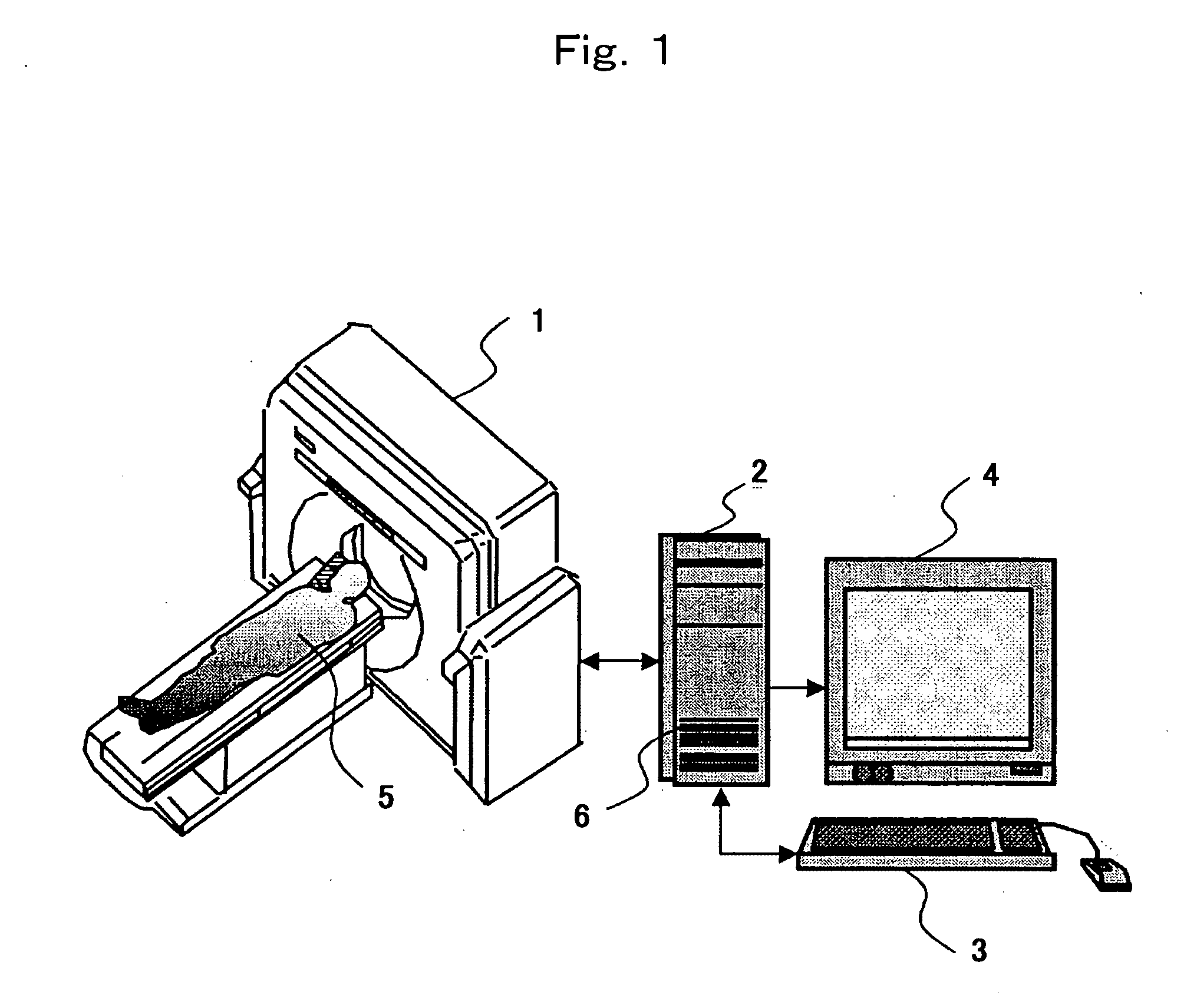
[0046]FIG. 1 is a view illustrating a method and an apparatus for displaying a functional image according to a preferred embodiment of the invention. The method and the apparatus for displaying a functional image of the invention comprises means 1 for collecting tomogram data such as X-ray attenuation signals and echo signals emitted from nuclear magnetic resonance, such as CT apparatus or MRI apparatus, a computer 2 for controlling the acquisition means 1 and for executing various operations, a console 3 such as a mouse or a keyboard, and display means 4 such as a display. The computer 2 is mounting a program for controlling the acquisition means 1, a program for forming a tomogram, such as reconstituting the image, a program for analyzing and mapping the biological function data, and a program for forming a composite image. In constituting the method and apparatus for displaying functional image according to the present invention, the above programs may be mounted in one computer ...

embodiment 2

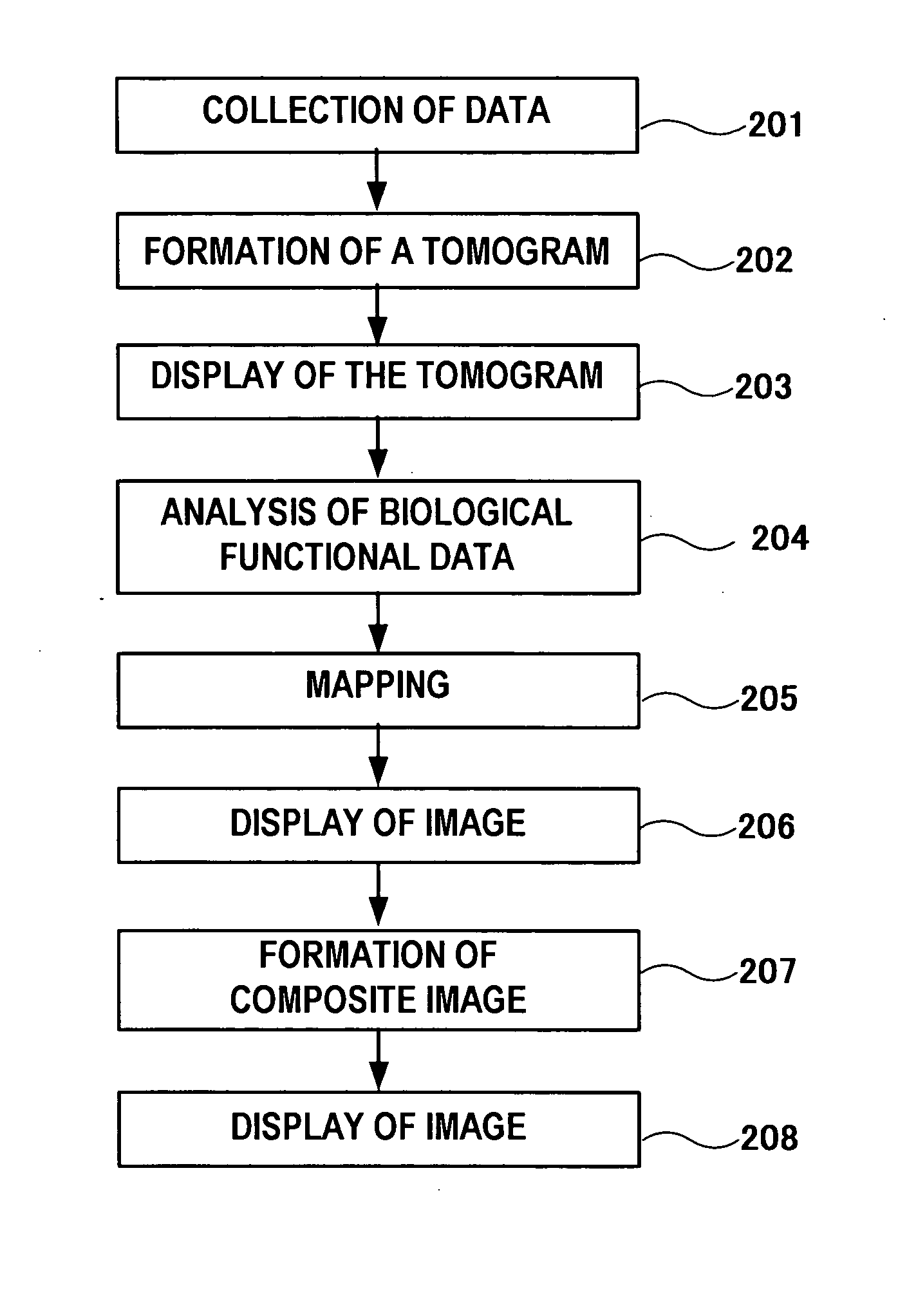
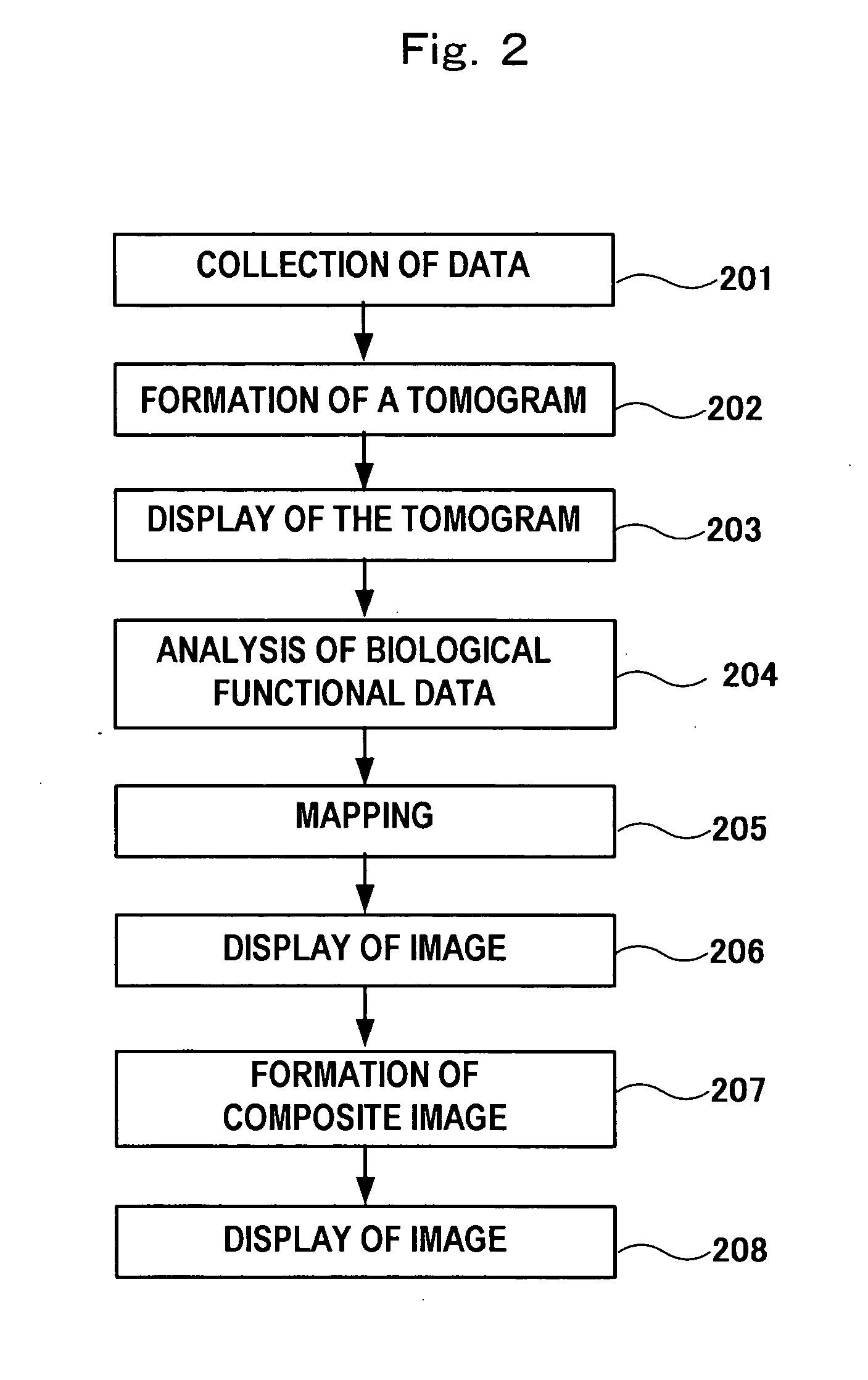
[0087] Embodiment 2 utilizes the constitution of FIG. 1 like in the embodiment 1. The constituent elements are as described in the embodiment 1 and their description is not repeated. In the embodiment 2, too, the image processing apparatus 2 is, for example, a computer mounting a program for controlling the data collection means 1, a program for forming a tomogram, such as reconstituting the image, a program for analyzing and mapping the biological function data, and a program for forming a composite image. The above programs may be mounted in one computer or may be mounted in a divided manner in a plurality of computers depending upon the kinds of operations.

[0088]FIG. 9 is a flowchart from collecting the data through up to displaying a composite image using a program in the image diagnosing apparatus according to the embodiment. The process according to the embodiment will now be described according to the flowchart. At step 301, acquisition means 1 (see FIG. 1) controlled by a c...


PUM


Abstract

There are provided a function image display method and device capable of performing analysis of biological function information by acquiring from a single image, information obtained from a tomogram and information obtained from a plurality of function images without need of observing the plurality of function images and the tomogram by successively moving the eye line, and easily making judgment about the danger degree of the biological function abnormality. A plurality of function images each displayed by a unique and arbitrary gradation color scale are combined with an arbitrary weight or an image obtained by calculating the inter-function images is displayed or these are combined with the tomogram with an arbitrary weight. Furthermore, an operator can arbitrarily set and modify the range to which weight is applied, the range for displaying the gradation color scale, and the range to be combined.

Description

TECHNICAL FIELD [0001] This invention relates to analyzing and evaluating biological function data in an image diagnostic apparatus such as a CT apparatus or an MRI apparatus relying upon a tomogram obtained thereby. FIELD OF THE INVENTION [0002] In analyzing the biological function data, it is often attempted to totally diagnose a plurality of function data. In the analysis of cerebral perfusion function data, for example, the diagnosis is carried out, usually, by totally observing the data obtained from a plurality of functional images such as cerebral blood flow (CBF) image, cerebral blood volume (CBV) image, mean transit time (MTT) image, as well as data (e.g., early CT sign and anatomical impression such as running of blood vessels, positions of tissues, etc.) obtained from a tomogram. [0003] An image display method for displaying biological function data has been disclosed in JP-A-2002-282248 wherein a piece of functional image is superposed on a tomogram and is displayed as a...

Claims


Application Information

Patent Timeline
Patent Type & Authority Applications(United States)
IPC IPC(8): G06K9/00A61B6/03
CPCA61B6/032A61B6/463A61B6/507
Inventor OMI, YASUOMIYAZAKI, OSAMUYASUDA, SUSUMU
Owner FUJIFILM HEALTHCARE CORP
Why Eureka
Try Eureka