Radiography apparatus and radiography method

a radiography and apparatus technology, applied in the field of radiography apparatus and radiography method, can solve the problems of difficult to improve diagnostic efficiency, difficult to minimize etc., and achieve the effect of improving diagnostic efficiency and reducing an amount of contrast medium or a patient dos

Inactive Publication Date: 2006-10-05
GE MEDICAL SYST GLOBAL TECH CO LLC
View PDF4 Cites 10 Cited by
  • Summary
  • Abstract
  • Description
  • Claims
  • Application Information

AI Technical Summary

Benefits of technology

[0011] According to the present invention, there are provided a radiography apparatus and a radiography method which make it easy to reduce an amount of a contrast medium or a patient dose and make it possible to improve diagnostic efficiency.

Problems solved by technology

Therefore, it may be hard to minimize an amount of a contrast medium or a patient dose.
Moreover, since an examination lasts over a long period of time, it may be hard to improve diagnostic efficiency.

Method used


Image

  • Radiography apparatus and radiography method
  • Radiography apparatus and radiography method
  • Radiography apparatus and radiography method

Examples


Embodiment Construction

[0019] An embodiment of the present invention will be described below.

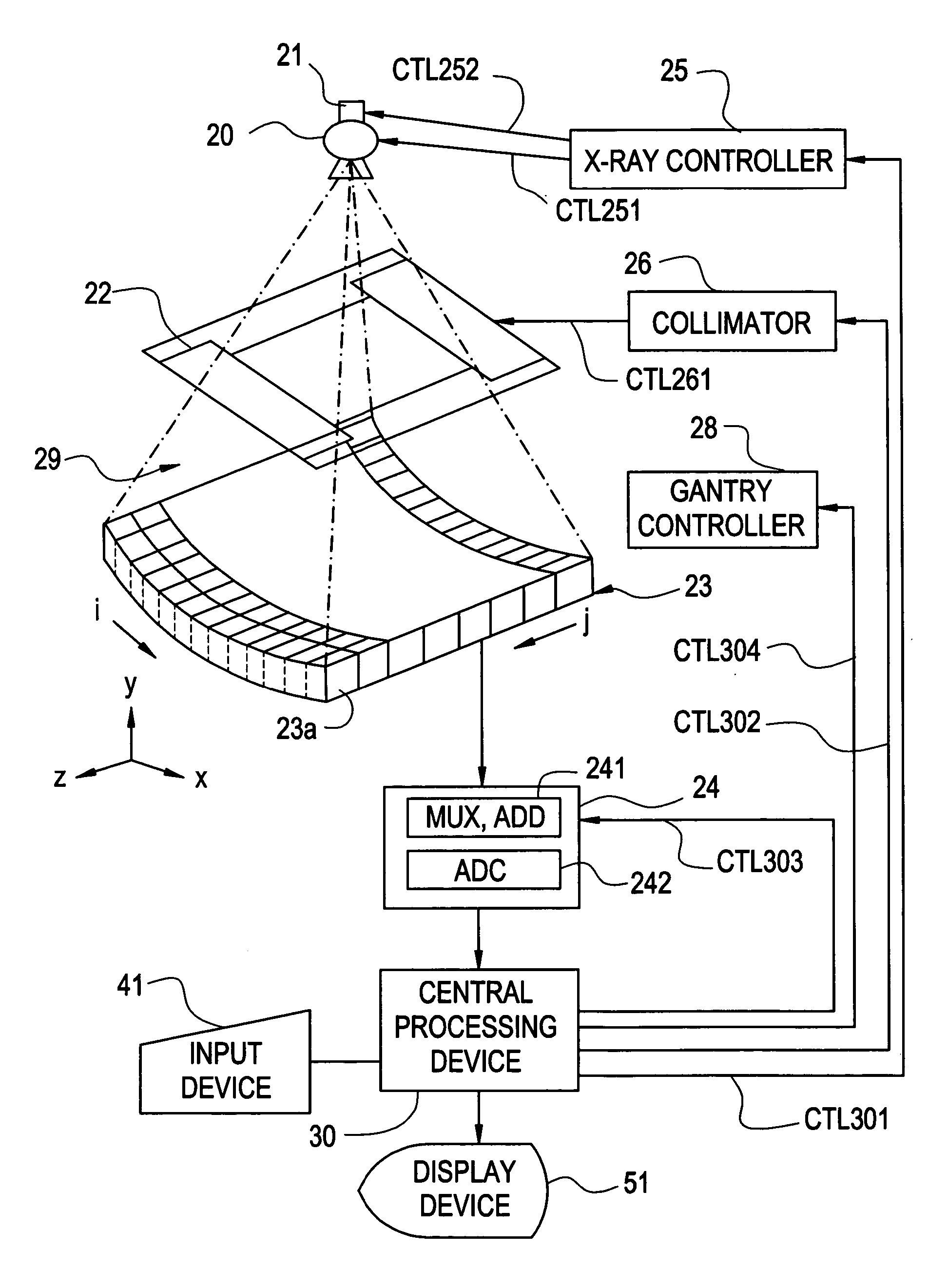
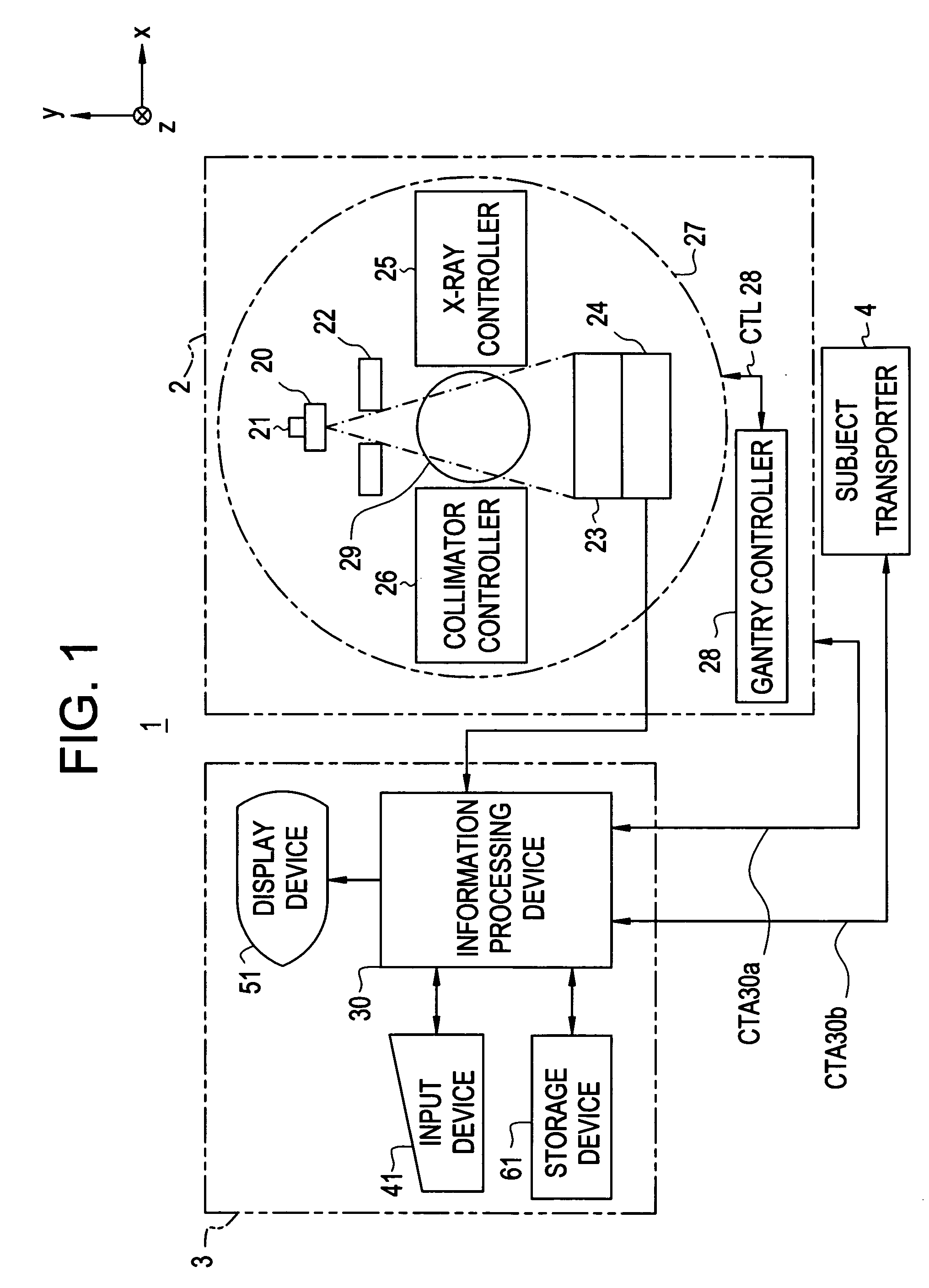
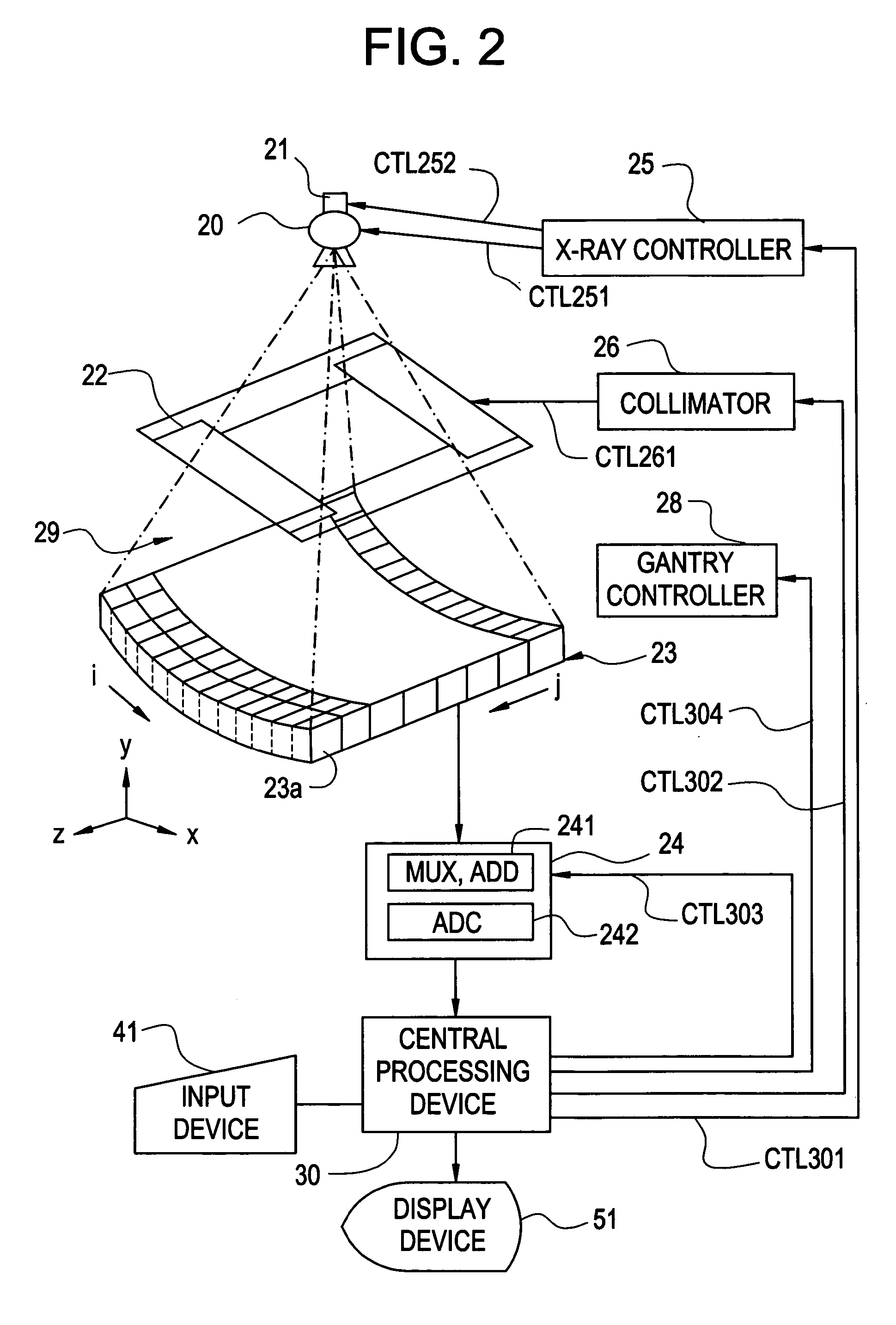
[0020]FIG. 1 is a block diagram showing the overall configuration of an X-ray CT apparatus 1 in accordance with an embodiment of the present invention. FIG. 2 shows the configuration of a major portion of the X-ray CT apparatus 1 in accordance with the embodiment.

[0021] As shown in FIG. 1, the X-ray CT apparatus 1 includes a scanner gantry 2, an operator console 3, and a subject transporter 4. The X-ray CT apparatus 1 uses projection data items of a subject, which are produced by scanning the subject with X-rays according to the conditions for a scan, to reconstruct images of the subject's tomographic layer.

[0022] The scanner gantry 2 will be described below.

[0023] The scanner gantry 2 scans a subject, who is moved to an imaging space 29 by the subject transporter 4, according to a control signal CTL30a sent from the operator console 3, and produces projection data items of the subject. The scanner gantry 2 in...


PUM

PropertyMeasurementUnit
tube currentaaaaaaaaaa
scan timeaaaaaaaaaa

Abstract

The present invention attempts to reduce an amount of a contrast medium or a patient dose and improve diagnostic efficiency. The conditions for a main scan are designated so that a first scan of scanning a subject with a tube current of a first tube current value fed to an X-ray tube will be performed in order to examine the blood flow in a subject's blood vessel into which a contrast medium is injected. Moreover, the conditions for a main scan are designated so that a second scan of scanning the subject with a tube current of a second tube current value fed to the X-ray tube will be performed in order to image the subject's blood vessel into which the contrast medium is injected. Images of the subject are constructed based on projection data items produced by scanning the subject under the sets of conditions for a main scan.

Description

BACKGROUND OF THE INVENTION [0001] The present invention relates to a radiography apparatus and a radiography method. More particularly, the present invention is concerned with a radiography apparatus and a radiography method which construct images of a subject according to projection data items of the subject produced by performing a main scan in which radiation is irradiated from a radiation tube to the subject and radiation transmitted by the subject is detected. [0002] A radiography apparatus including an X-ray computed tomography (CT) apparatus reconstructs images of a subject's tomographic layer according to projection data items produced by scanning a subject with radiation. The radiography apparatus is widely adopted for medical application and industrial application. [0003] In the radiography apparatus, for example, a blood flow examination called CT perfusion or angiography called CT angiography is performed using a contrast medium for the purpose of diagnosis of an ischem...

Claims


Application Information

Patent Timeline
Patent Type & Authority Applications(United States)
IPC IPC(8): H05G1/60A61B6/00G01N23/00G21K1/12
CPCA61B6/032A61B6/504A61B6/481A61B6/4085
Inventor KUDO, MASAYUKI
Owner GE MEDICAL SYST GLOBAL TECH CO LLC
Why Eureka
Try Eureka