
Image analysis of the golgi complex

a technology of golgi complex and image analysis, which is applied in the field of image analysis of golgi complex, can solve the problems of insufficient realization of the value of golgi complex as an indicator, and no simple, consistent technique for characterizing

Inactive Publication Date: 2006-10-12
CYTOKINETICS INC
View PDF34 Cites 2 Cited by
  • Summary
  • Abstract
  • Description
  • Claims
  • Application Information

AI Technical Summary

Benefits of technology

[0014] These and other features and advantages of the present invention w...

Problems solved by technology

Unfortunately, the value of the Golgi complex as an indicator has not been fully realized.
This is because no simple consistent technique has been developed for characterizing the condition of Golgi complex in a high throughput manner.

Method used


Image

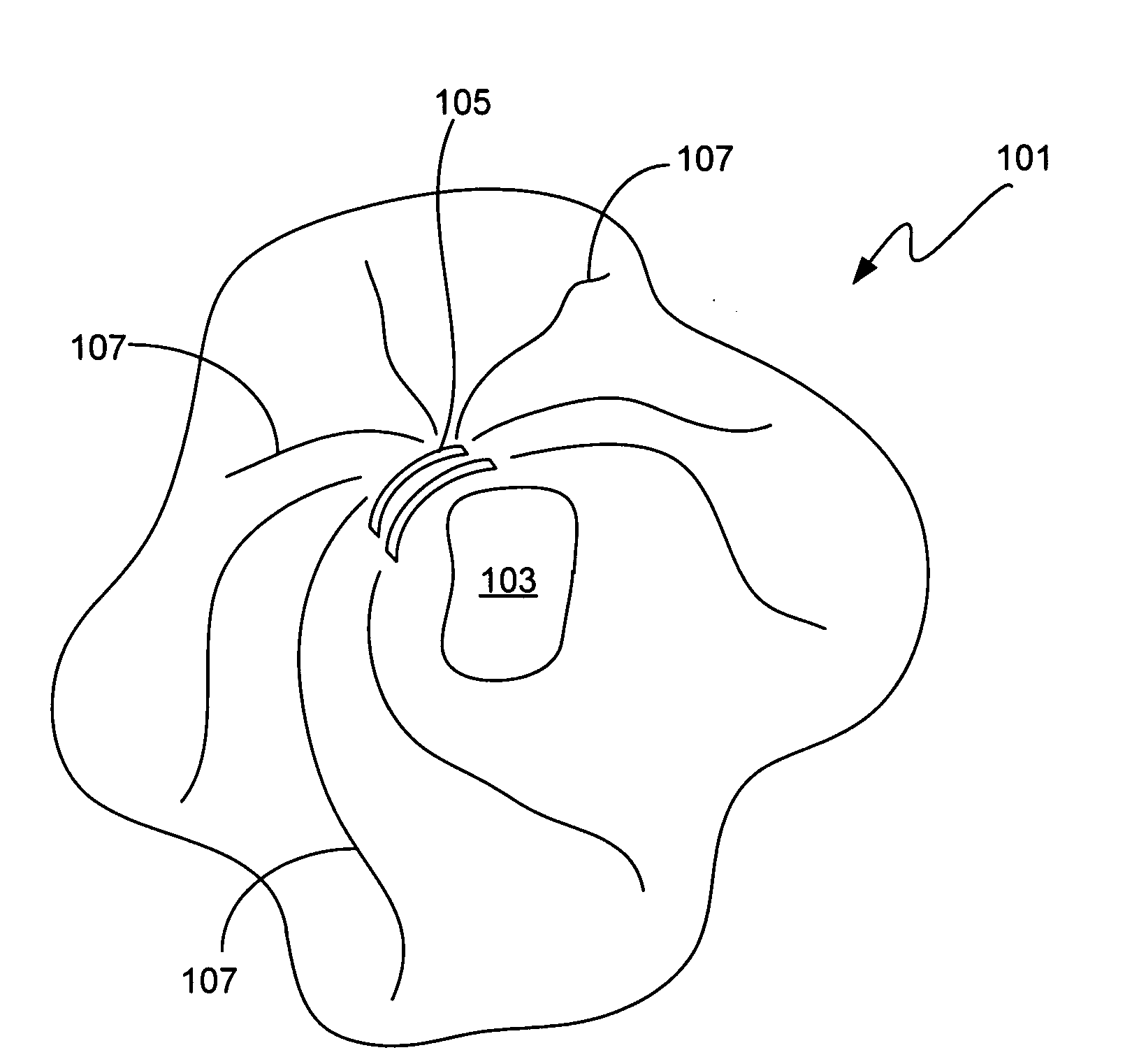
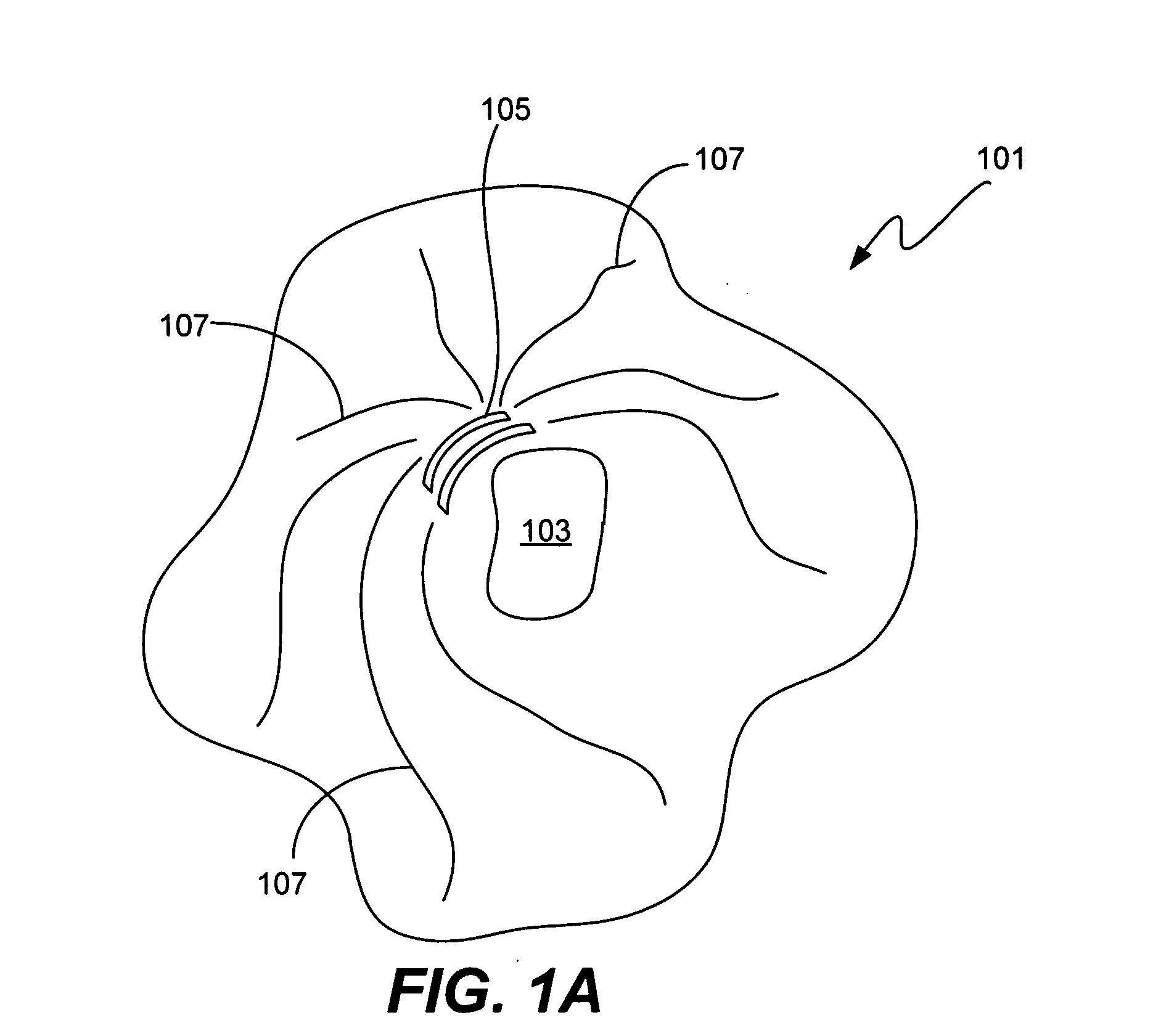
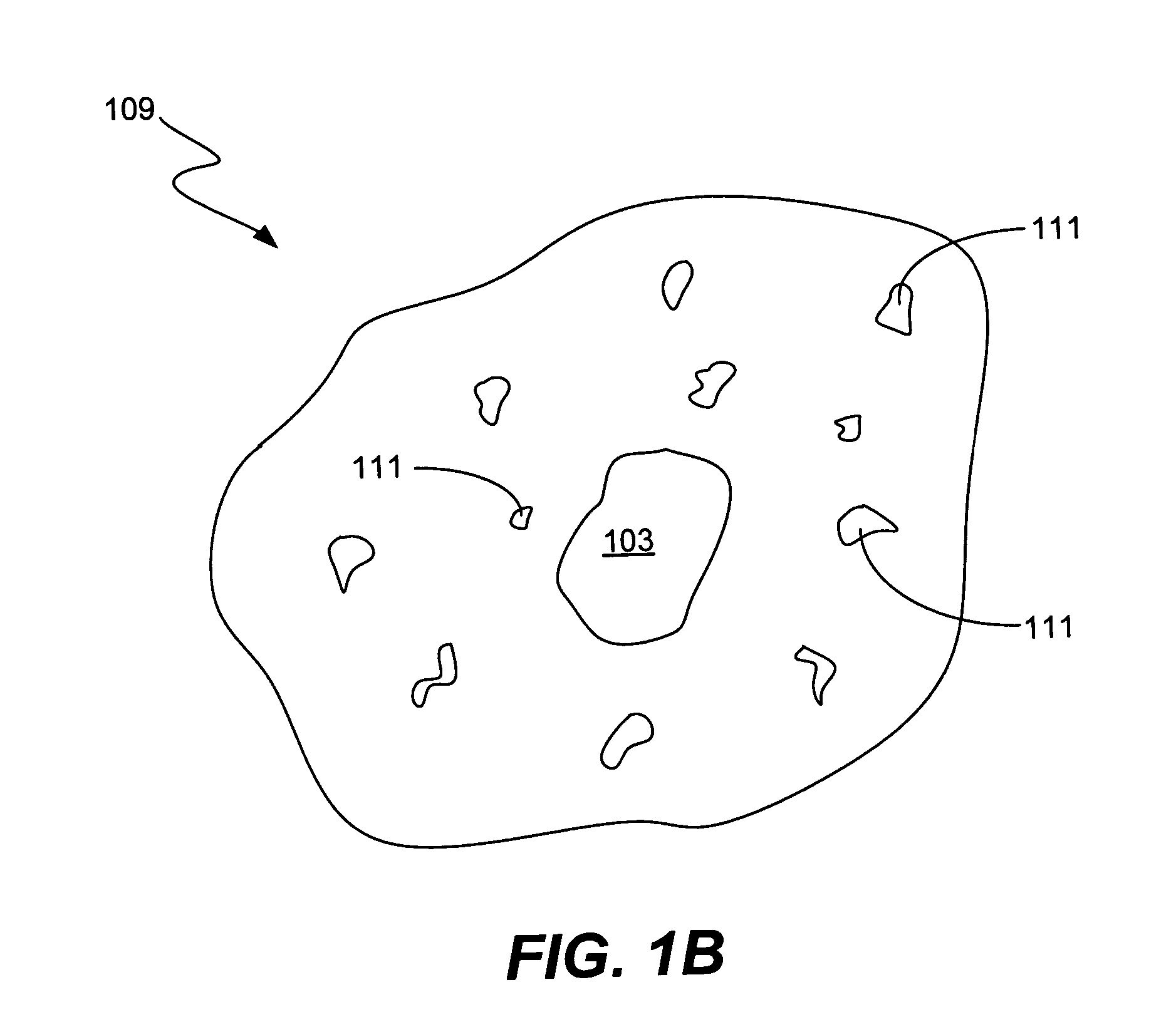
  • Image analysis of the golgi complex
  • Image analysis of the golgi complex
  • Image analysis of the golgi complex

Examples


examples

[0096] The following examples each involved generation and analysis of images obtained from SKOV3 cells treated with a Lens culinaris lectin marker. The cells were incubated with each drug for 24 hours before fixation and staining. In each case, ring regions were obtained by the Golgi segmentation algorithm described above. In addition, the Golgi complex within those ring regions was predicted, using a CART model as described above, as either normal, diffuse, dispersed, or diffuse and dispersed.

[0097] In a first example, SKOV3 cells that were treated with a low concentration (0.6 nanomolar) of the anti-microtubule drug Taxol. In this example, the Golgi complex remains normal as determined by the model. An expert independently examined the images and concluded that the Golgi complex was normal. In a second example, SKOV3 cells treated with a high concentration (2.6 micromolar) of microtubule-de polymerizing drug Nocodazole. Using the same model, the Golgi complex was found to be dis...


PUM


Abstract

Methods, code and apparatus analyze cell images to automatically identify and characterize the Golgi complex in individual cells. This is accomplished by first locating the cells in the image and defining boundaries of those cells that subsume some or all of the Golgi complex of those cells. The Golgi complex in the images typically have intensity values corresponding to the concentration of a Golgi component in the cell (e.g. a polysaccharide associated with the Golgi complex). The method / system then analyzes the Golgi components of the image (typically on a pixel-by-pixel basis) to mathematically characterize the Golgi complex of individual cells. This mathematical characterization represents phenotypic information about the cells' Golgi complex and can be used to classify cells. From this information, mechanism of action and other important biological information can be deduced.

Description

CROSS-REFEERNCE TO RELATED PATENT APPLICATIONS [0001] This application is a continuation of U.S. patent application Ser. No. 09 / 792,012 iled Feb. 20, 2001 by Vaisberg et al., and titled IMAGE ANALYSIS OF THE GOLGI COMPLEX, which is a continuation-in-part of U.S. patent application Ser. No. 09 / 729,754 now U.S. Pat. No. 6,876,760 issued Apr. 5, 2005 by Vaisberg et al., and titled CLASSIFYING CELLS BASED ON INFORMATION CONTAINED IN CELL IMAGES filed on Dec. 4, 2000, which is in turn is continuation in part of the following U.S. patent applications: U.S. patent application Ser. No. 09 / 310,879 by Vaisberg et al., and titled DATABASE METHOD FOR PREDICTIVE CELLULAR BIOINFORMATICS; U.S. patent application Ser. No. 09 / 311,996 by Vaisberg et al., and titled DATABASE SYSTEM INCLUDING COMPUTER FOR PREDICTIVE CELLULAR BIOINFORMATICS; U.S. patent application Ser. No. 09 / 311,890, now U.S. Pat. No. 6,743,576, issued Jun. 1, 2004 by Vaisberg et al., and titled DATABASE SYSTEM FOR PREDICTIVE CELLULAR...

Claims


Application Information

Patent Timeline
IPC IPC(8): G06K9/00G01N15/10G01N15/14
CPCG01N15/1475G01N2015/1006G06K9/0014G01N2015/149G06K9/00127G01N2015/1472G06V20/69G06V20/695G01N15/1433G01N15/149
Inventor VAISBERG, EUGENI A.CONG, GEWU, HSIEN-HSUN
Owner CYTOKINETICS INC
Learn More