Article, system, and method for securing medical device to tissue or organ

a technology for securing medical devices and organs, applied in the field of medicine, can solve the problems of insufficient in certain cases, inability to describe the use of securing medical devices, and inability to achieve the effect of precise placement and stapling, and good elasticity

Inactive Publication Date: 2006-10-19
GODIN NORMAN
View PDF7 Cites 46 Cited by
  • Summary
  • Abstract
  • Description
  • Claims
  • Application Information

AI Technical Summary

Benefits of technology

[0015] In the case of the GARD, the device is preferably inserted though the mouth and the esophagus, and when located at the intersection of the esophagus and stomach, a ring portion is stapled to a hiatus hernia with double tilt tag staples using ultrasound real time visualization and a flexible endoscope, which together facilitate precise placement and stapling.
[0016] The tilt-tag staple comprises an elongated joining member having a first end and a second end, a first tilt-tag joined to the first end of the joining member and a second tilt-tag joined to the second end of the joining member, each of the first and second tilt-tags normally aligned perpendicularly to the joining member, the tilt-tags bendable under resistance into a position substantially parallel to the joining member and returning to the normal position when resistance is removed. The tilt-tag staple is preferably constructed of nickel-titanium alloy, and tilt-tag staples of such material are novel and advantageous for several reasons. For temporary applications where temporary tilt-tag staples are needed, the tilt-tag staples can be constructed of biodegradable material, as long as the material is elastic enough to return to normal position after bending the tags to a substantially parallel position during the insertion steps. Such temporary applications include stapling to the esophagus or gastro-intestinal tract of captors monitoring pressure, pH and / or temperature or any other physical parameter. The biodegradable tilt-tags can be made of absorbable materials such as lactide, glycolide, or e-caprolactone for such temporary placement.
[0017] For permanent applications such as with a GARD, the joining material can be made in implant grade steel or preferably nitinol, surgical thread, or implant grade polymer such as nylon. The tilt-tags at each end of the joining member can also be made of steel, nitinol, implant-grade polymers such as nylon or any other biocompatible implantable material. In a preferred embodiment, the tilt-tags and joining segment are all made of a non-absorbable material, preferably in nitinol so as to have the flexibility for the tilt-tag to resume an angle from a parallel position when released.
[0018] Nitinol in its austenitic configuration has good elasticity properties which tend to restore the original right angle position between the joining member and the tags after release from their parallel insertion position. When the nitinol temperature is cooled as when spraying ice-water directly through the working channel of the endoscopic with ice water, the austenitic harder hyperelastic configuration can be changed into a martensitic much softer configuration. This new configuration makes it much less traumatic to pull on the tilt-tag (note see FIG. 4) to pull out the tilt-tag and remove the GARD allowing replacement.

Problems solved by technology

For certain types of devices placed through the mouth, conventional stapling with a rigid stapler is not possible and so various alternatives using flexible instruments have been devised.
While the aforementioned GARD ring device and method worked for some patients, it was insufficient in certain cases.
Furthermore, the Fritscher-Ravens, et al., system was not described for use for securing medical devices, for example a GARD, to the gastrointestinal tract or any other organ.

Method used


Image

  • Article, system, and method for securing medical device to tissue or organ
  • Article, system, and method for securing medical device to tissue or organ
  • Article, system, and method for securing medical device to tissue or organ

Examples


Embodiment Construction

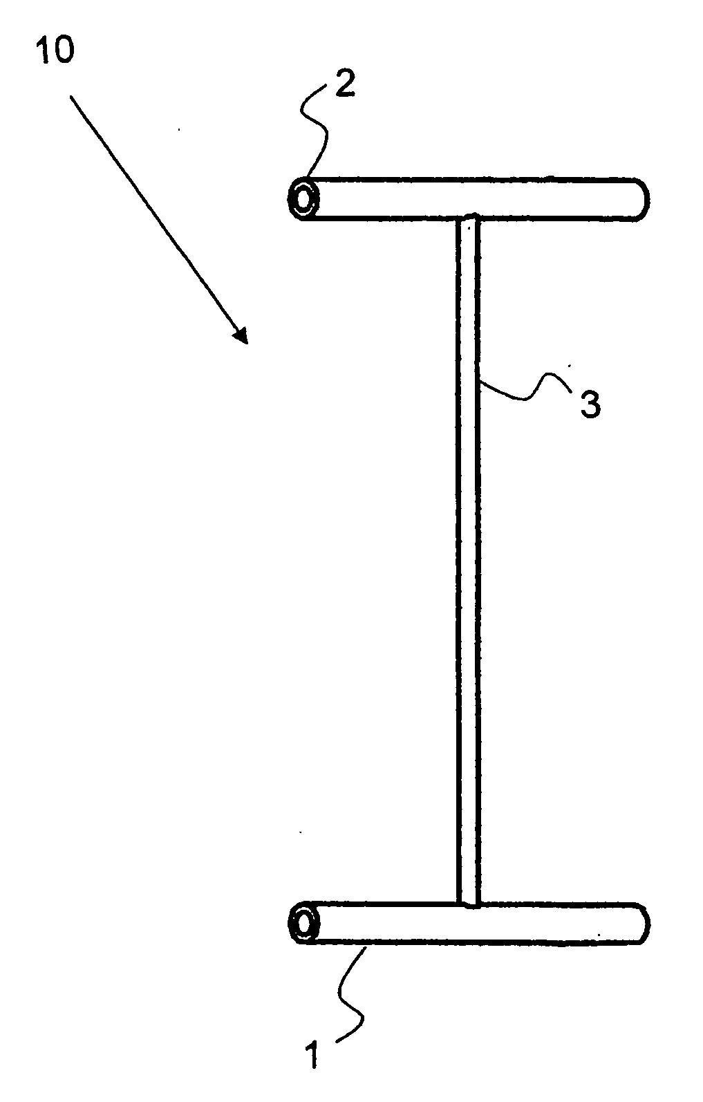
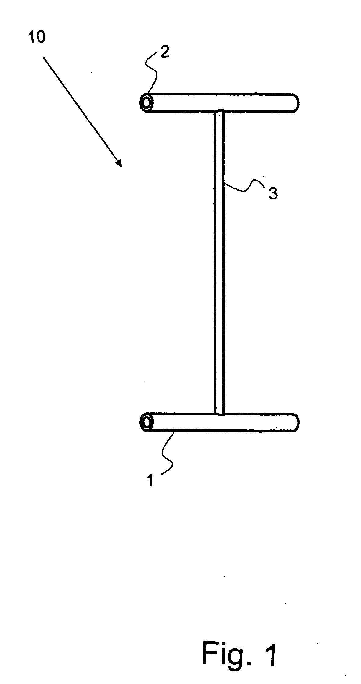
[0025] Referring first to FIG. 1, an example of a tilt-tag staple 10 of the invention is shown wherein a first tilt tag 1 and a second tilt tag 2 are joined perpendicularly at respective ends of joining member 3. In the illustrated embodiment, the tilt-tag staple 10 is molded in one piece from Nitinol, a commercially available nickel-titanium alloy.

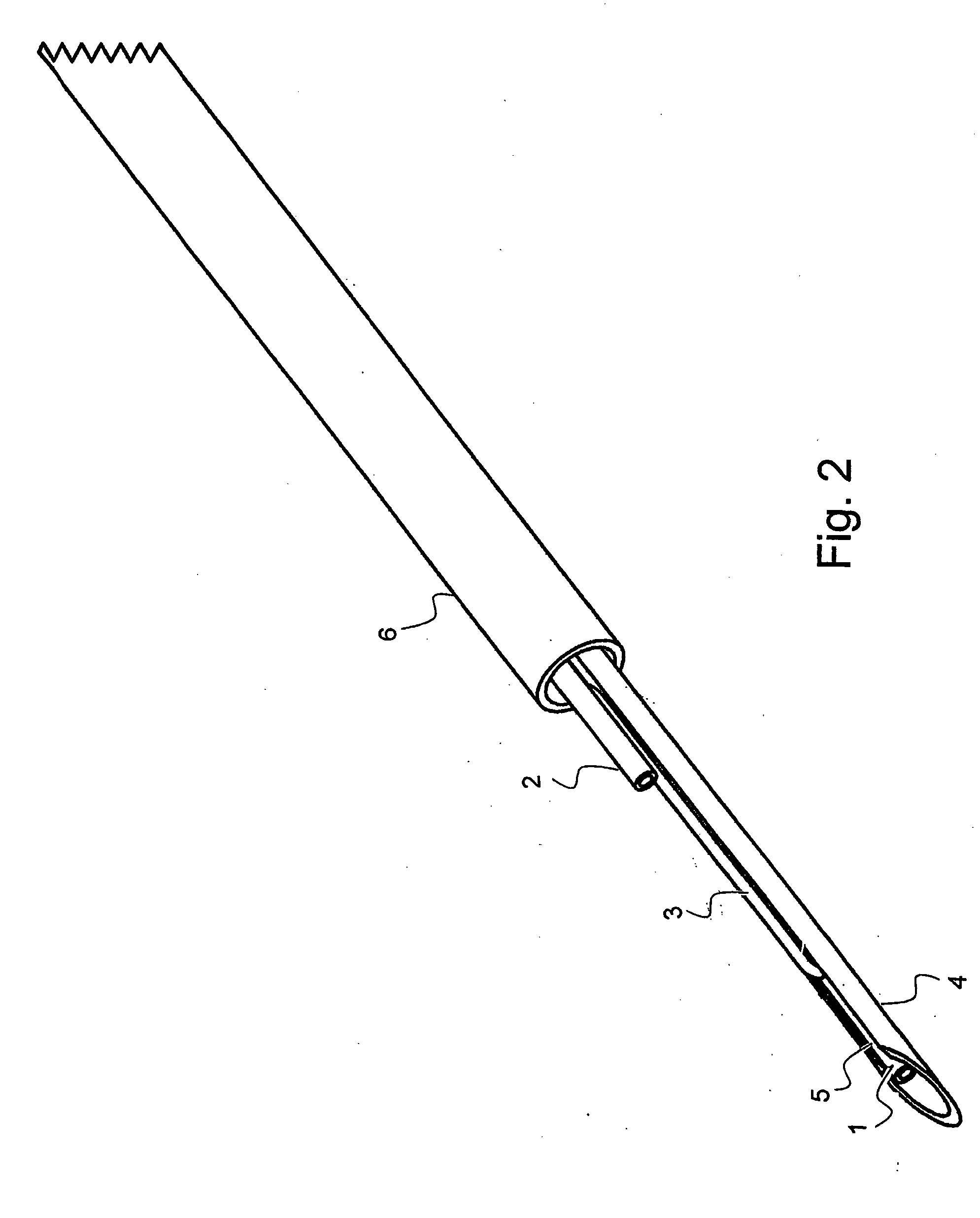
[0026] Referring now to FIG. 2, the first tilt tag 1 is inserted in a needle 4 having a slit 5 which is slightly wider than the diameter of the joining member 3 but is less wide than the diameter of the first tilt tag 1 so as to allow the first tilt tag 1 to slide in the channel of the needle 4 and for the joining member 3 to bend in a direction parallel or very close to parallel with the first tilt tag 1. The second tilt tag 2 is bent substantially parallel to the joining member 3 but is outside of the hollow needle channel. The positions of the second tilt tag 2 and the joining member 3 are maintained by the flexible tube 6, and the ne...


PUM


Abstract

An surgical tilt-tag staple comprising an elongated joining member having a first end and a second end, a first tilt-tag joined to the first end of the joining member and a second tilt-tag joined to the second end of the joining member, each of the first and second tilt-tags normally aligned perpendicularly to the joining member, the tilt-tags bendable under resistance into a position substantially parallel to the joining member and returning to the normal position when resistance is removed, is used to attach a medical device to the inner mucosa of a gastro-intestinal tract of a patient using ultrasound real time visualization and a flexible endoscope. Tilt-tag staples constructed of nickel-titanium alloy are preferred. Removal of such tilt-tag staples can be facilitated by cooling a tilt-tag. Medical devices with an electrical lead can be implanted without conventional suturing by using one terminal tilt tag on the lead to implant it.

Description

CROSS-REFERENCE TO RELATED APPLICATIONS [0001] Benefit of provisional application No. 60 / 672,135 filed Apr. 14, 2005 is claimed, the contents of which are incorporated by reference.BACKGROUND OF THE INVENTION [0002] The present invention relates to the field of medicine, particularly to suturing and securing medical devices to tissue or organs. [0003] The most common way to secure medical tissues to tissues or organs is by stapling with conventional surgical staples formed from a single length of wire in an approximate U-shape. For example, McGarry, et al., in U.S. Pat. No. 5,366,479, disclose endoscopic application of staples for attaching surgical mesh to body tissue in laparoscopic hernia surgery. In this patent, the stapler is a rigid instrument and cannot be used through the mouth into the esophagus where a flexible instrument is needed. [0004] For certain types of devices placed through the mouth, conventional stapling with a rigid stapler is not possible and so various altern...

Claims


Application Information

Patent Timeline
Patent Type & Authority Applications(United States)
IPC IPC(8): A61B17/08
CPCA61B17/0401A61B17/0469A61B17/0482A61B2017/0409A61B2017/081A61B2017/0419A61B2017/06052A61B2017/061A61B2017/0417
Inventor GODIN, NORMAN
Owner GODIN NORMAN
Why Eureka
Try Eureka