Lymphatic endothelial cells materials and methods

a technology of lymphatic endothelial cells and materials, applied in the field of lymphatic endothelial cell materials and methods, can solve the problems of insufficient methods for obtaining isolated lymphatic endothelial cells, the therapeutic and diagnostic implications of the dysfunction of these interactions remain elusive, and the inability to ascertain the mechanism of action of these important regulators of lymphatic endothelial cell function, etc., to achieve the effect of improving the ability to manipula

Inactive Publication Date: 2006-11-30
LUDWIG INST FOR CANCER RES
View PDF59 Cites 10 Cited by
  • Summary
  • Abstract
  • Description
  • Claims
  • Application Information

AI Technical Summary

Benefits of technology

[0009] The present invention, provides improvements in the ability to manipulate endothelial cells and lymphatic and vascular systems that have numerous practical uses in medicine and molecular biology. More particularly, the present invention provides a method for isolating lymphatic endothelial cells from a biological sample of comprising lymphatic endothelial cells, the method comprising contacting said biological sample with an antibody that preferentially recognizes lymphatic endothelial cells as compared to other endothelial cells, under conditions where the antibody binds lymphatic endothelial cells, and isolating lymphatic endothelial cells bound to said antibody. As used herein the term “antibody” is intended to refer to any antibody agent that specifically binds a target antigen (e.g., lymphatic endothelial cells) or any polypeptide that comprises an antigen binding fragment that specifically recognizes the antigen. More particularly, the antibody is one that is immunologically reactive with an epitope on the extracellular domain of VEGFR-3 that is specific for lymphatic endothelial cells. In the context of the present invention, “preferential” or “specific” means that the antibody binds the target antigen e.g., VEGFR-3 on lymphatic endothelial cells) with greater affinity or avidity than it binds similar antigens on other cells (e.g., VEGFR-3 on blood vascular endothelial cells). This differential binding permits the isolation of one cell type from another.
[0018] The present invention further provides a method of ameliorating a lymphatic disorder, wherein the method comprises ex vivo therapy comprising obtaining a biological sample from the patient in need of the therapy, wherein the biological sample comprises microvascular endothelial cells; contacting the microvascular endothelial cells with an antibody that preferentially binds to lymphatic endothelial cells as compared to other endothelial cells, wherein the antibody is an antibody that is immunologically reactive with the extracellular domain of VEGFR-3; isolating lymphatic endothelial cells that are bound by the antibody, transfecting the lymphatic endothelial cells with an expression construct comprising a nucleic acid encoding a protein operably linked to a promoter, in an amount effective to produce the expression of the protein in the cells; and reintroducing the transfected cells to the patient. The encoded protein can be any protein that one might wish to express in lymphatic endothelial cells (e.g., to treat a disease, palliate the symptoms of a disease, or to permit better diagnosis or imaging)
[0019] The present invention also provides a method of promoting the growth of lymphatic endothelial cells in culture comprising obtaining the lymphatic endothelial cells according to a method of the present invention; and stimulating the cells with a VEGFR-3 ligand; wherein stimulating the growth of the cells with the VEGFR-3 ligand promotes the survival of the cells in culture as compared to growth in the absence of the stimulation. In particularly preferred embodiments, the VBGFR-3 ligand is VEGF-C, VEGF-C156S or VEGF-D. The method may further comprise stimulating the cells with a VEGFR-2 ligand. In specific embodiments, it is contemplated that the stimulation of the cells protects the cells from apoptosis. In preferred embodiments, the protection of the cells from apoptosis is mediated through the activation of Akt or p42 / MAPK signaling molecules. In preferred embodiments, the stimulation of the cells allows the cells to maintain differentiated endothelial cell characteristics.

Problems solved by technology

However, the biochemical signaling pathways activated via VEGFR-3 are less well characterized than those of VEGFR-2, making it difficult to ascertain the mechanism of action of these important regulators of lymphatic endothelial cells function.
In the absence of such information, therapeutic and diagnostic implications of dysfunctions of these interactions remain elusive.
However, despite the availability of these markers, at present, there are no adequate methods of obtaining isolated lymphatic endothelial cells.

Method used


Image

  • Lymphatic endothelial cells materials and methods
  • Lymphatic endothelial cells materials and methods
  • Lymphatic endothelial cells materials and methods

Examples


example 1

Materials and Methods

[0219] The present example provides details of materials and methods employed throughout the application and in the Examples presented herein below.

[0220] Antibodies and growth factors. The primary antibodies used in immunofluorescence were mouse mAbs against human CD31 (Dako), vWF (Dako) or VEGFR-3 (clones 9D9F9, 2E11D11 and 7B3F9; Jussila et at., Cancer Res. 58:1599-1604, 1998), rabbit antiserum against human LYVE-1 (Banerji et al., J. Biol. Chem., 144(4)789-801, 1999), affinity purified rabbit anti-human podoplanin (Breiteneder-Geleff et al., et al., Am. J. Path., 154(2) 385-394, 1999) or rabbit anti-human VEGF-C (882; Joukov et al., EMBO J., 15:290-298, 1996). Monoclonal antibody against proliferating cell nuclear antigen (clone PC10) was from Santa Cruz Biotechnology. FITC- or TRITC-conjugated goat anti-rabbit IgG, goat anti-mouse IgG and donkey anti-mouse IgG were obtained from Jackson Immunoresearch. The rabbit antiserum against human VEGFR-2 was a kind...

example 2

Human Dermal Microvascular Endothelial Cells Consist of Distinct Populations of Blood Vascular and Lymphatic Endothelial Cells

[0229] The functions of different VEGF receptors have been extensively studied in transfected cell lines, but the lack of an appropriate cellular background can compromise results obtained from such studies. Therefore, the inventors set out to separate microvascular endothelial cells into specific constituent populations of endothelial cells of one type which were substantially free of other types of endothelial cells. More particularly, the inventors generated populations of lymphatic endothelial cells that were substantially free of blood vascular endothelial cells and vice versa. In order to pursue this endeavor and to elucidate VEGFR-3 signaling pathways promoting endothelial cell survival, the inventors used primary endothelial cells, human dermal microvascular endothelial cells (HMVEC) and human umbilical vein endothelial cells (HUVEC). It was determin...

example 3

Analysis of VEGFR Specific Ligands Used for the Cell Survival Experiments

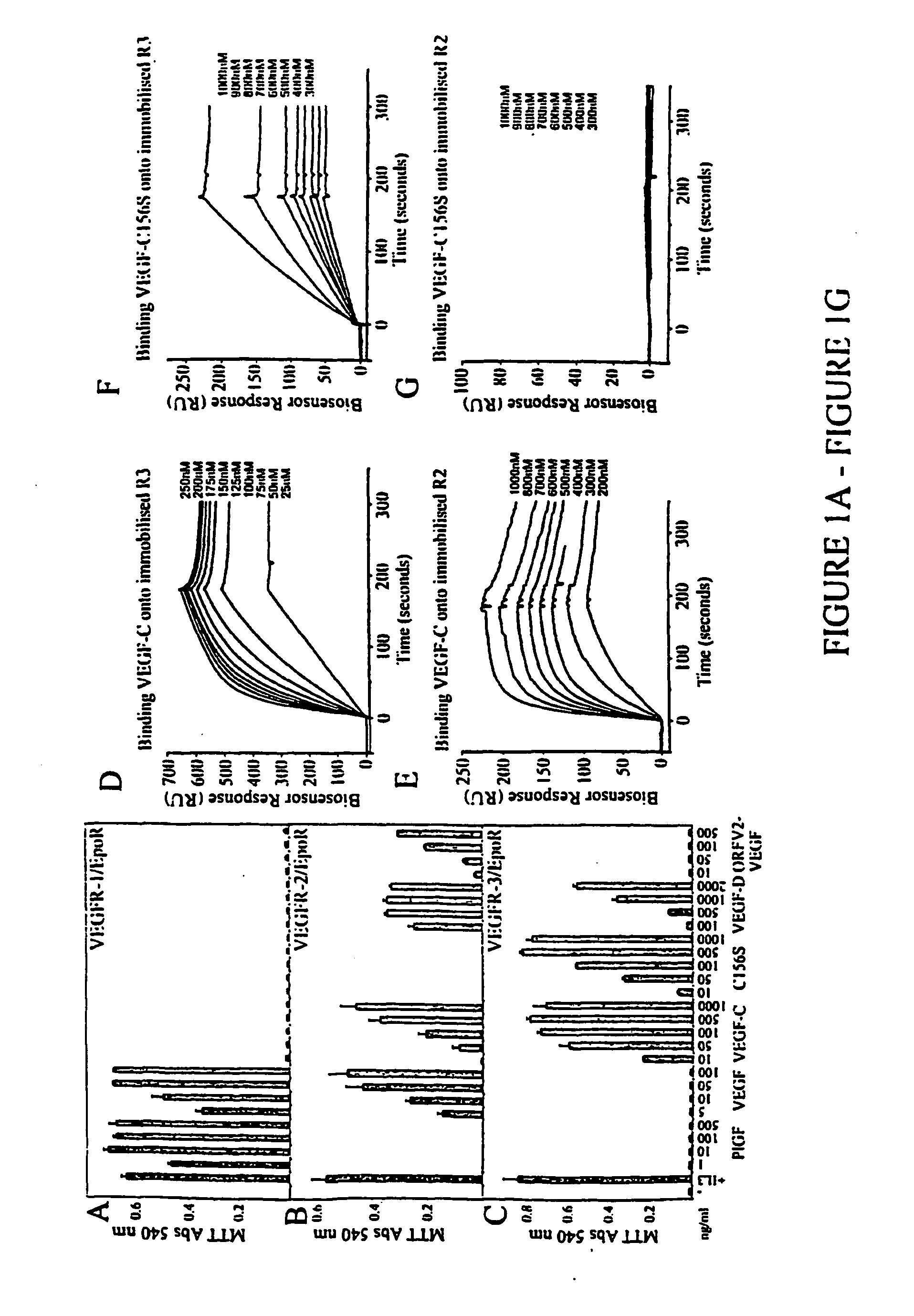
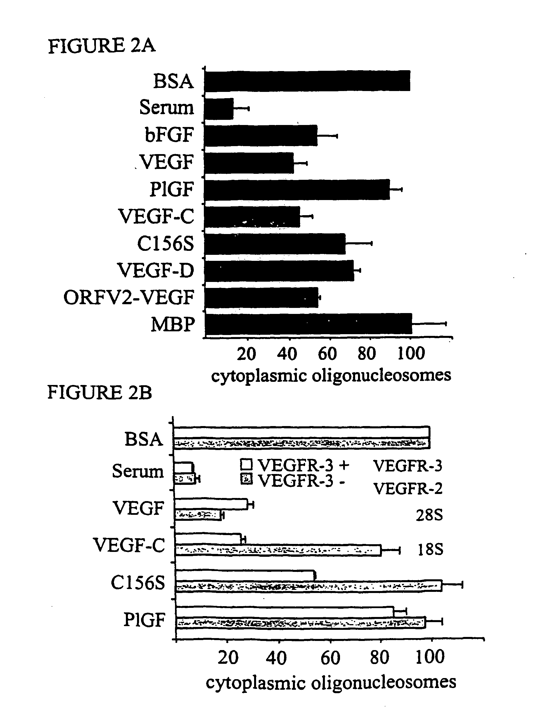
[0233] VEGF is an endothelial cell mitogen which has been also shown to protect endothelial cells from starvation and TNF-a induced apoptosis via activation of VEGFR-2 (Gerber et al., J. Biol Chem., 273:30336-30343, 1998; Spyridopoulos et al., J. Mol. Cell. Cardiol., 29:1321-1330, 1997). The abilities of the different VEGFRs to promote endothelial cell survival were compared by using VEGFR specific VEGFs. The specificities of the growth factors used were determined using a cell survival bioassay. For the bioassay, Ba / F3 pre-B cells were stably transfected with a chimeric receptor containing the extracellular domain of human VEGFR-1, VEGFR-2 or VEGFR-3 fused with the transmebrane and cytoplasmic domains of the mouse erythropoietin receptor.

[0234] As expected, only VEGF and PIGF were able to induce the survival of VEGFR-1 / EpoR cells (FIG. 1A). VEGF, VEGF-C, VEGF-D and orf viral NZ2 (ORFV2-VEGF) were able to sup...


PUM

PropertyMeasurementUnit
flow rateaaaaaaaaaa
concentrationsaaaaaaaaaa

Abstract

The present invention is directed to methods and compositions for isolating lymphatic endothelial cells from a mixed population of cells. More particularly, the inventors have found that certain antibodies that recognize the extracellular domain of VEGFR-3 can be used to specifically isolated lymphatic endothelial cells substantially free of other contaminating non-lymphatic endothelial cells. Methods and compositions for producing such cells and using such cells are described.

Description

FIELD OF THE INVENTION [0001] The present invention relates generally to materials and methods relating to the isolation of endothelial cells and, cells isolated according to the present invention. More specifically, the present invention is directed to obtaining populations of isolated lymphatic endothelial cells. BACKGROUND OF THE INVENTION [0002] The lymphatic system is a complex structure organized in parallel fashion to the circulatory system. In contrast to the circulatory system, which utilizes the heart to pump blood throughout the body, the lymphatic system pumps lymph fluid using the inherent contractility of the lymphatic vessels. The lymphatic vessels are not interconnected in the same manner as the blood vessels, but rather form a set of coordinated structures including the initial lymphatic sinuses [Jeltsch et al., Science, 276:1423-1425 (1997); and Castenholz, A., in Olszewski, W. L. (ed.), Lymph Stasis: Pathophysiology, Diagnosis, and Treatment. CRC Press: Boca Raton...

Claims


Application Information

Patent Timeline
Patent Type & Authority Applications(United States)
IPC IPC(8): A61K48/00A61K39/395G01N33/567C12N5/08C12N15/09G01N33/50A61K38/00A61P17/06A61P27/02A61P29/00A61P35/00C12N5/071C12N5/10G01N33/15G01N33/53G01N33/543G01N33/566G01N33/569
CPCC12N5/069G01N2333/71G01N33/56966C12N2501/165A61P7/10A61P17/06A61P27/02A61P29/00A61P35/00A61P43/00
Inventor ALITALO, KARIMAKINEN, TAIJA
Owner LUDWIG INST FOR CANCER RES
Why Eureka
Try Eureka