Methods and apparatus for securing and deploying tissue anchors

a technology of tissue anchors and methods, applied in the field of methods and apparatus for securing and deploying tissue anchors, can solve the problems of atypical diarrhea, electrolytic imbalance, and many life-threatening problems

Active Publication Date: 2006-11-30
USGI MEDICAL
View PDF99 Cites 126 Cited by
  • Summary
  • Abstract
  • Description
  • Claims
  • Application Information

AI Technical Summary

Benefits of technology

[0014] The tissue manipulation assembly may be delivered into the patient while in a low-profile configuration, e.g., transorally, through the shape-lockable endoscopic assembly, through an endoscope, an endoscopic device, or directly. Once desirably positioned, the launch tube may be urged proximally via its proximal end at handle. Because the jaw assembly pivot and the relative positioning of the upper jaw pivot along lower jaw member and launch tube pivot along upper jaw member, the proximal movement of the launch tube may effectively articulate upper jaw into an expanded jaw configuration. Proximally urging the launch tube may also urge the lower jaw member to pivot about the assembly pivot and form an angle relative to a longitudinal axis of the tubular body. The opening of the upper jaw relative to the lower jaw creates a jaw opening for grasping or receiving tissue. Moreover, the tissue manipulation assembly may also include a stop located adjacent to the jaw assembly pivot or within the pivot itself.

Problems solved by technology

However, many conventional surgical procedures may present numerous life-threatening post-operative complications, and may cause atypical diarrhea, electrolytic imbalance, unpredictable weight loss and reflux of nutritious chyme proximal to the site of the anastomosis.
Furthermore, the sutures or staples that are often used in surgical procedures for gastrointestinal disorders typically require extensive training by the clinician to achieve competent use, and may concentrate significant force over a small surface area of the tissue, thereby potentially causing the suture or staple to tear through the tissue.
One problem with conventional gastrointestinal reduction systems is that the anchors (or staples) should engage at least the muscularis tissue layer in order to provide a proper foundation.
In other words, the mucosa and connective tissue layers typically are not strong enough to sustain the tensile loads imposed by normal movement of the stomach wall during ingestion and processing of food.
This problem of capturing the muscularis or serosa layers becomes particularly acute where it is desired to place an anchor or other apparatus transesophageally rather than intra-operatively, since care must be taken in piercing the tough stomach wall not to inadvertently puncture adjacent tissue or organs.
One of the problems associated with these types of procedures is the time and number of intubations needed to perform the various procedures endoscopically.
Another problem is the time required to complete a plication from the surrounding tissue with the body lumen.
Another problem with conventional methods involves ensuring that the staple, knotted suture, or clip is secured tightly against the tissue and that the newly created plication will not relax under any slack which may be created by slipping staples, knots, or clips.
However, many of these types of devices are typically large and unsuitable for low-profile delivery through the body, e.g., transesophageally.
Moreover, when grasping or clamping onto or upon the layers of tissue with conventional anchors, sutures, staples, clips, etc., many of these devices are configured to be placed only after the tissue has been plicated and not during the actual plication procedure.

Method used


Image

  • Methods and apparatus for securing and deploying tissue anchors
  • Methods and apparatus for securing and deploying tissue anchors
  • Methods and apparatus for securing and deploying tissue anchors

Examples


Embodiment Construction

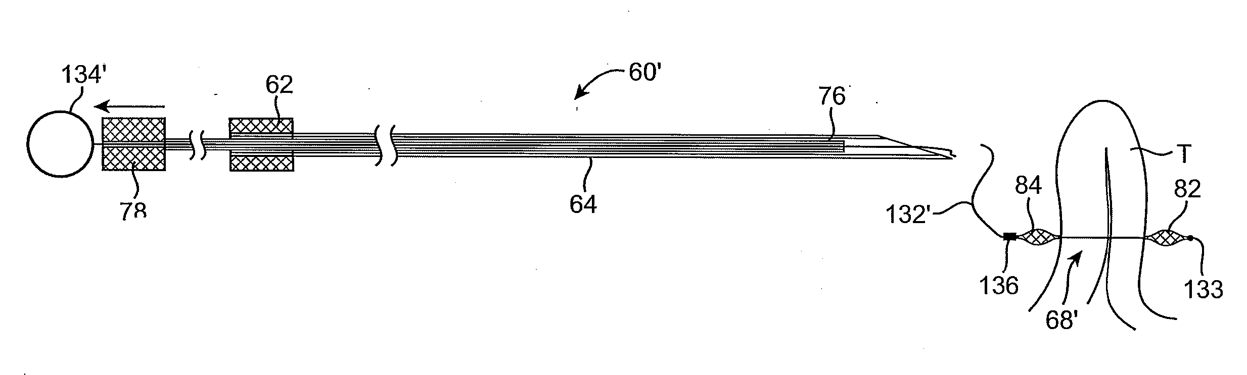
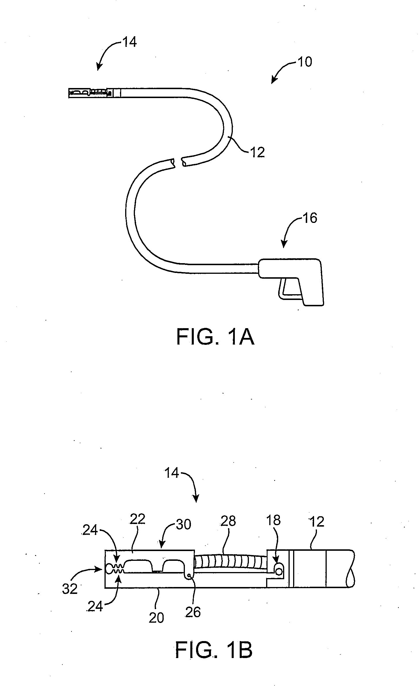
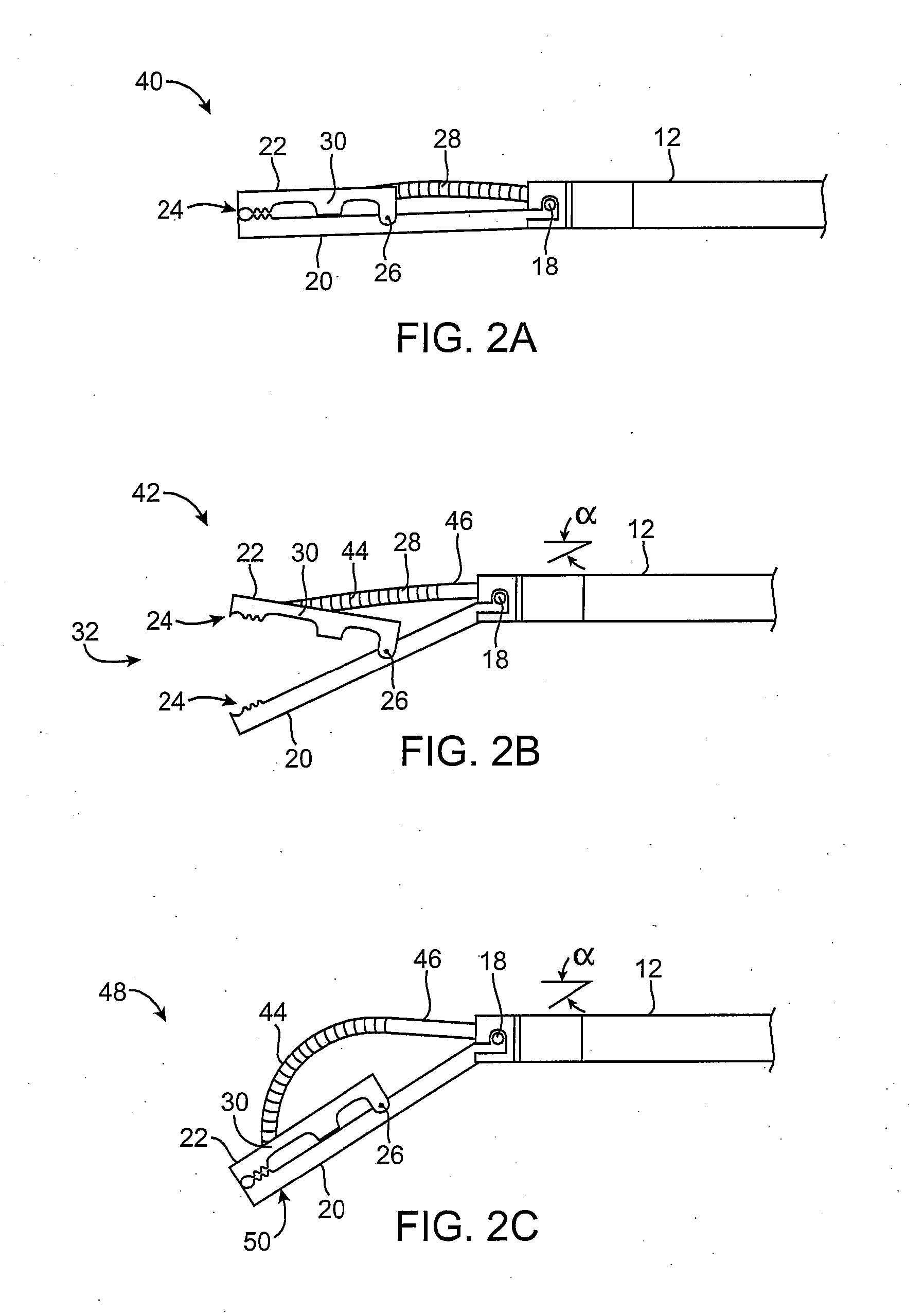
[0061] In manipulating tissue or creating tissue folds, a having a distal end effector may be advanced endoluminally, e.g., transorally, transgastrically, etc., into the patient's body, e.g., the stomach. The tissue may be engaged or grasped and the engaged tissue may be manipulated by a surgeon or practitioner from outside the patient's body. Examples of creating and forming tissue plications may be seen in further detail in U.S. patent application Ser. No. 10 / 955,245 filed Sep. 29, 2004, which has been incorporated herein by reference above, as well as U.S. patent application Ser. No. 10 / 735,030 filed Dec. 12, 2003, which is incorporated herein by reference in its entirety.

[0062] In engaging, manipulating, and / or securing the tissue, various methods and devices may be implemented. For instance, tissue securement devices may be delivered and positioned via an endoscopic apparatus for contacting a tissue wall of the gastrointestinal lumen, creating one or more tissue folds, and dep...


PUM


Abstract

Methods and apparatus for securing and deploying tissue anchors are described herein. A tissue manipulation assembly is pivotably coupled to the distal end of a tubular member. A reconfigurable launch tube is also pivotably coupled to the tissue manipulation assembly, which may be advanced through a shape-lockable endoscopic device, a conventional endoscope, or directly by itself into a patient. A second tool can be used in combination with the tissue manipulation assembly to engage tissue and manipulate the tissue in conjunction with the tissue manipulation assembly. A deployment assembly is provided for securing engaged tissue via one or more tissue anchors, the deployment assembly also being configured to disengage the anchors endoluminally or laparoscopically by applying thermal energy through at least one suture cutting element disposed along the deployment assembly.

Description

CROSS-REFERENCE TO RELATED APPLICATIONS [0001] The present application is a continuation-in-part of U.S. patent application Ser. No. 11 / 238,543 filed Sep. 30, 2005, which is a continuation-in-part of U.S. patent application Ser. No. 11 / 139,920 filed May 26, 2005, each of which is incorporated herein by reference in its entirety.FIELD OF THE INVENTION [0002] The present invention relates to methods and apparatus for securing and deploying tissue anchors. More particularly, the present invention relates to methods and apparatus for endoluminally or laparoscopically securing and deploying anchors within or against tissue, for instance, to form and / or secure tissue folds or to approximate regions of tissue, etc. BACKGROUND OF THE INVENTION [0003] A number of surgical techniques have been developed to treat various gastrointestinal disorders. One such example of a pervasive disorder is morbid obesity. Conventional surgical treatment for morbid obesity typically includes, e.g., bypassing ...

Claims


Application Information

Patent Timeline
Patent Type & Authority Applications(United States)
IPC IPC(8): A61B17/04
CPCA61B17/0401A61B17/0467A61B17/0469A61B17/0482A61B17/29A61B2017/003A61B2017/2927A61B2017/00867A61B2017/0419A61B2017/0458A61B2017/0464A61B2017/0496A61B2017/06052A61B2017/00349
Inventor EWERS, RICHARD C.LAM, CANG C.
Owner USGI MEDICAL
Why Eureka
Try Eureka