
Minimally Invasive Methods and Apparatus for Accessing and Ligating Uterine Arteries with Sutures

a uterine artery and minimally invasive technology, applied in the field of minimally invasive methods and apparatus for accessing and ligating uterine arteries with sutures, can solve the problems of fibroid shrinkage and cessation or diminution of clinical symptoms, excessive blood loss, and prolonged convalescence,

Inactive Publication Date: 2006-12-07
AMS RES CORP
View PDF29 Cites 65 Cited by
  • Summary
  • Abstract
  • Description
  • Claims
  • Application Information

AI Technical Summary

Benefits of technology

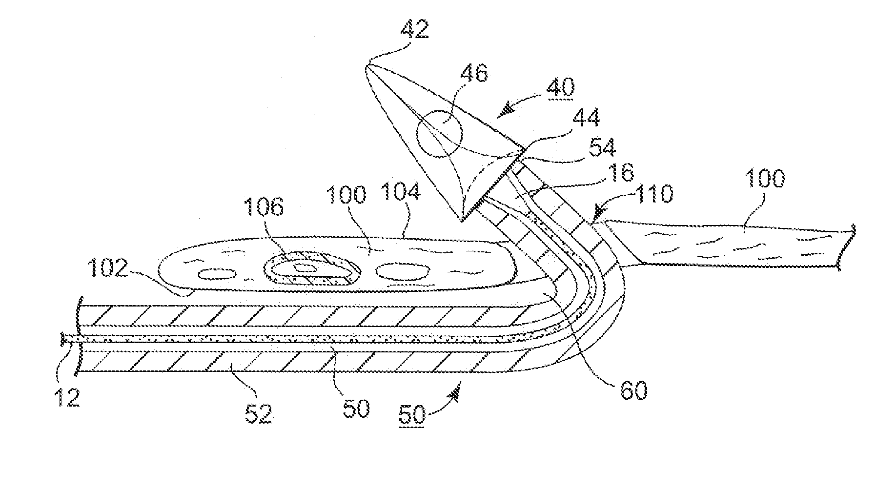
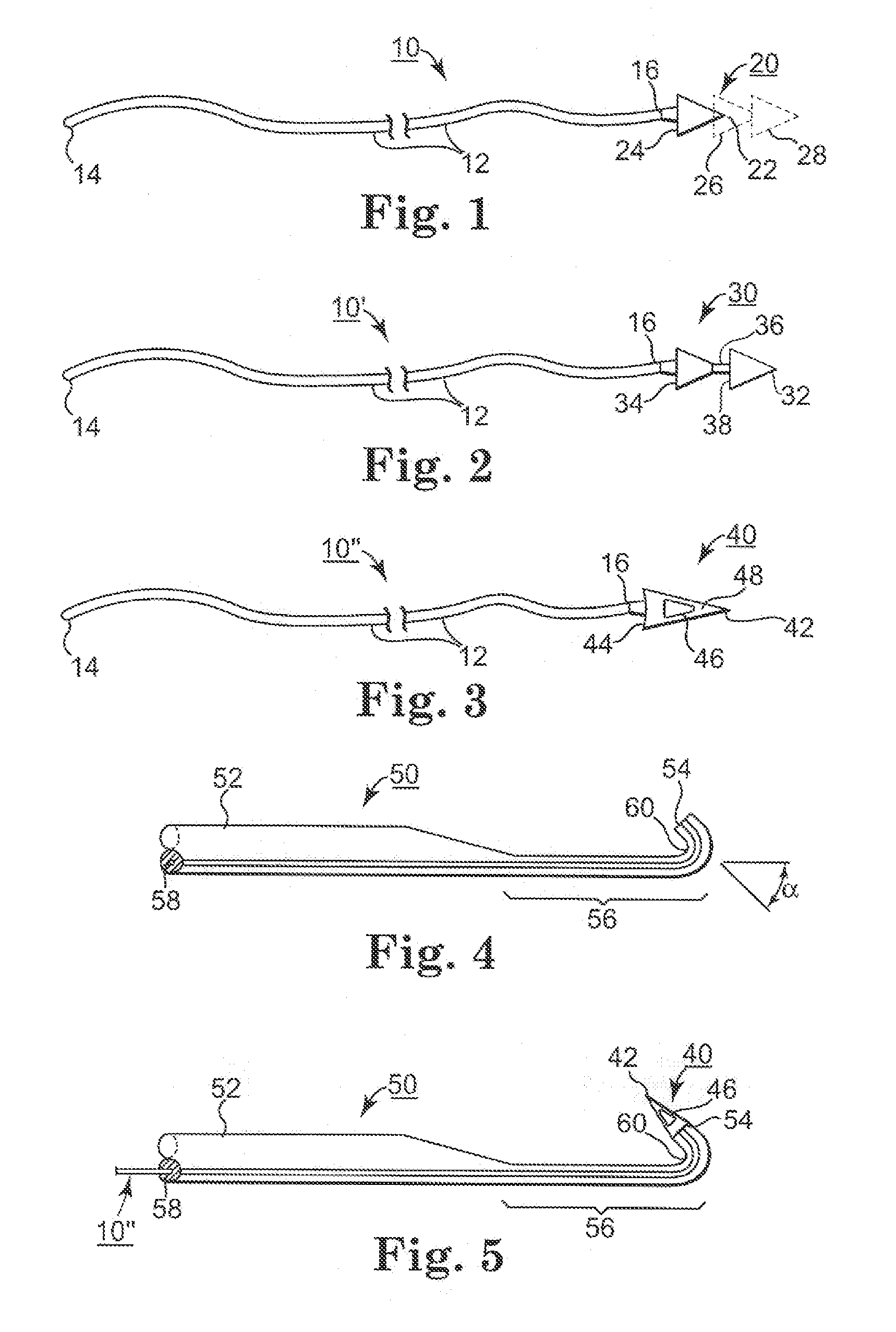
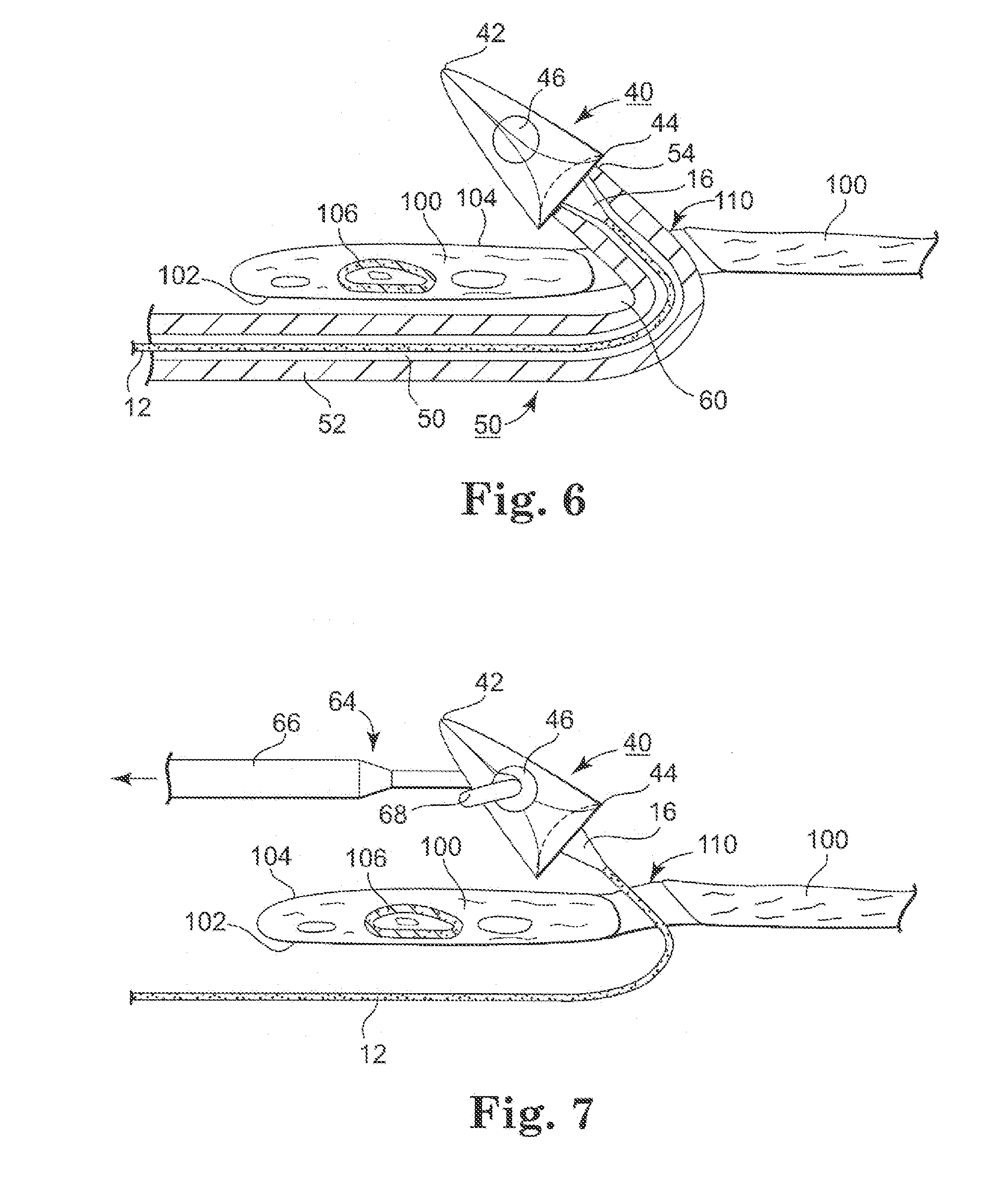
[0019] Preferably, the suture distal end is coupled to a tissue-penetrating tip, e.g., a needle or barb or hook or cone or dart, (hereinafter referred to as a “tissue-penetrating barb” that facilitates passage through ligamentous tissue supporting the uterine artery so that the suture distal end can be engaged on the second ligament side beyond the fornix incision and withdrawn proximally. For convenience, the suture coupled to a tissue-penetrating barb is referred to herein as a suture assembly.
[0025] Advantageously, procedures for occluding uterine arteries are simplified through the use of the instruments of the present invention to shorten the surgical time and trauma to the patient. The instruments of the present invention may be advantageously employed in ligating uterine arteries exposed through a conventional trans-vaginal surgical approach surrounding the cervix that is well known to and practiced by many surgeons. Moreover, it is not necessary in the practice of the present invention to employ instruments or equipment for locating the uterine arteries and verifying successful occlusion of the uterine arteries blocking blood flow.

Problems solved by technology

Thus, in addition to the loss of reproductive capability, a hysterectomy requires major invasive surgery that can involve excessive blood loss, prolonged convalescence, attendant pain and discomfort, and economic costs.
In most instances, the relatively faster growing fibroid cells require a higher volume of blood oxygen and die when starved of oxygen, leading to fibroid shrinkage and cessation or diminution of clinical symptoms.

Method used


Image

  • Minimally Invasive Methods and Apparatus for Accessing and Ligating Uterine Arteries with Sutures
  • Minimally Invasive Methods and Apparatus for Accessing and Ligating Uterine Arteries with Sutures
  • Minimally Invasive Methods and Apparatus for Accessing and Ligating Uterine Arteries with Sutures

Examples


Embodiment Construction

[0049] In the following detailed description, references are made to illustrative embodiments of methods and apparatus for carrying out the invention. It is understood that other embodiments can be utilized without departing from the scope of the invention. The surgical procedures, tools and kits of the present invention are not necessarily limited to but are particularly suited to effect uterine artery ligation in a minimally invasive manner to treat uterine fibroids or other conditions of the uterus. Thus, preferred methods and apparatus are described for controlling uterine arterial blood flow by diminishing or blocking arterial blood flow to the uterus to treat diseases and disorders, e.g., uterine fibroids and uterine bleeding.

[0050] The methods of the preferred embodiments of the present invention involve making incisions through the fornix to access the uterine arteries and cardinal ligaments. One suitable approach to accessing the right and left uterosacral and cardinal lig...


PUM


Abstract

Surgical procedures, tools, and kits particularly suited to effect uterine artery ligation in a minimally invasive manner to treat uterine fibroids or other conditions of the uterus are disclosed. An incision through the fornix is made to access the uterine arteries supported by the cardinal and uterosacral ligaments. A distal end of one or more sutures is passed around each uterine artery and a portion of the ligament supporting and enclosing the uterine artery, wherein the portion may include a uterine vein. The passage is effected by directing a suture distal end along a first side of the portion of the ligament beyond to the fornix incision, then through the ligament, and then back proximally along the second ligament side toward the fornix incision.

Description

[0001] The present application claims the benefit of U.S. Provisional Application No. 60 / 687,678 filed Jun. 6, 2005 (the entire contents incorporated by reference herein).FIELD [0002] The present invention pertains to surgical procedures, tools, and kits particularly suited to effect uterine artery ligation in a minimally invasive manner to treat uterine fibroids or other conditions of the uterus. BACKGROUND [0003] Each year, many women undergo a surgical removal of the uterus (hysterectomy) due to the growth of muscular tumors of the uterus (leiomyoma or uterine fibroids) or for uterine cancer, adenomyosis, menorrhagia, uterine prolapse, and dysfunctional uterine bleeding (abnormal menstrual bleeding that has no discrete anatomic explanation such as a tumor or growth). The uterus has a pear-shaped, uterine body extending between a fundus extending right and left to junctions with the right and left Fallopian tubes and a uterine neck (cervix) that extends to the vagina. The uterus h...

Claims


Application Information

Patent Timeline
Patent Type & Authority Applications(United States)
IPC IPC(8): A61B17/04
CPCA61B17/0469A61B17/0483A61B17/12013A61B2017/06042A61B2017/0475A61B2017/06028A61B2017/047
Inventor ARNAL, KEVIN R.MUJWID, JAMES R.
Owner AMS RES CORP
Learn More