Glaucoma stent system

a stent system and glaucoma technology, applied in the field of improved medical devices and, can solve the problems and achieve the effects of blurred vision, headache, and untreated blindness

Inactive Publication Date: 2007-01-11
GLAUKOS CORP
View PDF99 Cites 134 Cited by
  • Summary
  • Abstract
  • Description
  • Claims
  • Application Information

AI Technical Summary

Benefits of technology

[0023] The trabecular meshwork and juxtacanilicular tissue together provide the majority of resistance to the outflow of aqueous and, as such, are logical targets for surgical removal in the treatment of open-angle glaucoma. In addition, minimal amounts of tissue are altered and existing physiologic outflow pathways are utilized. Trabecular bypass surgery has the potential for much lower risks of choroidal hemorrhage, infection and uses existing physiologic outflow mechanisms. This surgery could be performed under topical anesthesia in a physician's office with rapid visual recovery.
[0042] One of the advantages of trabecular bypass surgery, as disclosed herein, and the use of a stent implant to bypass diseased trabecular meshwork at the level of trabecular meshwork and thereby use existing outflow pathways is that the treatment of glaucoma is substantially simpler than in existing therapies. A further advantage of some preferred embodiments is the utilization of simple microsurgery that may be performed on an outpatient basis with rapid visual recovery and greatly decreased morbidity. Finally, a distinctly different approach is used than is found in existing implants. Physiological outflow mechanisms are used or re-established by the implant of some preferred embodiments, in contra-distinction with previously disclosed methodologies.

Problems solved by technology

Glaucoma is a group of eye diseases that causes pathological changes in the optic disk and corresponding visual field loss resulting in blindness if untreated.
However, there are secondary open-angle glaucomas that may include edema or swelling of the trabecular spaces (from steroid use), abnormal pigment dispersion, or diseases such as hyperthyroidism that produce vascular congestion.
However, these various drug therapies for glaucoma are sometimes associated with significant side effects, such as headache, blurred vision, allergic reactions, death from cardiopulmonary complications and potential interactions with other drugs.
However, long-term review of surgical results showed only limited success in adults.
In retrospect, these procedures probably failed secondary to repair mechanisms and a process of “filling in”.
The filling in is the result of a healing process which has the detrimental effect of collapsing and closing in of the created opening throughout the trabecular meshwork.
Once the created openings close, the pressure builds back up and the surgery fails.
However, the relatively small hole created by this trabeculopuncture technique exhibits a filling in effect and fails.
Although morbidity was zero in both trials, success rates did not warrant further human trials.
Failure again was from filling in of created defects in trabecular meshwork by repair mechanisms.
The risk of placing a glaucoma drainage implant also includes hemorrhage, infection and postoperative double vision that is a complication unique to drainage implants.
All of the above techniques and variations thereof have numerous disadvantages and moderate success rates.
They involve substantial trauma to the eye and require great surgical skill by creating a hole over the full thickness of the sclera / cornea into the subconjunctival space.
Furthermore, normal physiological outflow pathways are not used.

Method used


Image

  • Glaucoma stent system
  • Glaucoma stent system
  • Glaucoma stent system

Examples


Embodiment Construction

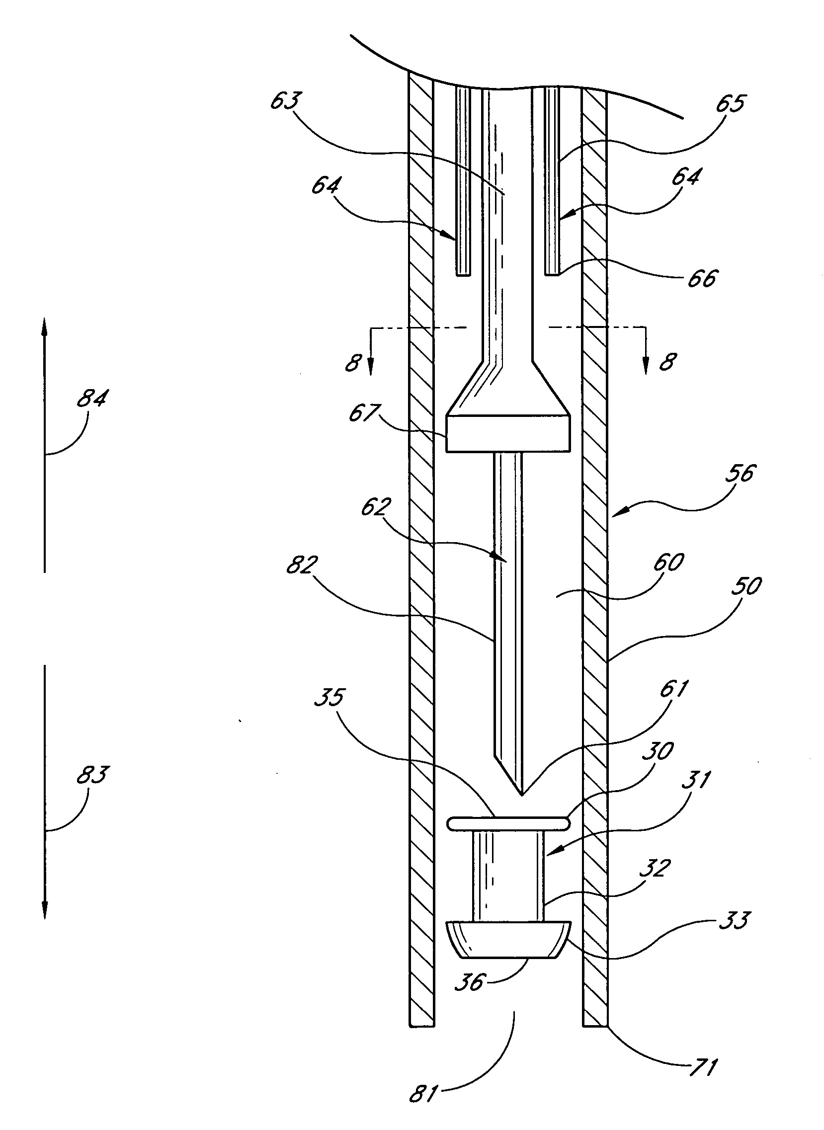
[0070] The drawings generally illustrate a method for the treatment of glaucoma by trabecular bypass surgery. In particular, a stent implant is used to bypass diseased or deficient trabecular meshwork at the level of trabecular meshwork to use or restore existing outflow pathways and methods thereof are disclosed.

[0071] While the description sets forth various embodiment specific details, it will be appreciated that the description is illustrative only and should not be construed in any way as limiting the invention. Furthermore, various applications of the invention, and modifications thereto, which may occur to those who are skilled in the art, are also encompassed by the general concepts described herein and below.

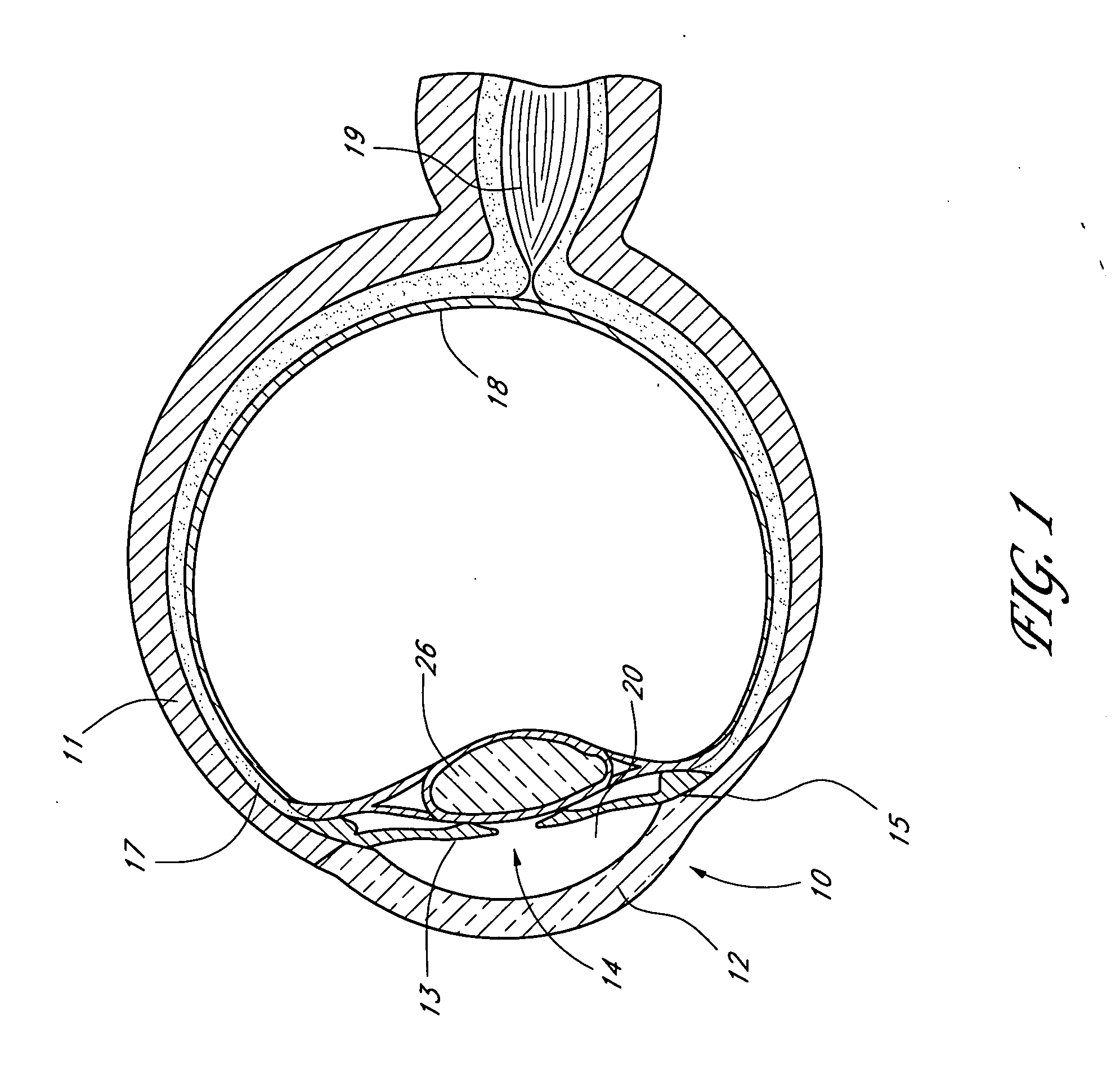
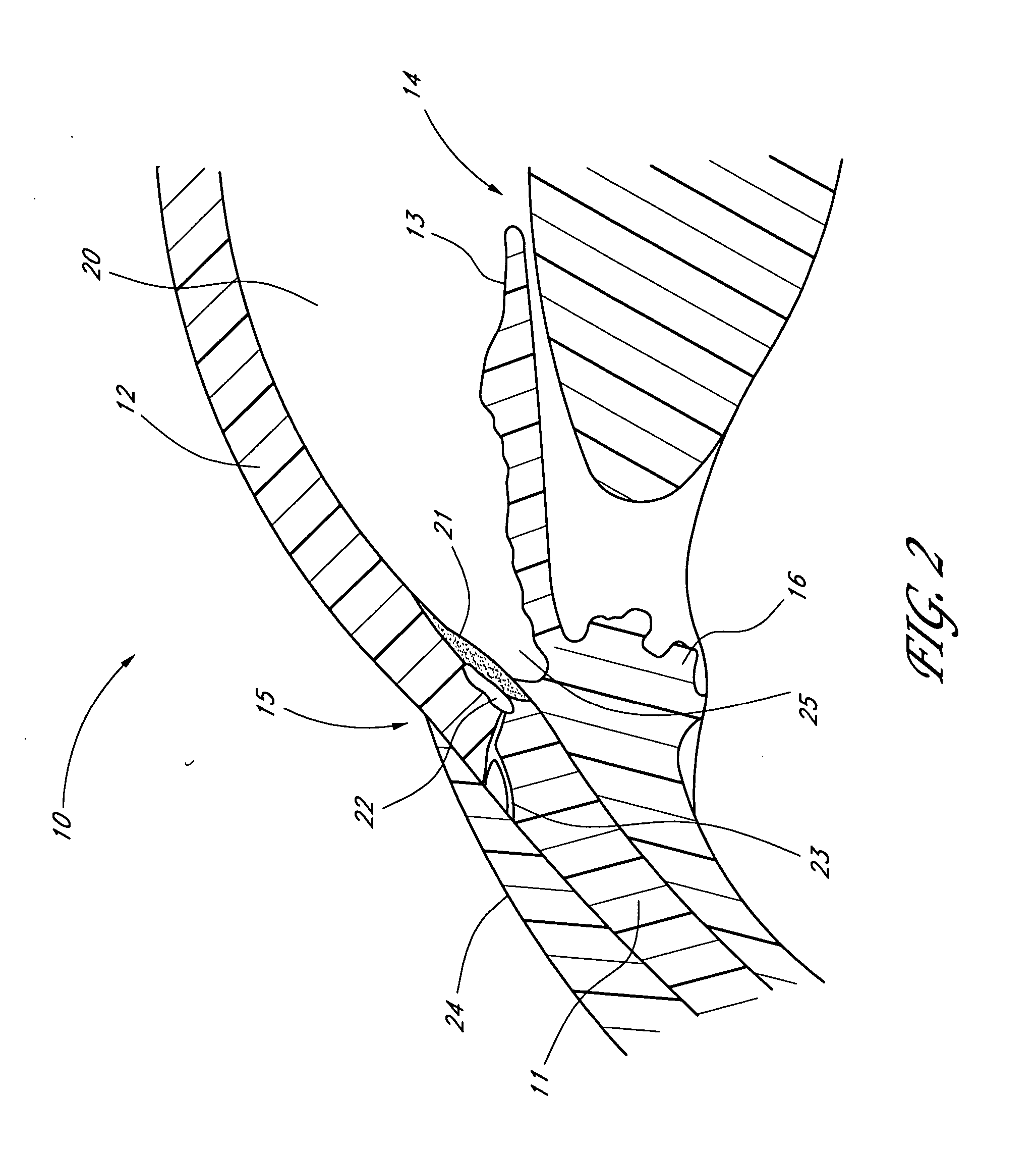
[0072] For background illustration purposes, FIG. 1 shows a sectional view of an eye 10, while FIG. 2 shows a close-up view, showing the relative anatomical locations of a trabecular meshwork 21, an anterior chamber 20, and Schlemm's canal 22. Thick collagenous tissue...


PUM


Abstract

Surgical methods and related medical devices for treating glaucoma are disclosed. The method comprises trabecular bypass surgery, which involves bypassing diseased trabecular meshwork with the use of a stent implant. The stent implant is inserted into an opening created in the trabecular meshwork by a piercing member that is slidably advanceable through the lumen of the stent implant for supporting the implant insertion. The stent implant is positioned through the trabecular meshwork so that an inlet end of the stent implant is exposed to the anterior chamber of the eye and an outlet end is positioned into fluid collection channels at about an exterior surface of the trabecular meshwork or up to the level of aqueous veins.

Description

RELATED APPLICATIONS [0001] This application is a continuation of U.S. application Ser. No. 10 / 231,342, filed Aug. 28, 2002, which claims the benefit of U.S. Provisional Application No. 60 / 315,463, filed Aug. 28, 2001, entitled “GLAUCOMA SHUNT FOR AB INTERNO USE” and U.S. Provisional Application No. 60 / 363,980, filed Mar. 14, 2002, entitled “MEANS AND PROCEDURES FOR IMPLANTING A GLAUCOMA SHUNT AB INTERNO”, the entirety of each one of which is hereby incorporated by reference herein.BACKGROUND OF THE INVENTION [0002] 1. Field of the Invention [0003] The invention generally relates to improved medical devices and methods for the reduction of elevated pressure in organs of the human body. More particularly, the invention relates to the treatment of glaucoma by trabecular bypass surgery, which is a means for using an implant or stent, such as a micro stent, shunt or the like, to bypass diseased trabecular meshwork at the level of trabecular meshwork and use / restore existing outflow path...

Claims


Application Information

Patent Timeline
Patent Type & Authority Applications(United States)
IPC IPC(8): A61F11/00A61F9/007
CPCA61F9/00781A61F2/95A61F9/007A61F2002/9505A61F2002/9511A61M27/002
Inventor TU, HOSHENGHAFFNER, DAVID S.SMEDLEY, GREGORY T.NIKSCH, BARBARA A.GHARIB, MORTEZA
Owner GLAUKOS CORP
Why Eureka
Try Eureka