
Endovenous access and guidance system utilizing non-image based ultrasound

a non-image based ultrasound and access control technology, applied in the direction of angiography, catheters, applications, etc., can solve the problems of perforation of the heart and reduced effectiveness of the devi

Active Publication Date: 2007-01-18
TELEFLEX LIFE SCI LTD
View PDF103 Cites 212 Cited by
  • Summary
  • Abstract
  • Description
  • Claims
  • Application Information

AI Technical Summary

Benefits of technology

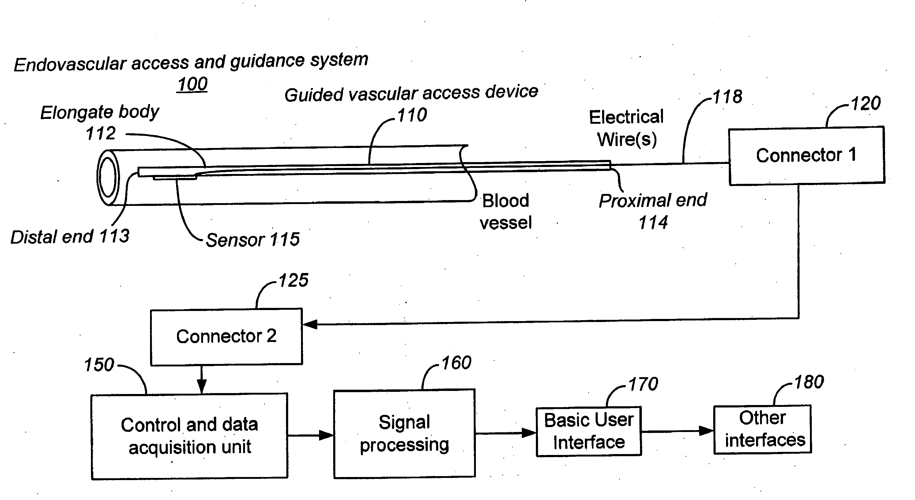
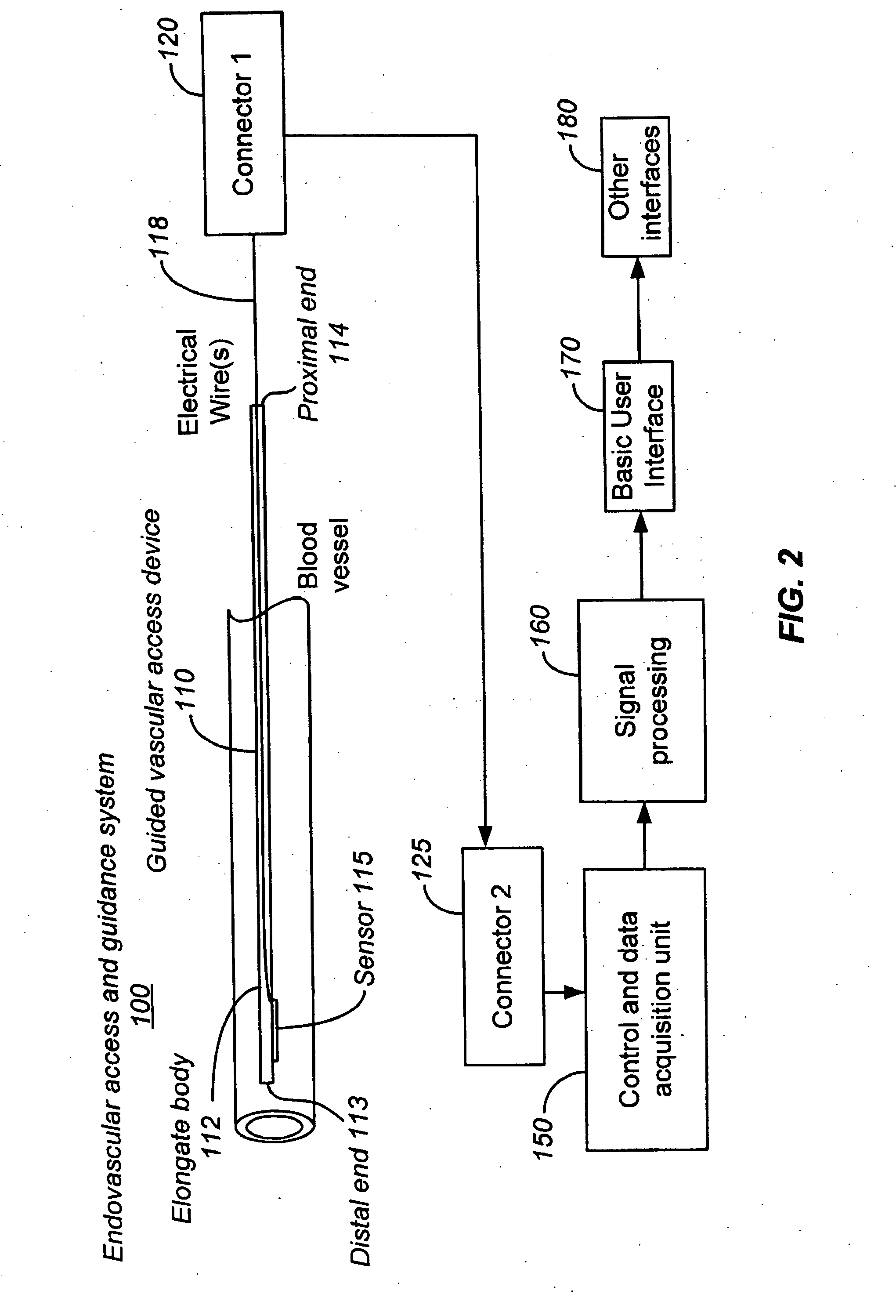
[0022] An aspect of the invention includes an endovenous access and guidance system. The system comprises: an elongate flexible member adapted and configured to access the venous vasculature of a patient; a sensor disposed at a distal end of the elongate flexible member and configured to provide in vivo non-image based ultrasound information of the venous vasculature of the patient; a processor configured to receive and process in vivo non-image based ultrasound information of the venous vasculature of the patient provided by the sensor and to provide position information regarding the position of the distal end of the elongate flexible member within the venous vasculature of the patient; and an output device adapted to output the position information from the processor. In some embodiments, the elongate flexible member is further adapted to provide a catheter, a guidewire, and / or a stylet. In other embodiments, the device is adapted to deliver therapy to a patient, or provide venous access for another device. In still another embodiment, the system is adapted to further comprise a sensor attachment mechanism adapted to removably detach the sensor from the elongate flexible member while the elongate flexible member remains in the vasculature of the patient. In yet another embodiment, the system is configured such that the processor processes in vivo non-image based ultrasound information of the vasculature system of the patient provided by the sensor to indicate in the output information the proximity of the sensor to a structure within the vasculature of the patient. In still other embodiments, the processor can be further configured to process in vivo non-image based ultrasound information of the vasculature system of the patient to indicate in the output information movement of the elongate flexible member in a desired direction within the vasculature of the patient. Alternatively, the processor is further configured to process in vivo non-image based ultrasound information of the vasculature system of the patient based on a parameter selected from a group consisting of: a venous blood flow direction, a venous blood flow velocity, a venous blood flow signature pattern, a pressure signature pattern, A-mode information and a preferential non-random direction of flow. In another aspect of the invention, the system further comprises a divergent lens associated with the sensor, or a plurality of lenses associated with the sensor. The divergent lens can be adapted to attach the sensor to the elongate flexible member. The sensor may be further configured such that it is a first sensor adapted to transmit an ultrasound signal, the system further comprising a second sensor adapted to receive non-image based ultrasound information. Additionally, a sensor drive mechanism adapted drive the sensor in a plurality of ultrasound transmission modes can be provided. In some embodiments, the sensor is a first sensor, the system further comprising a second sensor disposed at the distal end of the elongate flexible member and configured to provide in vivo non-image based ultrasound information of the venous vasculature of the patient to the processor. In other embodiments, a centering element adapted to substantially center the distal end of the elongate member within a vessel. In some instances, two or more additional sensors wherein the sensor and the two or more additional sensors are attached to the elongate flexible member in an arrangement that mimics an endovascular junction. Additionally, a steering element for directing the device tip in response to feedback information derived from the acquired data. Further embodiments can include a torque control element for directing the device tip in response to feedback information derived from the acquired data.

Problems solved by technology

One challenge common to all vascular-based therapies is health care provider access to the specific location or section of the vascular tree.
Even though it is routine, it is a very time and labor-intensive procedure for the hospital staff, which also makes it expensive.
If the catheter tip moves, not only its effectiveness diminished but, in some situations, it can perforate the heart.

Method used


Image

  • Endovenous access and guidance system utilizing non-image based ultrasound
  • Endovenous access and guidance system utilizing non-image based ultrasound
  • Endovenous access and guidance system utilizing non-image based ultrasound

Examples


Embodiment Construction

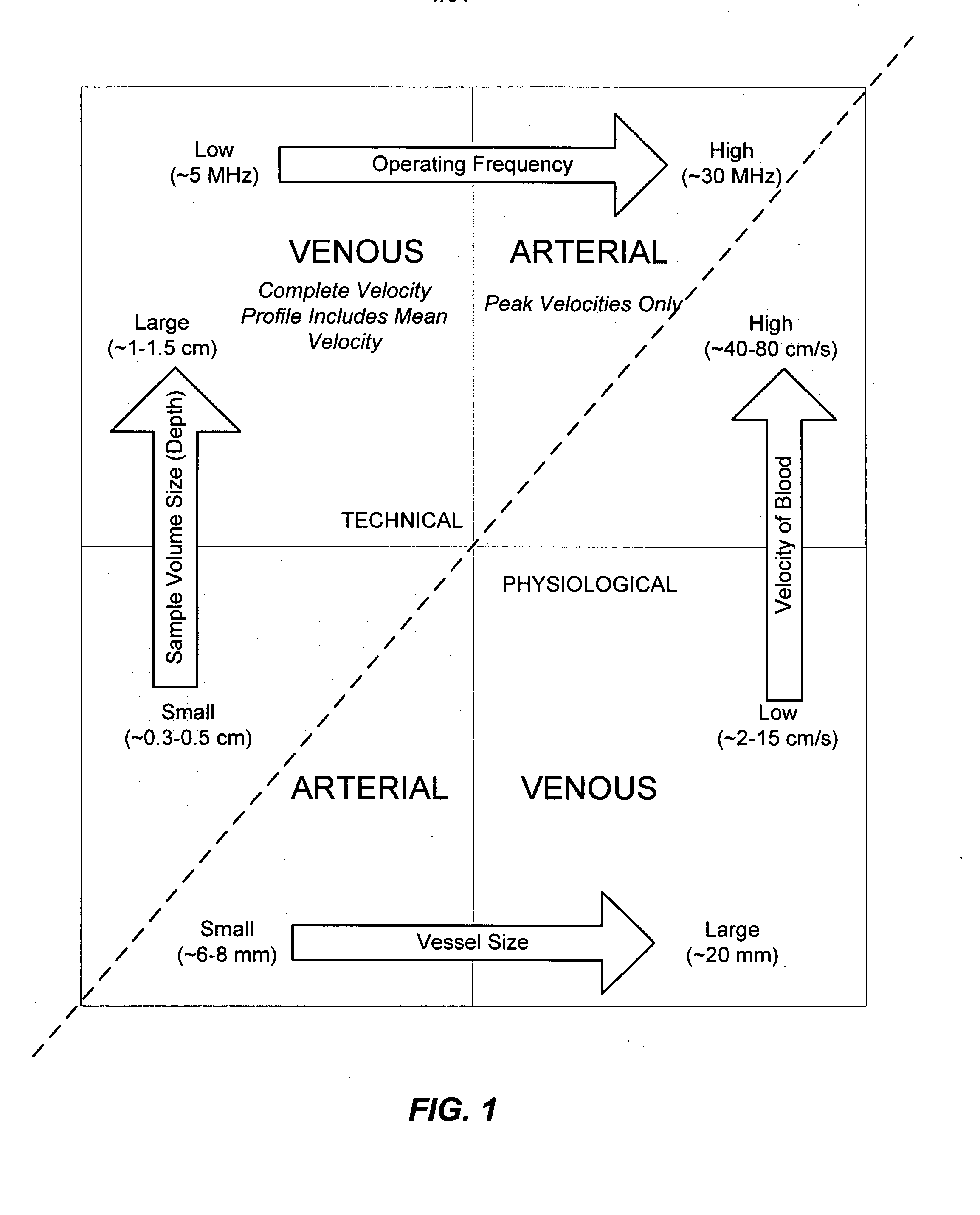
[0078] Embodiments of the present invention provide guided vascular access devices, systems for processing signals from the guided vascular access devices and user interface for providing information to a user based on outputs from the processing system. Other aspects of embodiments the invention relate to the use of intravascularly measured physiological parameters for locating, guiding, and placing catheters in the vasculature (see FIG. 1). In one aspect, the present invention relates to a catheter assembly with built-in sensors for measuring of physiological parameters such as blood flow, velocity, or pressure. In a different aspect, the present invention relates to data processing algorithms that can identify and recognize different locations in the vasculature based on the pattern of physiological parameters measured at that location. In a third aspect, the present invention relates to an instrument that has a user interface which shows guiding and positioning information. The ...


PUM


Abstract

The invention relates to the guidance, positioning and placement confirmation of intravascular devices, such as catheters, stylets, guidewires and other flexible elongate bodies that are typically inserted percutaneously into the venous or arterial vasculature. Currently these goals are achieved using x-ray imaging and in some cases ultrasound imaging. This invention provides a method to substantially reduce the need for imaging related to placing an intravascular catheter or other device. Reduced imaging needs also reduce the amount of radiation that patients are subjected to, reduce the time required for the procedure, and decrease the cost of the procedure by reducing the time needed in the radiology department. An aspect of the invention includes, for example, an endovenous access and guidance system. The system comprises: an elongate flexible member adapted and configured to access the venous vasculature of a patient; a sensor disposed at a distal end of the elongate flexible member and configured to provide in vivo non-image based ultrasound information of the venous vasculature of the patient; a processor configured to receive and process in vivo non-image based ultrasound information of the venous vasculature of the patient provided by the sensor and to provide position information regarding the position of the distal end of the elongate flexible member within the venous vasculature of the patient; and an output device adapted to output the position information from the processor.

Description

CROSS-REFERENCE [0001] This application claims the benefit of U.S. Provisional Patent Application No. 60 / 678,209 filed on May 6, 2005 by Sorin Grunwald, et al., entitled “Method and Apparatus for Intravascular Catheter Guiding and Positioning” and to U.S. Provisional Patent Application Ser. No. 60 / 682,002 filed on May 18, 2005, also by Sorin Grunwald, et al., entitled “Method and Apparatus for Intravascular Catheter Guiding and Positioning” each of which is incorporated herein by reference in its entirety.BACKGROUND OF THE INVENTION [0002] The invention relates to the guidance, positioning and placement confirmation of intravascular devices, such as catheters, stylets, guidewires and other elongate bodies that are typically inserted percutaneously into the venous or arterial vasculature, including flexible elongate bodies. Currently these goals are achieved using x-ray imaging and in some cases ultrasound imaging. This invention provides a method to substantially reduce the need for...

Claims


Application Information

Patent Timeline
Patent Type & Authority Applications(United States)
IPC IPC(8): A61B8/14
CPCA61B5/02152A61B5/06A61B5/1459A61B8/06A61B8/0841A61B8/12A61B2019/5276A61B8/445A61B8/4472A61B8/461A61B19/5244A61B2017/00106A61B8/42
Inventor GRUNWALD, SORINHILL, BRADLEY
Owner TELEFLEX LIFE SCI LTD
Learn More