
System and method for the identification and quantification of a biological sample suspended in a liquid

a biological sample and liquid technology, applied in the field of system and method for the identification and quantification of biological samples suspended in liquid, can solve the problems of difficulty still present in such methods, inability to identify all possible species, and inability to perform identification with high confidence, so as to reduce the cost of material

Inactive Publication Date: 2007-02-15
POCARED DIAGNOSTICS
View PDF38 Cites 148 Cited by
  • Summary
  • Abstract
  • Description
  • Claims
  • Application Information

AI Technical Summary

Benefits of technology

[0013] Accordingly, it is an object of the present invention to provide a system that identifies and quantifies a biological sample in a fluid. It is a further object of the present invention to provide such a system that performs the analysis in a rapid manner. Another object of the present invention is to provide a system for identifying and quantifying a biological sample in a fluid that does not require the use of reagents in order to reduce the cost of material.

Problems solved by technology

Chromogenic media may be used to isolate and identify some of the microorganisms involved in human pathology, but it cannot identify all the possible species.
However, the great difficulty that still exists with such methods is the time of bacterial identification, which, for standard chemical methods using automated equipments, is between 18 and 24 hours having an isolated organism (which takes approximately an additional 24 hours).
However, the identification was not performed with high confidence.
However, each of the methods and / or systems discussed above involve either the use of reagents or requires sophisticated operator sample preparation that make the methods and / or systems more difficult to operate and more prone to operator mistakes.
The time which is needed for such sample preparation also makes the methods and / or systems discussed above unsuitable for rapid diagnostics.

Method used


Image

  • System and method for the identification and quantification of a biological sample suspended in a liquid
  • System and method for the identification and quantification of a biological sample suspended in a liquid
  • System and method for the identification and quantification of a biological sample suspended in a liquid

Examples


example 1

[0050] The following tables provide the excitation-emission matrices for a Phosphate Buffer Solution without (Table 1) and with (Table 2) Klebsiella pneumoniae at a concentration of 1.6×10⁷ CFU / mL. These excitation-emission matrices were produced using the front-face configuration of system 1 as illustrated in FIG. 3.

TABLE 1(Without Klebsiella pneumoniae)Fluorescence Intensity, Integrated CountsEmissionWavelength,Excitation Wavelengthnm2502602702802903003043.98E+023.19E+028.26E+025.01E+022.51E+023087.33E+027.46E+028.51E+023.30E+033.93E+023129.02E+027.85E+029.24E+027.13E+034.25E+021.38E+033167.92E+028.25E+028.98E+022.73E+03580E+021.31E+033209.61E+028.21E+028.42E+028.96E+022.44E+031.26E+033249.98E+028.87E+028.22E+027.64E+026.17E+031.23E+033289.49E+029.69E+028.58E+028.31E+022.72E+031.49E+033321.00E+039.47E+028.70E+028.66E+027.19E+023.65E+033369.90E+028.04E+027.66E+028.31E+026.62E+025.95E+033409.28E+027.60E+027.49E+027.12E+025.83E+023.67E+033449.92E+027.38E+026.67E+026.27E+025.90E+021...

example 2

[0053] The following tables provide the excitation-emission matrices for water (Table 3) and water with E. Coli (Table 4) at a concentration of 3.9×10⁷ CFU / mL. These excitation-emission matrices were produced using the right-angle configuration of system 1 as in FIG. 2.

TABLE 3(Water)Fluorescence Intensity, Integrated CountsEmissionWavelength,Excitation Wavelengthnm2502602702802903003009.90E+028.81E+024.30E+035.44E+023041.21E+031.04E+037.48E+021.71E+035.47E+023081.50E+031.21E+035.58E+026.75E+034.60E+023121.56E+031.27E+035.65E+026.12E+035.23E+025.80E+023161.62E+031.35E+036.40E+021.44E+031.65E+035.72E+023201.66E+031.39E+036.80E+028.85E+027.10E+035.29E+023241.52E+031.26E+037.38E+028.93E+027.16E+035.03E+023281.49E+031.24E+036.66E+028.55E+021.63E+031.54E+033321.36E+031.22E+026.32E+027.27E+027.27E+027.46E+033361.23E+039.97E+025.67E+026.88E+026.09E+027.92E+033401.09E+038.90E+025.79E+026.05E+025.61E+021.78E+033441.07E+028.68E+025.17E+025.31E+024.93E+025.87E+023489.80E+027.50E+025.15E+027.0...

example 3

[0056] The following tables provide the excitation-emission matrices for phosphate buffer solution without (Table 5) and with E. Coli (Table 6) at a concentration of 5.7×10⁷ CFU / mL. These excitation-emission matrices were produced using the front-face configuration of system 1 as illustrated in FIG. 3.

TABLE 5(Without E. Coli)Fluorescence Intensity, Integrated CountsEmissionWavelength,Excitation Wavelengthnm2702802903003049.31E+025.44E+022.75E+023088.68E+023.39E+034.00E+023128.75E+027.13E+034.13E+021.05E+033168.68E+022.58E+035.40E+028.58E+023208.46E+028.01E+022.40E+038.33E+023247.79E+027.28E+026.10E+037.83E+023287.89E+027.21E+022.60E+039.90E+023327.61E+026.93E+026.06E+022.86E+033366.90E+026.39E+025.25E+025.02E+033406.40E+025.69E+024.89E+022.77E+033445.89E+025.46E+024.52E+021.02E+033485.65E+025.49E+024.56E+028.77E+023525.82E+025.21E+024.37E+021.14E+033565.56E+025.10E+024.35E+021.07E+033605.80E+024.80E+024.31E+027.29E+023645.54E+024.71E+024.35E+027.62E+023685.29E+024.33E+023.99E+027....


PUM


Abstract

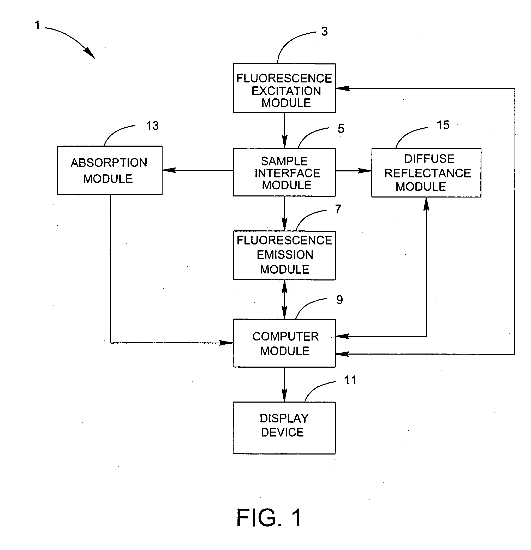
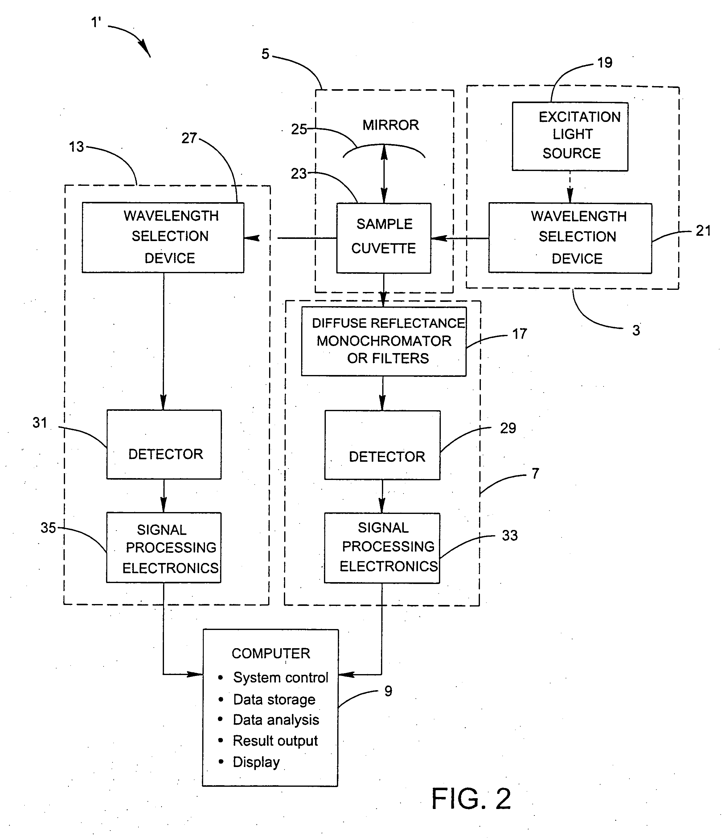
A system for the identification and quantification of a biological sample suspended in a liquid includes a fluorescence excitation module with at least one excitation light source; a sample interface module optically coupled to the fluorescence excitation module for positioning a biological sample to receive excitation light from the at least one excitation light source; a fluorescence emission module optically coupled to the sample interface module and comprising at least one detection device for detecting fluorescence excitation-emission matrices of the biological sample; and a computer module operatively coupled to the fluorescence emission module. The computer module performs multivariate analysis on the fluorescence excitation-emission matrices of the biological sample to identify and quantify the biological sample. The multivariate analysis may comprise extended partial least squared analysis for identification and quantification of the biological sample. A method for the identification and quantification of a biological sample suspended in a liquid is also provided.

Description

CROSS REFERENCE TO RELATED APPLICATIONS [0001] This application claims the benefit of U.S. Provisional Patent Application No. 60 / 706,489 entitled “System for the Identification and Quantification of Biological Sample Suspended in Liquids” filed Aug. 8, 2005, which is hereby incorporated by reference in its entirety.BACKGROUND OF THE INVENTION [0002] 1. Field of the Invention [0003] The present invention relates, in general, to a system and method for the identification and quantification of a biological sample suspended in a liquid. More specifically, the present invention relates to a system and method that utilizes multivariate analysis on fluorescence excitation-emission matrices of the biological sample to identify and quantify the biological sample. [0004] 2. Description of Related Art [0005] In bacteriology, staining methods are used to identify two general groups of bacteria, Gram positive and Gram negative, without identifying the species. Chromogenic media may be used to is...

Claims


Application Information

Patent Timeline
IPC IPC(8): C12Q1/00G06F19/00C12M1/34G01J3/42
CPCG01N21/21G01N21/31G01N21/51G01N21/645G01N21/6486G01N2201/1293G01N2021/6419G01N2021/6482G01N2021/6491G01N2201/0612G01N2201/129G01N2021/6417
Inventor BARNES, RUSSELL H.INGBER, GALGURFINKEL, JONATHAN
Owner POCARED DIAGNOSTICS
Learn More