EXAMPLE 1
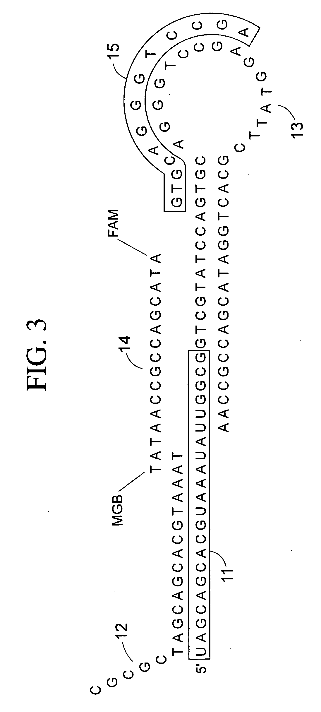
[0074] A protocol and reagents than can be used according to some embodiments of the present-teachings is shown in Table 1, proceeding from top to bottom in chronological order, (occasionally showing zeros were [reagent] is not applicable). Use of this method resulted in appropriately lower Ct values in a TaqMan® assay for miR-16 from a single stem cell, as compared to Ct values in a TaqMan® assay for miR-16 from two stem cells. The stem-loop reverse reverse transcription primer, forward primer, reverse primer, TaqMan® probe, that can be used to query miR-1 6 are: Stem-Loop Reverse Transcription Primer SEQ ID NO: 1 5′CTCAACTGGTGTCGTGGAGTCGGCAATTCAGTTGAGCGCCATA3′ Forward Primer SEQ ID NO: 2 5′ACACTCCAGCTGGGTAGCAGCACGTAATA3′ TaqMan Probe SEQ ID NO: 3 5′6-Fam-TTCAGTTGAGCCGCCAATA-MGB3′
[0075] TABLE 1 Reagent Volume (ul) [Stock] [Final] STEP1 RT 3× Mix 10× Applied Bio-systems 0.5 10 1 1.5 cDNA Archiving Kit buffer MMLV Reverse Transcriptase .335 50 3.35 (3.3 units/ul) 1.005 50 units/ul 100 mM dNTP 0.25 100 5 (100 mM/ul) 0.75 Applied Biosystems 0.065 20 0.26 (0.26 units/ul) 0.195 RNase Inhibitor 192-plex micro RNA stem- 0.5 50 5 (5 nM) 1.5 loop primer primers (50 mM) Total RNA 10 ng/ul samples 3 10 6 9 dH20 0.35 0 0 1.05 total Volume 5 15 16 C. 30 min, (42 C. 1 min-50 C. 1 sec) × 60 cycles, 85 C. 5 minutes STEP 2 PCR-based pre-amplification 2× universal master mix 12.5 2 1 37.5 (Applied Biosystems, No Ung) RT-template 5 0 0 15 192-plex new forward 2.5 500 50 (50 uM) 7.5 primer (500 nM) Universal Reverse 1.25 100 5 (5 uM) 3.75 Primer 100 uM AmpiTaq Gold (Applied 1.25 5 0.25 0.25 units/ul 3.75 Biosystems) 5 units/ul dNTP 100 mM 0.5 100 2 (2 mM) 1.5 MgCl2 100 mM 0.5 100 2 (2 mM) 1.5 dH20 1.5 0 0 4.5 total Volume 25 75 95 C. 10 min, 55 C. 2 min, (95 C. 1 sec, 65 C. 1 min) × 14 cycles 25 ul pre amplification PCR template + 75 ul dH20 = 100 ul STEP 3-Plurality of 210× Mix Decoding PCRs (TaqMans ®) 2× universal master mix 5 2 1 1050 (Applied Biosytems, No Ung) Forward Primer 5 uM + 2 10 2 1 uM FP, 200 nM 420 TaqMan ® probe (1 uM) TaqMan ® Probe Universal Reverse Primer 0.1 10 .1 (1 uM) 21 (100 uM) RT-template diluted 0.1 0 0 (1:4000) 21 following pre-amp PCR dH20 2.8 0 0 588 total Volume 10 2100 95 C. 10 min, (95 C. 15 sec, 60 C. 1 min) × 40
[0076] The present teachings further explored various cell lysis conditions effective for the isolation of micro RNA molecules. Generally, treatment at 95 C for 5 minutes was found to release micro RNAs from stem cells. Treatment for 99 C for 10 minutes was found to degrade about 50 percent of the micro RNAs released from the stem cells. Less than 1 percent of the micro RNAs were released from stem cells at 4 C. A variety of methods can be employed for isolating single cells prior to lysis, including for example aspiration with a micropipette tip, laser capture microdissection, fluorescent activated cell sorting of labeled cells (FACS), and other techniques readily available to one of ordinary skill in the art of molecular and cell biology.
[0077] In the context of stem cell identification, and of course other application areas, the present teachings contemplate embodiments in which small numbers of cells are analyzed, including a single cell. In some embodiments, the present teachings provide for analysis of one or more target micro RNA sequence molecules in a single cell. In some embodiments, the present teachings provide for analysis of one or more target micro RNA sequence molecules in five or fewer cells. In some embodiments, the present teachings provide for analysis of one or more target micro RNA sequence molecules in ten or fewer cells. In some embodiments, the present teachings provide for analysis of one or more target micro RNA sequence molecules in fifty or fewer cells. In some embodiments, the present teachings provide for analysis of one or more target micro RNA sequence molecules in one hundred and fifty or fewer cells. In some embodiments, the present teachings provide for analysis of one or more target micro RNA sequence molecules in greater than one hundred and fifty cells. As discussed supra, any of a variety of amplification strategies can be employed in the context of the present teachings for the analysis of small numbers of cells. The test samples from which such small numbers of cells can be recovered comprise conventionally fixed and stained histological and cytological preparations on microscope slides, single cells dissected from early-stage embryos generated by in vitro fertilization, microdissected needle-biopsy cores, blood samples, and forensics samples. Laser-capture microdissection is another attractive method of recovering diagnostic cells from histological preparations. Such laser-capture systems are commercially available from such sources as Arcturus (for example, the Veritas™ Microdissection Instrument).
[0078] Although the disclosed teachings have been described with reference to various applications, methods, kits, and compositions, it will be appreciated that various changes and modifications may be made without departing from the teachings herein and the claimed invention below. The foregoing examples are provided to better illustrate the disclosed teachings and are not intended to limit the scope of the teachings presented herein.
<160> NUMBER OF SEQ ID NOS: 3
<210> SEQ ID NO: 1
<211> LENGTH: 43
<212> TYPE: DNA
<213> ORGANISM: Mus musculus
<400> SEQENCE: 1
ctcaactggt gtcgtggagt cggcaattca gttgagcgcc ata 43
<210> SEQ ID NO: 2
<211> LENGTH: 29
<212> TYPE: DNA
<213> ORGANISM: Mus musculus
<400> SEQENCE: 2
acactccagc tgggtagcag cacgtaata 29
<210> SEQ ID NO: 3
<211> LENGTH: 19
<212> TYPE: DNA
<213> ORGANISM: Mus musculus
<400> SEQENCE: 3
ttcagttgag ccgccaata 19