Method and System for Automatic Lung Segmentation

a lung segmentation and automatic technology, applied in image enhancement, image analysis, instruments, etc., can solve the problems of increasing the difficulty of known conventional reconstruction methods to meet the stringent constraints of current imaging applications, requiring further manual correction, and unsatisfactory semi-automatic methods, so as to improve the efficiency of the segmentation algorithm and increase the efficiency of the lung segmentation technique.

Inactive Publication Date: 2007-06-07
SIEMENS MEDICAL SOLUTIONS USA INC
View PDF13 Cites 81 Cited by
  • Summary
  • Abstract
  • Description
  • Claims
  • Application Information

AI Technical Summary

Benefits of technology

[0011] This invention provides a systematic technique of automatically segmenting lung regions. To increase the efficiency of a lung segmentation technique, a region-based technique, such as region growing, is first performed by a computer on the middle slice of the CT volume. A contour-based technique is then used for a plurality of non-middle slices of the CT volume. This allows the implementation to be multithreaded and results in an improvement in the segmentation algorithm's efficiency

Problems solved by technology

However, it is becoming more challenging for known conventional reconstruction methods to meet the stringent constraints of current imaging applications.
The results of these semi-automatic methods can be unsatisfactory and may require further manual corrections (e.g., because of the presence of other regions of similar density.
This could bias the value of the automated threshold and, as a result, the lung may be over or under grown).
In these techniques, algorithm speed is often affected by region-growing of the lungs.
In particular, the growing of a lung region from a seed point is often computationally intensive and, as a result, the time required to perform the region growing is traditionally long relative to other operations.
Further, lung nodules typically cannot be correctly recovered by a set of morphological operations.
As a result, lung nodules are often improperly excluded from segmented lungs.

Method used


Image

  • Method and System for Automatic Lung Segmentation
  • Method and System for Automatic Lung Segmentation
  • Method and System for Automatic Lung Segmentation

Examples


Embodiment Construction

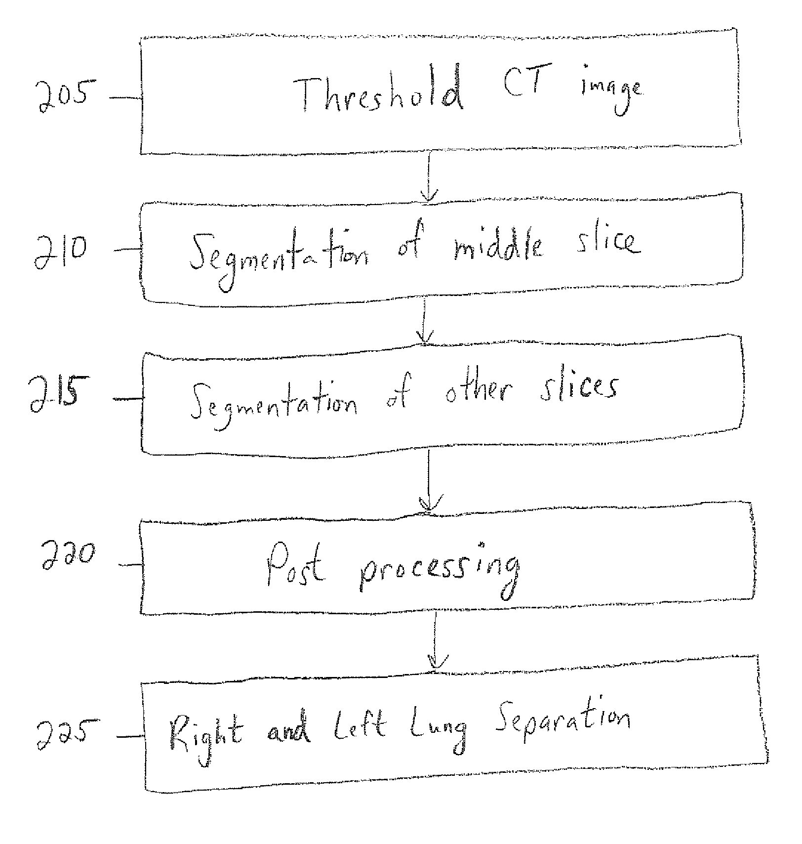
[0029] In accordance with an embodiment of the present invention, a method and apparatus for automatically segmenting lung regions are described. A contour-based method to extract lung regions in 2D includes performing region growing in a “middle slice” of the CT volume. Segmentation may be executed on a slice-by-slice basis, thereby enabling multiple slices to be segmented at substantially the same time.

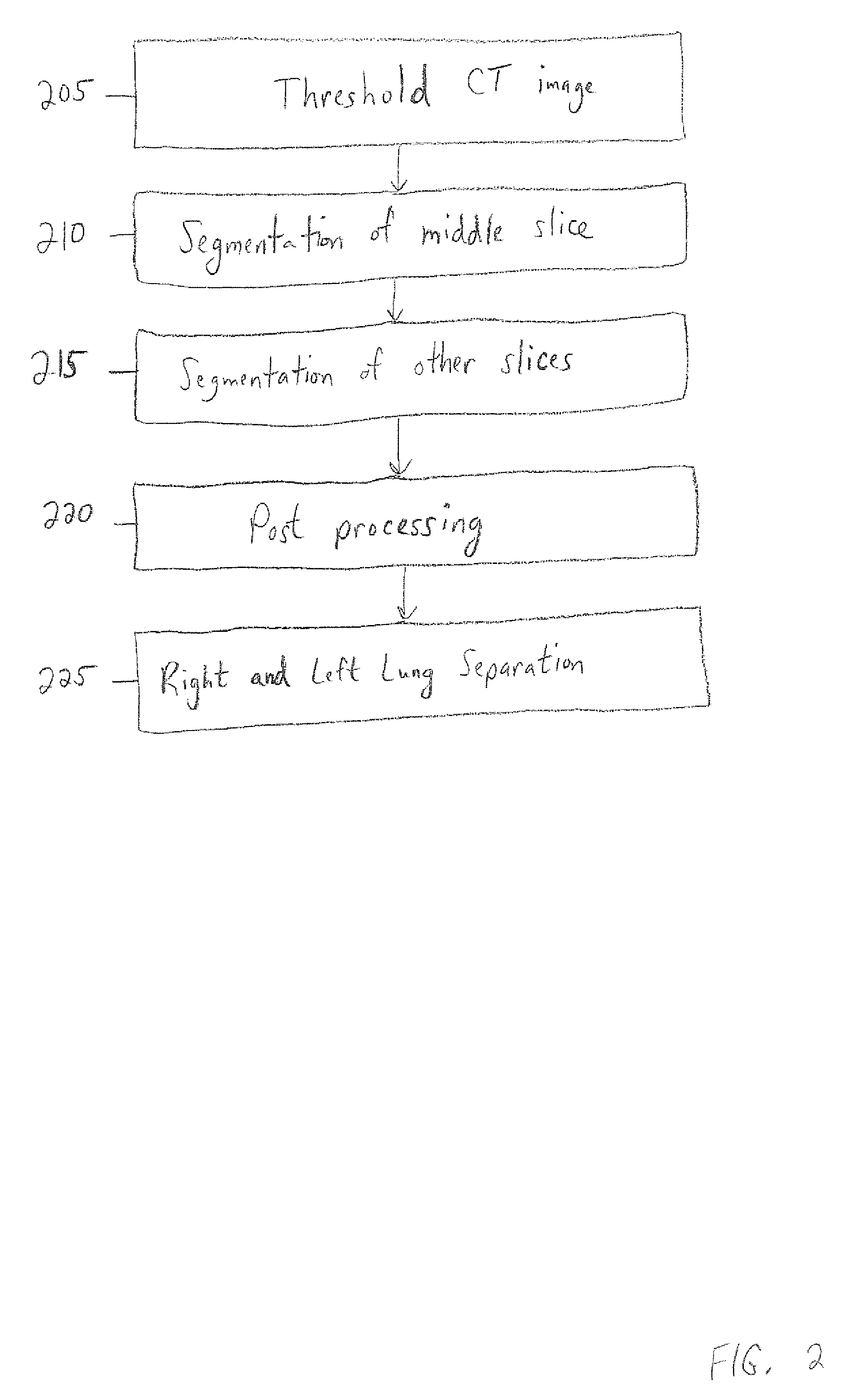
[0030]FIG. 2 is a flowchart illustrating the steps performed by a computer in accordance with an embodiment of the present invention. The computer first thresholds a CT image in step 205 using a predetermined threshold. Lung parenchyma tissues (i.e., the respiratory portions of the lung) have a density level in the range of −910 HU (i.e., Hounsfield units) to −500 HU, while the chest wall, vessel and bone are above this level. In one embodiment, the predetermined threshold is set to −400 HU. This threshold can be used to extract the lung regions (when segmenting a middle slice, as ...


PUM


Abstract

Disclosed is a systematic way of automatically segmenting lung regions. To increase the efficiency of a lung segmentation technique, a region-based technique, such as region growing, is performed by a computer on a middle slice of the CT volume. A contour-based technique is then used for a plurality of non-middle slices of the CT volume. This allows the implementation to be multithreaded and results in an improvement in the segmentation algorithm's efficiency.

Description

[0001] This application claims the benefit of U.S. Provisional Application No. 60 / 1742,439, filed Dec. 5, 2005, which is incorporated herein by reference. BACKGROUND OF THE INVENTION [0002] Computer Axial Tomography (CAT), sometimes known generally as Computerized Tomography (CT), is used in many applications, especially medical radiology, to obtain two or three dimensional views of the interior of three dimensional bodies (CT or CAT Scans). The technique involves subjecting a three dimensional body to radiation that enters the body from many different angles. The amount of radiation that is scattered or reflected by the body is then detected as a function of the angle of scattering. The scattered data is then analyzed to construct an image of the interior of the body. A two dimensional “slice” of the interior can be “reconstructed”, for example on a screen, and viewed. The slice can be reconstructed for any desired angle of intersection with the body. [0003] While computerized tomo...

Claims


Application Information

Patent Timeline
Patent Type & Authority Applications(United States)
IPC IPC(8): G06K9/00
CPCG06K9/4638G06K2209/05G06T7/0081G06T7/0083G06T2207/10081G06T2207/30061G06T7/11G06T7/12G06V10/457G06V2201/03
Inventor ODRY, BENJAMINQING, SHUPINGSHEN, HONG
Owner SIEMENS MEDICAL SOLUTIONS USA INC
Why Eureka
Try Eureka