Biomedical positioning and stabilization system

a positioning system and biomedical technology, applied in the field of medical devices, can solve the problems of significant failure rate, inability to contribute to the major goal of the surgical procedure, and left unsupported, and achieve the effects of increasing segment-to-segment friction, reducing labor intensity, and great flexibility

Inactive Publication Date: 2007-06-07
WELLAN MEDICAL SOLUTIONS +1
View PDF72 Cites 152 Cited by
  • Summary
  • Abstract
  • Description
  • Claims
  • Application Information

AI Technical Summary

Benefits of technology

[0022] In an exemplary embodiment of the present invention, a flexible armature is attached to a rigid stand placed upon the floor, or attached to another stable surface such as a bed rail, wall, ceiling or piece of equipment. A joint connects the armature to an instrument holder able to accommodate and rigidly attach an ultrasound sensing probe or other medical device. The medical device then remains rigidly attached to the described invention during the procedure. Furthermore, this set position is resistant to minor patient motion or other disturbances. If required, small alterations can be made by the operator during the procedure with minimal effort. Such adjustment may be desirable, for example, if access to a new anatomical structure is needed. In this manner, the primary operator is able to maintain a ‘hands-free’ approach.
[0023] In an illustrative embodiment, the armature section is constructed from discrete polymer segments each defining an inner lumen and interengaging hemispherical nose sections and tail sections. The hemispherical nature of the connection affords a degree of bending between segments in three dimensions as well as axial rotation between segments. In a length of approximately 6 inches to 4 feet, the armature is capable of great flexibility, allowing it to be repositioned at will against the patient's region of interest / treatment. The armature can be locked, once positioned by, use of mechanical, electrical or fluid (vacuum) mechanisms that increase segment-to-segment friction or otherwise fix the segments at their current orientation. The lumen of the armature can be provided with a conduit to allow internal routing of electronic and other leads from the probe / distal end. A ball joint can be provided at the proximal end of the armature that allows a degree of flotation to enable the armature to be lifted above the level of a recumbent patient to allow access to the probe, and to enable a moderate degree of weight-generated pressure to be applied to the region of the patient to ensure the probe tip remains effectively (but not distortingly) engaged against the region.
[0024] The holder can define a variety of structures some of which allow a wide variety of probe shapes and sizes to be removably engaged. One embodiment defines a quick-disconnect assembly with a connector formed directly on the probe and a receiving connector attached to the distal end of the armature. Another embodiment employs a clamping arrangement to secure the probe. Another embodiment employs a resilient material (such as memory foam) or inflated bladder to frictionally secure the probe. Another embodiment allows remote machine operation via controls located on the holder.
[0025] In a method for employing the support device, the practitioner (potentially a single individual) prepares the probe (an ultrasound probe in one example) and / or holder, which interconnects to a display device within view by orienting the armature near or over a recumbent patient. The practitioner can use one sterilized hand to now drape the probe and armature section with a sterile drape that is opened at a proximal end to slide over the armature and sealed at a distal end placed in engagement with a tip of the probe. At no time does the practitioner contact a non-sterile object. The practitioner now uses the sterile hand(s) to reorient the tip of the ultrasound probe into engagement with the skin region of the patient by flexing the armature to overcome predetermined friction between the interengaged nose sections and tail section of at least some of the segments to bend and rotate the segments so as to acquire a desired image at the display device. The armature section maintains a predetermined reoriented shape at least by the predetermined friction. This can be supplemented by activating a locking mechanism on the support device. The practitioner now guides a needle or other instrument to a subcutaneous target with at least one hand while viewing the image and while the ultrasound probe. During this guiding procedure, the instrument holder remains ungrasped by another hand, allowing the practitioner to maintain all focus on the guiding step while an image is maintained. If adjustment of the probe is needed, it is easily reoriented by grasping the holder, flexing and rotating the armature as needed and ungrasping the holder once proper orientation is reestablished. The guiding procedure, or another procedure can then continue with all attention focused thereon.

Problems solved by technology

Often, these devices are held by an assisting operator, but they may also be left unsupported.
The operator is therefore employed solely to support the measurement device in a given orientation, and is therefore unable to contribute to the major goal of the surgical procedure while the measurement is underway.
The major challenge for the clinician performing nerve blocks is related to finding the nerve of interest.
Because anatomy is variable, this technique results in significant failure rates, multiple needle passes, and significant potential for pain and injury to the nerve and adjacent structures.
Once a satisfactory image of the structure is acquired, subtle movements of the hand holding the probe may result in degradation of the image, requiring a repositioning of the probe.
Too much pressure tends to distort the underlying tissues, making for an inaccurate image and pinching of internal tissues that may lead to misdirection of the is needle.
Too little pressure yields a bad image.
The single-practitioner approach is rarely used in practice, both due to quality and safety concerns and also to prevailing medical practice rules and custom.
This process of covering device with a sterile “bag” adds further complexity as the practitioner is wearing sterile gloves that can become contaminated by the process of applying the sterile bag.
Thus, even this simple process requires further assistance that entails additional personnel, and hence, cost greater for the procedure.
In the medical field, an additional layer of complexity is added to the development of devices that create a hands-free environment, due to anatomical considerations, which differ from patient to patient, to clinician preference regarding the positioning and use of equipment, and the above-described need to maintain a sterile environment, which dictates the use of certain materials and form factors.
Nevertheless, each of these devices provides only limited user maneuverability and fine adjustment.
They do not provide for user manipulation in more than two planes / degrees of freedom, and / or do not provide for rotational motion about one or more axes at the distal end—a significant limitation in a surgical environment.
Furthermore, operation of these rigid, and often heavy, support systems is cumbersome, frequently requiring the use of more than two hands to properly position them without incurring damage to the article used or person undergoing a medical procedure.
However, the suction technology used to hold the target anatomy at a desired location is not easily transferred to the task of rigidly positioning surgical tools, including ultrasound probes or surgical retractors.
In some cases, the weight, size, or shape of the device to be held may not be accommodated by the prior devices.
In others, the level of rigidity and maneuverability of the armature system may not be sufficient.
These arrangements are cumbersome and difficult to operate quickly and efficiently when only one operator is involved.
Furthermore, these arrangements do not provide the ability for the user or operator to create fine adjustments after the device is locked into place.
This device makes single-handed operation of the medical device possible, but may not be responsive to minute changes in the system requiring readjustment of the distal end of the armature.
Furthermore, the interface portion of the system may be difficult for some users to operate effectively, mimicking fine adjustments normally made at the hand of a skilled operator, or in a timely manner, without an appreciable learning curve.
This device encompasses an armature system that is easy to use and accommodates fine adjustments from the user at any stage during the procedure for which it is being used, but it is limited in that it provides no mechanism for rigidly locking the armature into place, relying solely on the stiffness of the flexible arm for locking, and in its scope, being only applicable to camera-like devices.

Method used


Image

  • Biomedical positioning and stabilization system
  • Biomedical positioning and stabilization system
  • Biomedical positioning and stabilization system

Examples


Embodiment Construction

A. Overview and Operation of an Embodiment

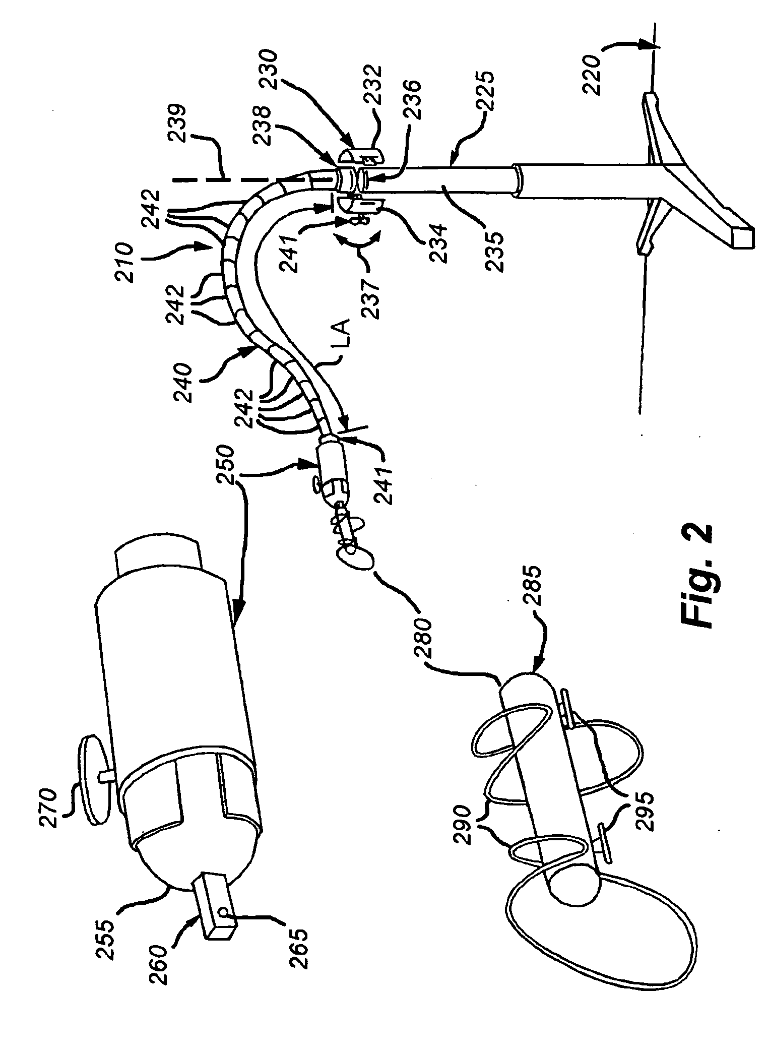
[0068] An illustrative embodiment of an apparatus or device for supporting, positioning and stabilizing diagnostic or therapeutic devices is shown in FIG. 2. The overall support device 210 is shown resting atop a rigid, generally flat surface 220 (e.g. a treatment room floor), and is securely mounted to a conventional stand 225, using an illustrative clamping subsystem 230. In this embodiment, the exemplary stand 225 is one normally employed for delivery of intravenous (IV) fluids. However, as described further below, a variety of base units can be used to elevate and suspend the support apparatus of this invention. For example, the support device 210 may alternatively be affixed to other stable or metastable devices or equipment for the duration of the procedure. These may include, but are not limited to, rigid attachment to a hospital bed, backboard, equipment table, or ceiling fixture (see below).

[0069] The clamping subsystem 230, in thi...


PUM


Abstract

This invention provides a support device that allows the adjustable, yet rigid placement of a probe or other medical instrument against a region of interest / treatment on a patient. The system and method of rigid fixation, positioning, and adjustment contemplated herein is useful for a broad array of medical procedures including, but not limited to, ultrasound-guided anesthetic delivery. In an exemplary embodiment of the present invention, a flexible armature is attached to a rigid stand placed upon the floor, or attached to another stable surface such as a bed rail, wall, ceiling or piece of equipment. A joint connects the armature to an instrument holder able to accommodate and rigidly attach an ultrasound sensing probe or other medical device. The medical device then remains rigidly attached to the described invention during the procedure. Furthermore, this set position is resistant to minor patient motion or other disturbances. If required, small alterations can be made by the operator during the procedure with minimal effort. Such adjustment may be desirable, for example, if access to a new anatomical structure is needed. In this manner, the primary operator is able to maintain a ‘hands-free’ approach.

Description

CROSS-REFERENCE TO RELATED APPLICATION [0001] The present application claims the benefit of U.S. Provisional Patent Application Ser. No. 60 / 727,319, which was filed on Oct. 17, 2005, by Brian D. Sites, et al. for a BIOMEDICAL POSITIONING AND STABILIZATION SYSTEM and is hereby incorporated by reference.BACKGROUND OF THE INVENTION [0002] 1. Field of the Invention [0003] The invention relates to medical devices and, in particular, to a device for positioning and stabilizing diagnostic or therapeutic devices used in medical procedures. [0004] 2. Background Information [0005] In clinical practice, there are many different procedures utilized for various diagnostic, therapeutic, or monitoring applications. These are typically conducted by a highly skilled operator who relies heavily on the ability to simultaneously perform multiple tasks, such as viewing a monitor while positioning a probe or dissecting tissue while exerting separation force upon the walls of an incision. Examples of such...

Claims


Application Information

Patent Timeline
Patent Type & Authority Applications(United States)
IPC IPC(8): A61B8/00A45D19/04E04G3/00A47J47/16F16D3/00F16M11/00F16D1/00F16D1/12F16C11/00F16C11/06A61B19/00
CPCA61B8/00Y10T403/32319A61B17/0206A61B19/081A61B19/26A61B2019/5276F16M11/14F16M11/18F16M11/2064F16M11/28F16M11/40F16M13/027F16M2200/022Y10T403/18A61B8/4209A61B8/4218A61B8/4444A61B90/50A61B46/10A61B2090/378A61B2090/571
Inventor HICKEY, KATHERINE M.PARASHAR, AMISHSITES, BRIAN D.SPENCE, BRIAN CONLEY
Owner WELLAN MEDICAL SOLUTIONS
Why Eureka
Try Eureka