Method and device for use in endoscopic organ procedures

a technology for endoscopic organs and procedures, applied in the field of medical equipment and methods, can solve the problems of leaving patients who are considered obese or moderately obese with few, if any, interventional options, and achieve the effects of facilitating the “dumping” syndrome, facilitating the alimentary flow of digestive secretions, and facilitating adhesion prevention

Active Publication Date: 2007-07-05
ETHICON ENDO SURGERY INC
View PDF25 Cites 23 Cited by
  • Summary
  • Abstract
  • Description
  • Claims
  • Application Information

AI Technical Summary

Benefits of technology

[0009] The present invention provides for improved methods and apparatus for the transoral, or endoscopic, restriction of a hollow body organ, such as the creation of a small stomach pouch. For purposes of the present invention, the hollow body organ shall include the entire gastrointestinal tract, including, but not limited to, the esophagus, stomach, portions of or the entire length of the intestinal tract, etc., unless specified otherwise. In the case of the present invention, the surgeon or endoscopist may insert devices as described below through the patient's mouth, down the esophagus and into the stomach or intestine as appropriate. The procedure can be performed entirely from within the patient's stomach or other organ, and does not require any external incision. The end result of the procedure is the formation of a variety of organ divisions or plications that serve as barriers or “partitions” or “pouches” that are substantially sealed off from the majority of the organ cavity. For example, in the case of dividing the stomach, the “pouch” or partitions that are created may seal a small portion of the stomach just below the esophagus to allow only small amounts of food or liquid to be consumed by the patient. This pouch or partition will mimic the section of stomach sealed off from the majority of the organ in a traditional obesity surgery heretofore described; however, it can be formed and secured entirely from inside the stomach endoscopically, obviating the need for a prolonged procedure, external incisions, minimizing the risk of infections, and in some cases, general anesthesia.
[0010] The methods and tools of the present invention may also be used in treating GERD in that stomach folds just below the esophagus can be acquired and fastened to create a desired “pleat”, thereby effectively extending the length of the esophagus and preventing reflux. Preferably, multiple folds of tissue can be acquired to effect this end. Further, features of the present invention would assist in the longevity of the GE Junction (GEJ) / AEsophageal pleat as compared to current devices and techniques as the plication would include a more significant amount of muscular tissue. In addition, the devices and methods of the present invention may be used to revise or repair failures seen in current surgical procedures, such as dilation of the pouch and / or stoma (stomata) formed in a traditional Roux-En-Y gastric bypass, or VBG. In these cases, when the stoma dilates or shifts, the tools of the present invention would be useful to circumferentially gather tissue at the site of dilation to narrow it, thereby making the stoma functional again, or by further reducing the volume of an existing pouch which has dilated.
[0013] In the procedures of the present invention relating to treatment of gastric disorders such as gastroesophageal reflux disease (GERD), or in cases of treating obesity, a flexible circular stapler may be inserted transorally down the patient's esophagus and into the stomach at the region of the GEJ. Tissue may then be acquired circumferentially about the stapler device, or at least partially about the circumference of the stapler device at some point less than 360 degrees (possibly in a 180 degree formation) relative to a longitudinal axis of the device such that the tissue acquisition creates a “waist” within the organ volume. Subsequently, the tissue fixation element may then be deployed to fix the tissue in a manner to promote healing.
[0015] Any of the fastening devices described herein may employ, e.g., bioabsorbable or biofragmentable staples or fixation element. Such fastening devices would typically dissolve or otherwise degrade leaving only the fixation region once the desired tissue healing has occurred. The remaining healed tissue, now a tissue “ring” (TR), would be sufficiently adhered or healed together to maintain the integrity of the pouch and stoma. In addition, the fastening devices may include coatings or other secondary features to aid healing, such as resorbable meshes, sclerosing agents, surgical felt, or tissue grafts.
[0016] The pouch or partitions may be created by a procedure of the present invention to remain permanently within the stomach to restrict it indefinitely. Alternatively, the creation of the pouch or partitions may be reversible (e.g., once weight loss is achieved, or reflux minimized) or revised (in the event pouch side needs to be modified). Reversal can also be achieved via various methods such as dilation of the restricted section, or, e.g., using an electro-surgical device such as a bovie to cut the restricted section to free the tissue folds. Further, if the physician so desires, techniques of the present invention may be augmented or assisted by the use of other techniques such as laparoscopy. Optionally, techniques of the present invention may be combined with other procedures such as for the treatment of GERD or the transoral placement of a bypass prosthesis or other type of liner in the intestine to bypass the hormonally active portion of the small intestine, typically between the stoma to just proximal of the jejunum. Such a liner may be placed within the orifice of a stoma created by devices described herein or within stomas created by various conventional procedures, as also described herein. For present purposes, a stoma refers simply to an artificial or “man made” narrowing within a body organ. The liner may be tubular in construction and made to match the diameter of the stoma created by the present invention such that they can be hooked together to achieve the desired clinical effect. Additionally, the distal end of the liner may also be anchored to tissue distally located from the stoma or it may be left unanchored relying on its resilient physical structure to avoid kinking or twisting.
[0018] In either case, the liner may define one or more fenestrations or channels on the portion of the liner in communication with the gastric remnant, and / or at or near the site of the common bile ducts so as to allow fluids to drain from the organ or ducts. The liner and the fluid conduit may be made separately and attached together or they may be made integrally from the same material. Also, the liner and / or the fluid conduit may be made of a braided design to inhibit kinking as the device reacts to the peristalsis motion of the intestines. Another alternative may utilize a singular liner having one or more channels defined longitudinally along the outer surface of the liner rather than as a separate fluid conduit. These channels may form spaces between the tissue and the liner itself to allow for the flow of fluids within the channels. In another variation of the singular liner, the liner may have fenestrations or openings positioned along its length near or at the zones of active secretion in the intestines to permit fluid flow from the organ or bile ducts into the lumen of the liner (so as to prevent blockage thereof), while still maintaining a barrier to the majority of the intestine to achieve malabsorption and to facilitate “dumping” syndrome upon ingestion of high fat or high caloric foods. An alternative variation of this singular liner may have multiple valved openings along its length to allow for the unidirectional flow of secretions into the liner, but prohibiting contact between the intestines and the food contents within the liner.

Problems solved by technology

Although the outcome of these stomach reduction surgeries leads to patient weight loss because patients are physically forced to eat less due to the reduced size of their stomach, several limitations exist due to the invasiveness of the procedures, including time, general anesthesia, healing of the incisions and other complications attendant to major surgery.
In addition, these procedures are only available to a small segment of the obese population (morbid obesity, Body Mass Index ≧40) due to their complications, leaving patients who are considered obese or moderately obese with few, if any, interventional options.

Method used


Image

  • Method and device for use in endoscopic organ procedures
  • Method and device for use in endoscopic organ procedures
  • Method and device for use in endoscopic organ procedures

Examples


Embodiment Construction

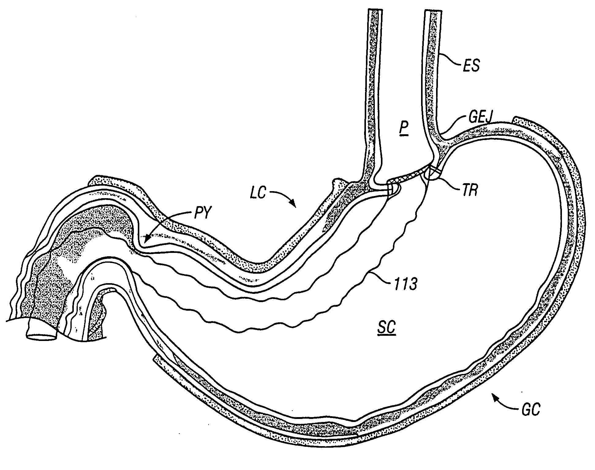
[0045] The present invention provides, in part, for methods and devices for hollow organ division and restriction, more particularly providing methods and devices to perform a transoral, endoscopically mediated stomach reduction for purposes of, e.g., treating obesity. For purposes of the present invention, the hollow body organ shall include the entire gastrointestinal tract, including, but not limited to, the esophagus, stomach, portions of or the entire length of the intestinal tract, etc., unless specified otherwise.

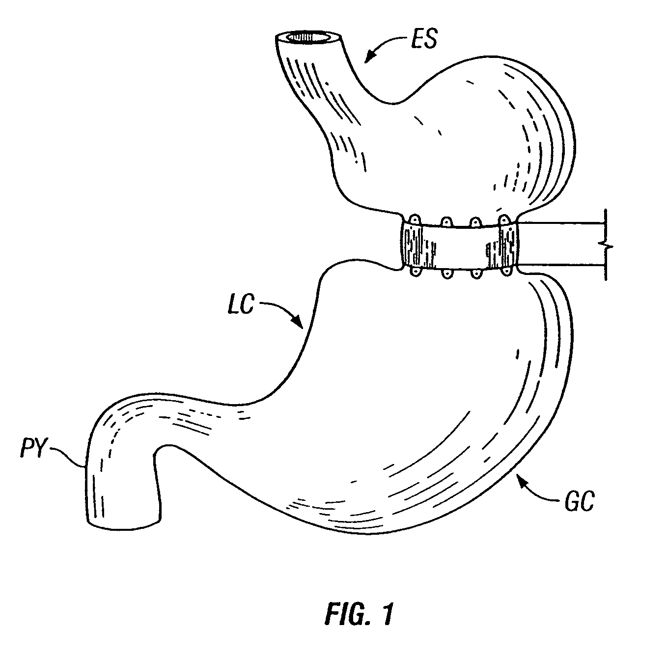
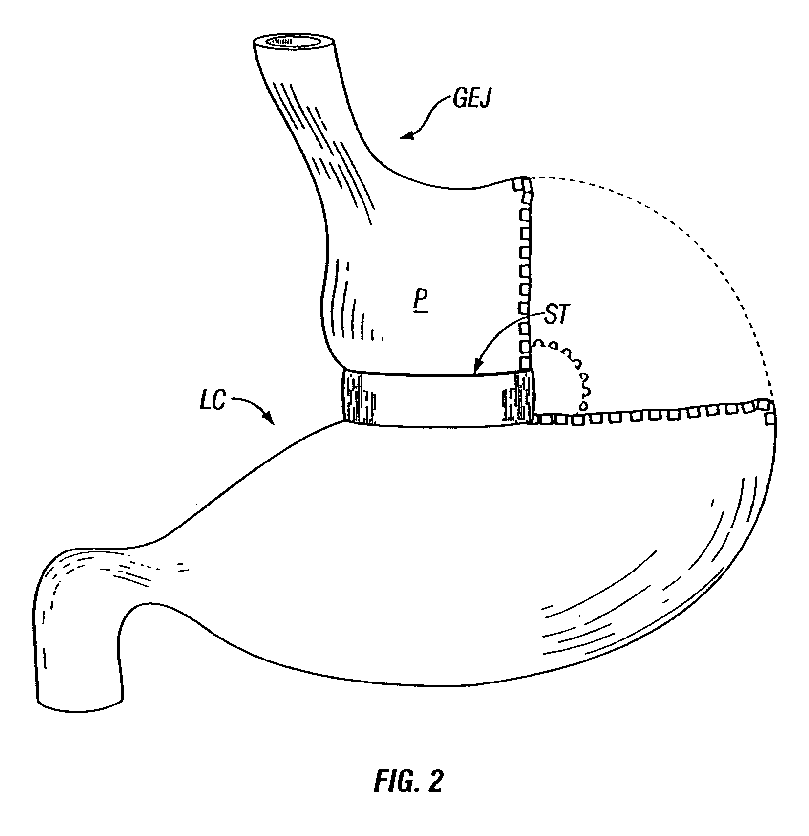
[0046] As previously discussed, the results of some clinical procedures of the prior art are shown in FIGS. 1-3, from a perspective external to the stomach. An example of a result of the procedure in one variation of the present invention is shown in FIG. 4A, which depicts an external anterior view of a stomach organ 100, having an esophagus 101 (cut away to reveal the esophageal lumen 102), and further depicting a circumferential orifice or stoma 103, configured fr...


PUM


Abstract

Methods and devices for use in tissue approximation and fixation are described herein. The present invention provides, in part, methods and devices for acquiring tissue folds in a circumferential configuration within a hollow body organ, e.g., a stomach, positioning the tissue folds for affixing within a fixation zone of the stomach, preferably to create a pouch or partition below the esophagus, and fastening the tissue folds such that a tissue ring, or stomas, forms excluding the pouch from the greater stomach cavity. The present invention further provides for a liner or bypass conduit which is affixed at a proximal end either to the tissue ring or through some other fastening mechanism. The distal end of the conduit is left either unanchored or anchored within the intestinal tract. This bypass conduit also includes a fluid bypass conduit which allows the stomach and a portion of the intestinal tract to communicate.

Description

CROSS-REFERENCE TO RELATED APPLICATIONS [0001] This application is a continuation-in-part of U.S. patent application. Ser. No. 10 / 279,257 filed Oct. 23, 2002, which is incorporated herein by reference in its entirety.FIELD OF THE INVENTION [0002] The present invention relates generally to medical apparatus and methods and more particularly to devices and methods for dividing a hollow body organ or otherwise restricting or partitioning a certain section of that organ, such as a stomach, intestine or gastrointestinal tract as well as devices and methods for placing a liner within or partially within the hollow body organ. BACKGROUND OF THE INVENTION [0003] In cases of severe obesity, patients may currently undergo several types of surgery either to tie off or staple portions of the large or small intestine or stomach, and / or to bypass portions of the same to reduce the amount of food desired by the patient, and the amount absorbed by the gastrointestinal tract. The procedures currentl...

Claims


Application Information

Patent Timeline
Patent Type & Authority Applications(United States)
IPC IPC(8): A61B17/08A61B17/072A61B17/11A61B17/28A61B17/30A61F5/00
CPCA61B17/072A61B17/1114A61B17/1155A61B2017/00323A61F5/0079A61B2017/2905A61B2017/306A61F5/0076A61F5/0086A61B2017/07214
Inventor GANNOE, JAMYGERBI, CRAIGWELLER, GARYCOLLIER, MATTHEW J.
Owner ETHICON ENDO SURGERY INC
Why Eureka
Try Eureka