Automatic multi-dimensional intravascular ultrasound image segmentation method

an intravascular ultrasound and image segmentation technology, applied in the field of image segmentation, can solve the problems of poor quality image, no ivus edge detection method found widespread acceptance by clinicians, and important constraints against the clinical use of ivus

Inactive Publication Date: 2007-07-19
VAL CHUM PARTNERSHIP +1
View PDF13 Cites 122 Cited by
  • Summary
  • Abstract
  • Description
  • Claims
  • Application Information

AI Technical Summary

Problems solved by technology

1676-1688, 1995) that conventional stent implantation resulted in incomplete apposition and expansion causing thrombosis, which had the result of changing the clinical practice.
However, a typical IVUS acquisition generally contains several hundreds of images, which has the effect of making analysis of the data a long and fastidious task that is further subject to an important variability between intra-observers and inter-observers when non-automatic methods are used.
These aspects generate important constraints against the clinical use of IVUS.
Other constraints related to the use of IVUS include poor quality image due to speckle noise, imaging artifacts, and shadowing of parts of the vessel wall by calcifications.
Still, in 2001, the clinical expert consensus from the American College of Cardiology in the hereinabove cited document by Mintz et al. reported that no IVUS edge detection method had found widespread acceptance by clinicians.
However, these new models were again using local pixel or edge information and they were not taking advantage of the statistical information of IVUS data (speckle texture).
Further, the fact that a fast marching method propagates interfaces in the direction of the boundaries through an exhaustive analysis of the propagation region has the effect of decreasing the variability of segmentation results.

Method used


Image

  • Automatic multi-dimensional intravascular ultrasound image segmentation method
  • Automatic multi-dimensional intravascular ultrasound image segmentation method
  • Automatic multi-dimensional intravascular ultrasound image segmentation method

Examples


Embodiment Construction

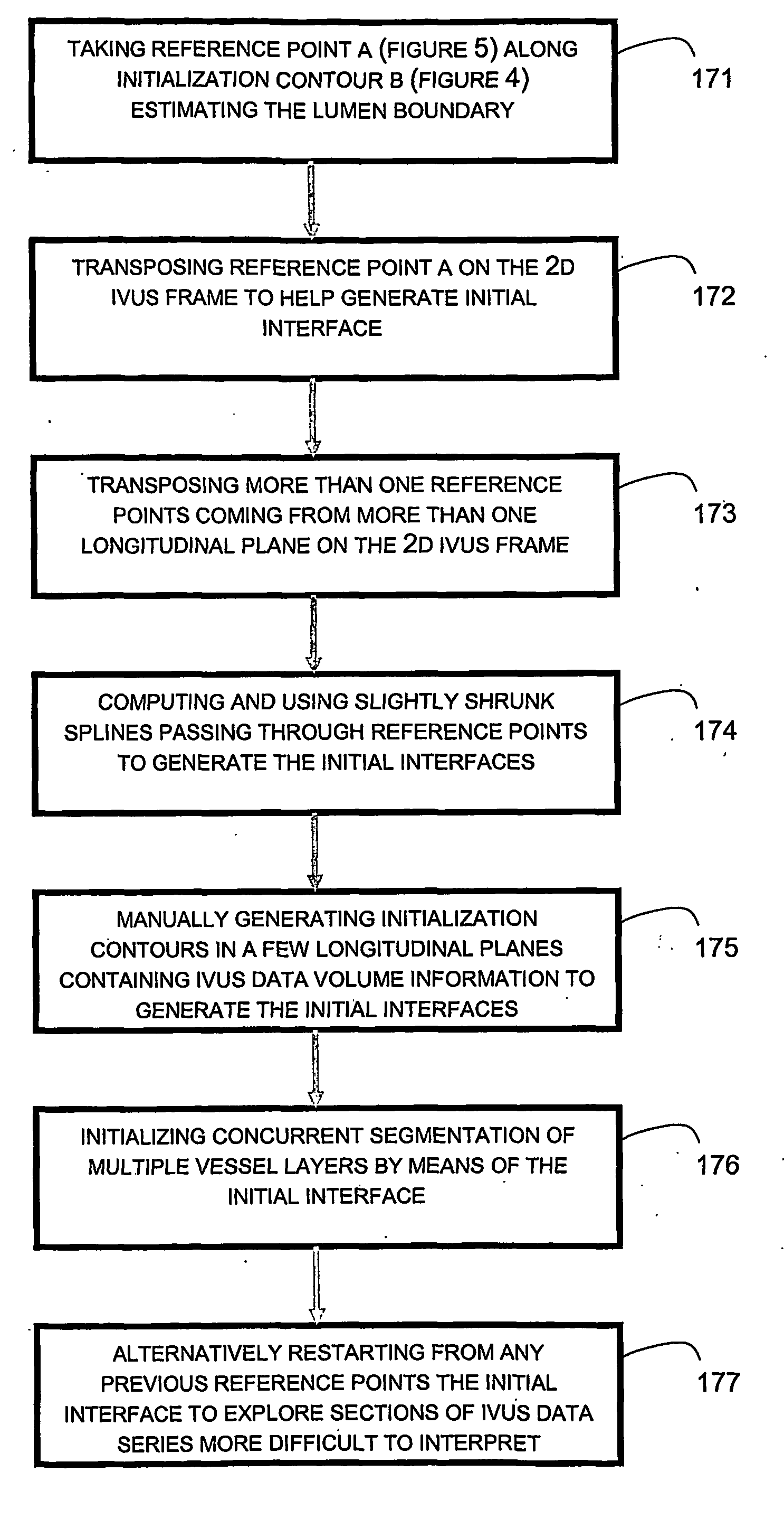
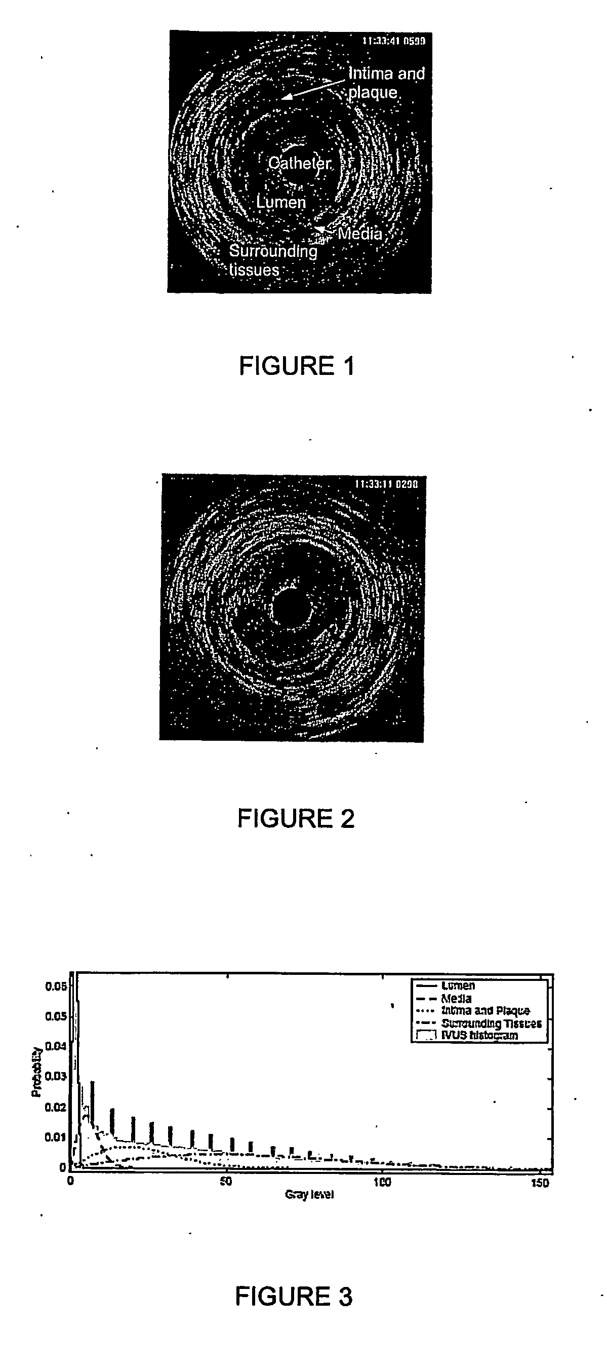
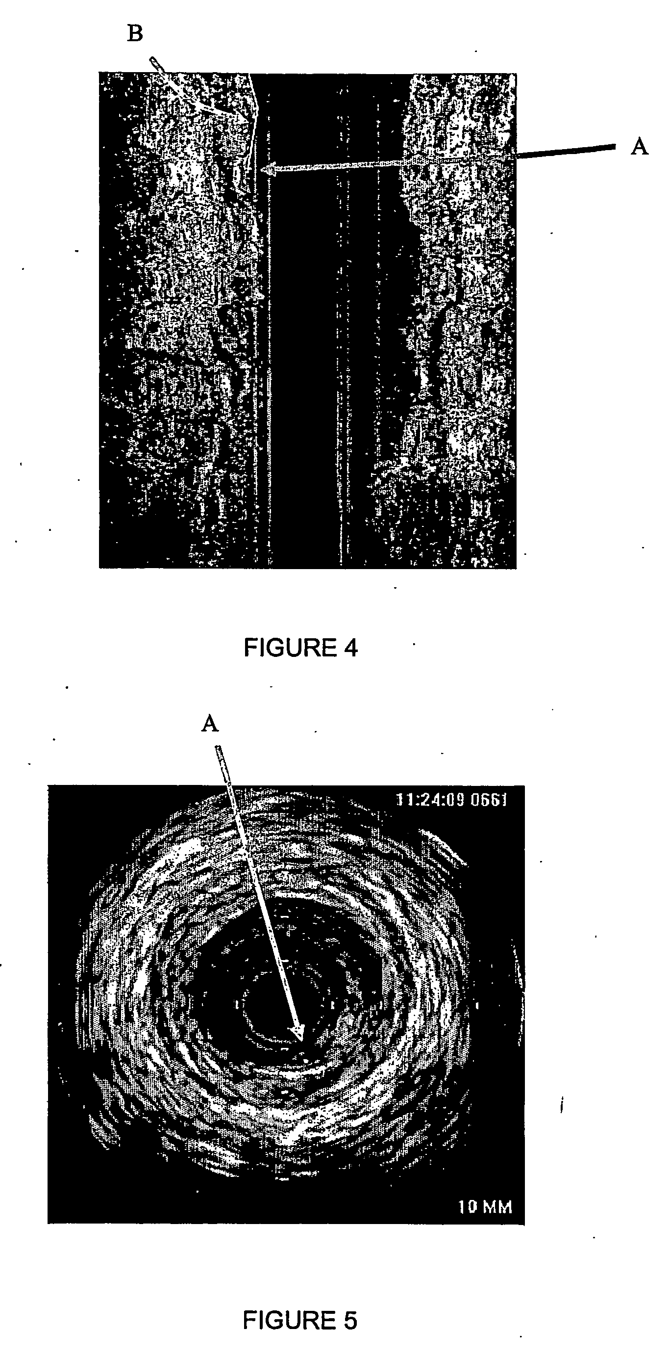
[0063] The non-restrictive illustrative embodiments of the present invention relate to a method and device for concurrently estimating boundaries between the plurality of layers of a blood vessel from IVUS image data. The method and device involve a segmentation of the IVUS image data by propagating interfaces in each layer to be estimated from initial interfaces that are generated from the IVUS image data. The technique for estimating the boundaries of the various layers uses a fast marching method based on probability functions, such as for example a probability density function (PDF) or gradient function to estimate the distribution color map of images, such as for example to estimate the gray levels or the multi-colored levels in images.

[0064] The following description is organized as follows. First of all, a PDF estimation technique for the different vessel layers will be presented. Then, an IVUS 3D fast marching method based on the estimated PDFs and based on the gray level g...


PUM


Abstract

The present invention generally relates to intravascular ultrasound (IVUS) image segmentation methods, and is more specifically concerned with an intravascular ultrasound image segmentation method for characterizing blood vessel vascular layers. The proposed image segmentation method for estimating boundaries of layers in a multi-layered vessel provides image data which represent a plurality of image elements of the multi-layered vessel. The method also determines a plurality of initial interfaces corresponding to regions of the image data to segment and further concurrently propagates the initial interfaces corresponding to the regions to segment. The method thereby allows to estimate the boundaries of the layers of the multi-layered vessel by propagating the initial interfaces using a fast marching model based on a probability function which describes at least one characteristic of the image elements.

Description

FIELD OF THE INVENTION [0001] The present invention generally relates to image segmentation. More specifically but not exclusively, the present invention is concerned with an intravascular ultrasound image segmentation technique for characterizing blood vessel vascular layers from intravascular ultrasound image sequences. BACKGROUND OF THE INVENTION [0002] Over the past few years, intravascular ultrasound (IVUS) technology has become very useful for studying atherosclerotic disease. IVUS is a medical imaging technique that produces cross-sectional images as a catheter is pulled-back inside a blood vessel. These images show the lumen but also the layered structure of the vascular wall. IVUS provides quantitative assessment of the vascular wall, information about the nature of atherosclerotic lesions as well as the plaque shape and size such that in clinic, IVUS was rapidly recognized as a valuable tool in diagnosis and in pre-intervention analysis of atherosclerosis. [0003] The abili...

Claims


Application Information

Patent Timeline
Patent Type & Authority Applications(United States)
IPC IPC(8): G06K9/00A61B8/08G06T5/00G06V10/26G06V10/764
CPCA61B5/02007A61B8/0858G06K9/34G06K9/6278G06T2207/30101G06T7/0083G06T7/0089G06T2207/10132G06T2207/20161G06K2209/05G06T7/12G06T7/149G06V10/26G06V2201/03G06V10/764G06F18/24155
Inventor CLOUTIER, GUYROY-CARDINAL, MARIE-HELENEMEUNIER, JEANSOULEZ, GILLESTHERASSE, ERIC
Owner VAL CHUM PARTNERSHIP
Why Eureka
Try Eureka