
Ultrasound activated medical device

a technology of activated medical devices and ultrasound, applied in the direction of coatings, prostheses, pharmaceutical delivery mechanisms, etc., can solve the problem of restnosis, the stented vessel is again blocked

Inactive Publication Date: 2007-08-09
BOSTON SCI SCIMED INC
View PDF14 Cites 34 Cited by
  • Summary
  • Abstract
  • Description
  • Claims
  • Application Information

AI Technical Summary

Problems solved by technology

In many instances, however, the stented vessel becomes blocked again (known as restenosis) due to various biological processes, including tissue healing and regeneration, scar formation, irritation, and immune reactions that lead to an excess proliferation of the cells.

Method used


Image

  • Ultrasound activated medical device
  • Ultrasound activated medical device
  • Ultrasound activated medical device

Examples


Embodiment Construction

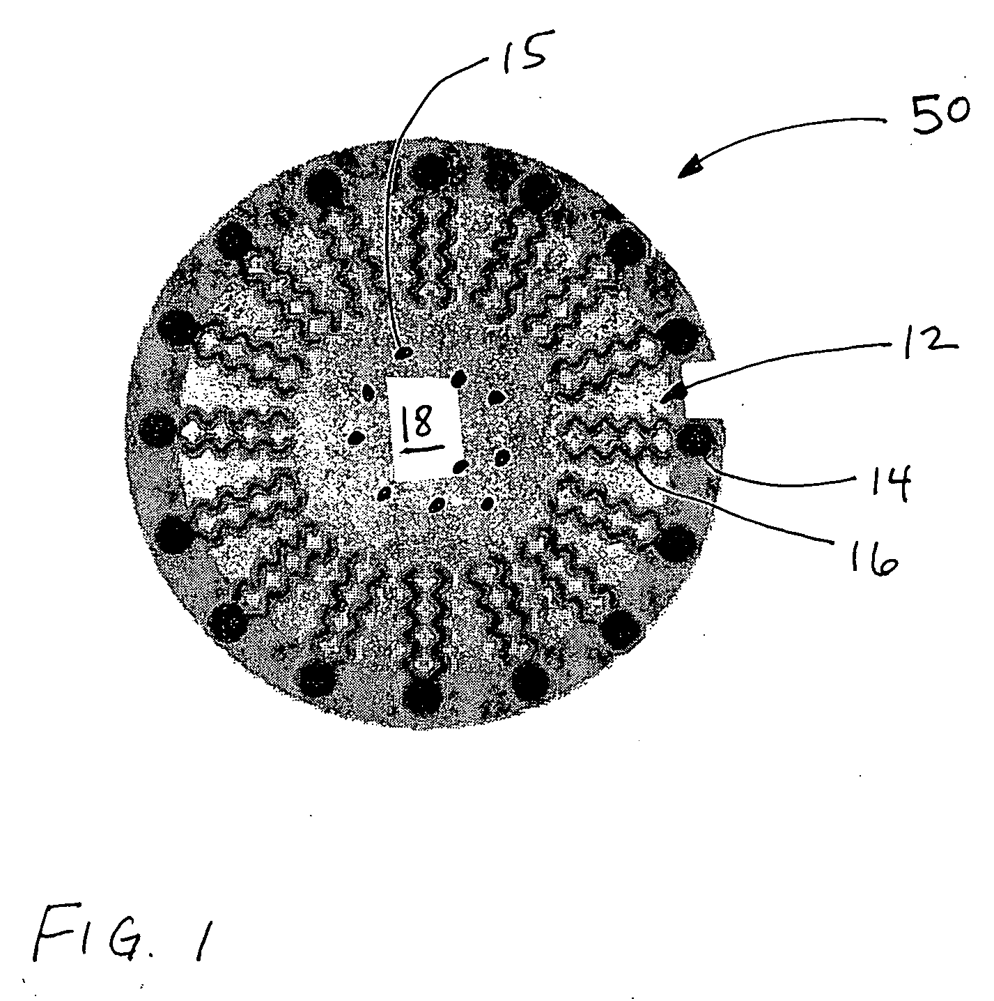
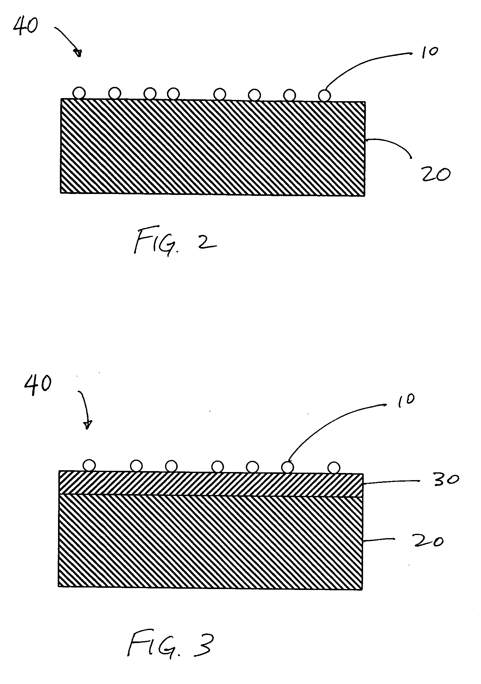
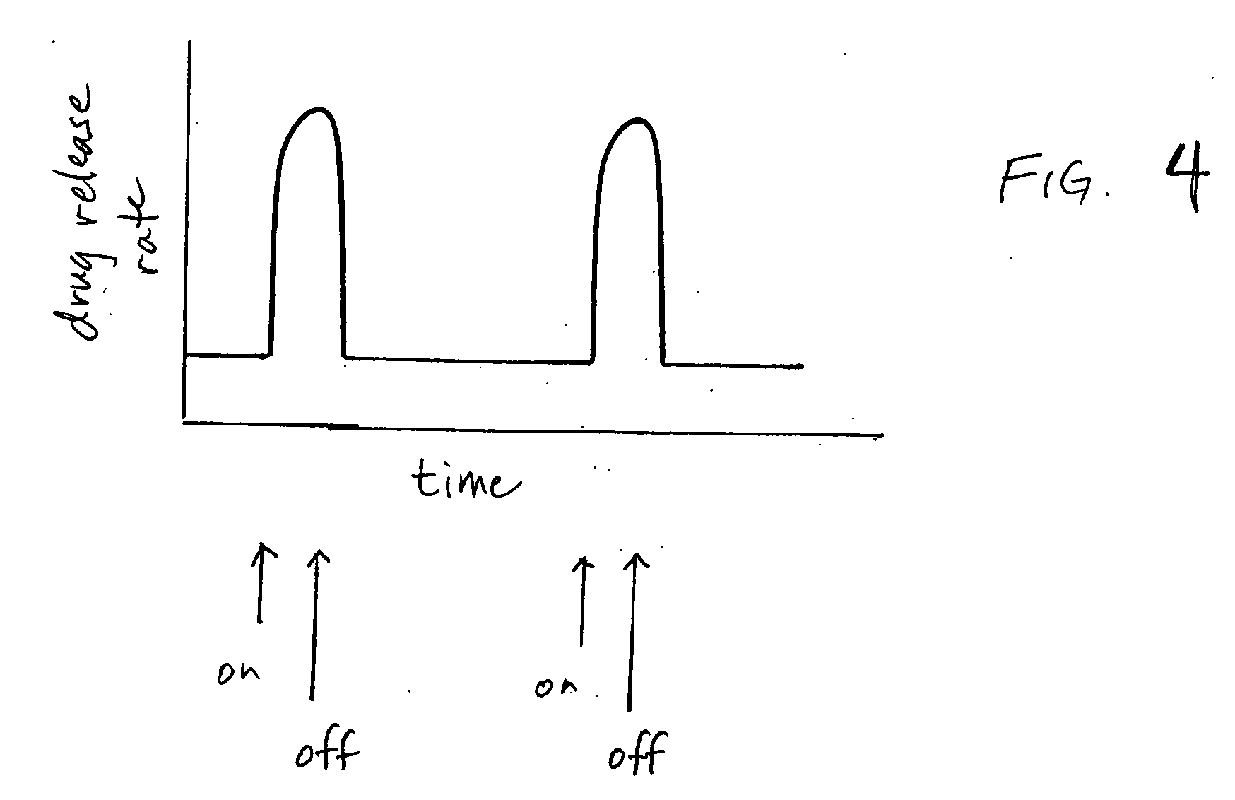
[0022] The present invention provides a medical device comprising a medical device body having a plurality of drug-containing vesicles disposed thereon (unless otherwise indicated, the terms “drug” and “therapeutic agent” are used interchangeably herein). According to the present invention, the vesicles are ultrasound-sensitive drug carriers that release the drug contained therein when exposed to ultrasound energy. The vesicles have sufficient structural stability to retain the drug contained therein under non-exposed conditions (i.e., when not exposed to ultrasound energy) yet are able to become destabilized and release the retained drug upon exposure to ultrasound energy. The vesicles can be any type of carrier that can retain a drug such as, for example, a micelle, liposome, nanoparticle, bubble, microbubble, microsphere, microcapsule, clathrate bound vesicle, or hexagonal H II phase structure and can be manufactured of any ultrasonic-sensitive material such as, for example, ultr...


PUM

PropertyMeasurementUnit
radiiaaaaaaaaaa
amphiphilicaaaaaaaaaa
metallicaaaaaaaaaa

Abstract

A medical device comprising a medical device body having drug-loaded vesicles thereon. The vesicles are ultrasound sensitive and release the drug upon ultrasound stimulation. Also provided is a method for controlling drug release from a medical device using drug-loaded vesicles that are ultrasound sensitive.

Description

TECHNICAL FIELD [0001] The present invention relates to drug-coated medical devices and methods of controlling drug release from the same. BACKGROUND OF THE INVENTION [0002] Many implantable medical devices are coated with a drug or therapeutic agent that acts to improve the effectiveness of the device. One such example of a drug-coated implantable medical device is a stent. Stents are tubular structures formed in a mesh-like pattern that are designed to be inserted into an organ or vessel. For example, a coronary artery stent is placed in a coronary artery across an area of blockage after it has been opened by an angioplasty procedure. The stent serves as a permanent scaffolding for the newly widened coronary artery. In many instances, however, the stented vessel becomes blocked again (known as restenosis) due to various biological processes, including tissue healing and regeneration, scar formation, irritation, and immune reactions that lead to an excess proliferation of the cells...

Claims


Application Information

Patent Timeline
Patent Type & Authority Applications(United States)
IPC IPC(8): A61F2/02
CPCA61F2250/0068A61K9/0009A61K9/1075A61L27/34A61L27/50A61L2300/604A61L2300/236A61L2300/256A61L2300/414A61L2300/432A61L2300/45A61L27/54
Inventor RADHAKRISHNAN, RAJESHCHANDRASEKARAN, CHANDRU
Owner BOSTON SCI SCIMED INC
Learn More