X-ray beam calibration for bone mineral density assessment using mammography system

a mammography system and x-ray beam technology, applied in the field of mammography imaging, can solve the problems of increased risk of factures, high cost, hunched backs, etc., and achieve the effect of adequate image capture quality

Inactive Publication Date: 2007-08-16
CARESTREAM HEALTH INC
View PDF16 Cites 3 Cited by
  • Summary
  • Abstract
  • Description
  • Claims
  • Application Information

AI Technical Summary

Benefits of technology

[0016] The present invention provides an x-ray beam calibration apparatus and a method for BMD screening and analysis when using a mammography imaging system to acquire extremity x-ray images, such as a hand. The apparatus and the method described can be employed prior to the acquisition of any patient image on a mammography imaging system to determine the proper x-ray techniques for an average patient. The selection of proper x-ray techniques promotes adequate image capture quality suitable for subsequent image analysis.

Problems solved by technology

It can result in increased risk to factures, height loss, hunched backs, and pain.
Such a procedure carries risk.
While some consider DEXA to be accurate, the apparatus is bulky and expensive.
Thus, sonography is considered by some to be not as accurate or sensitive as DEXA in the determination of bone mineral density.

Method used


Image

  • X-ray beam calibration for bone mineral density assessment using mammography system
  • X-ray beam calibration for bone mineral density assessment using mammography system
  • X-ray beam calibration for bone mineral density assessment using mammography system

Examples


Embodiment Construction

[0027] The following is a detailed description of the preferred embodiments of the invention, reference being made to the drawings in which the same reference numerals identify the same elements of structure in each of the several figures.

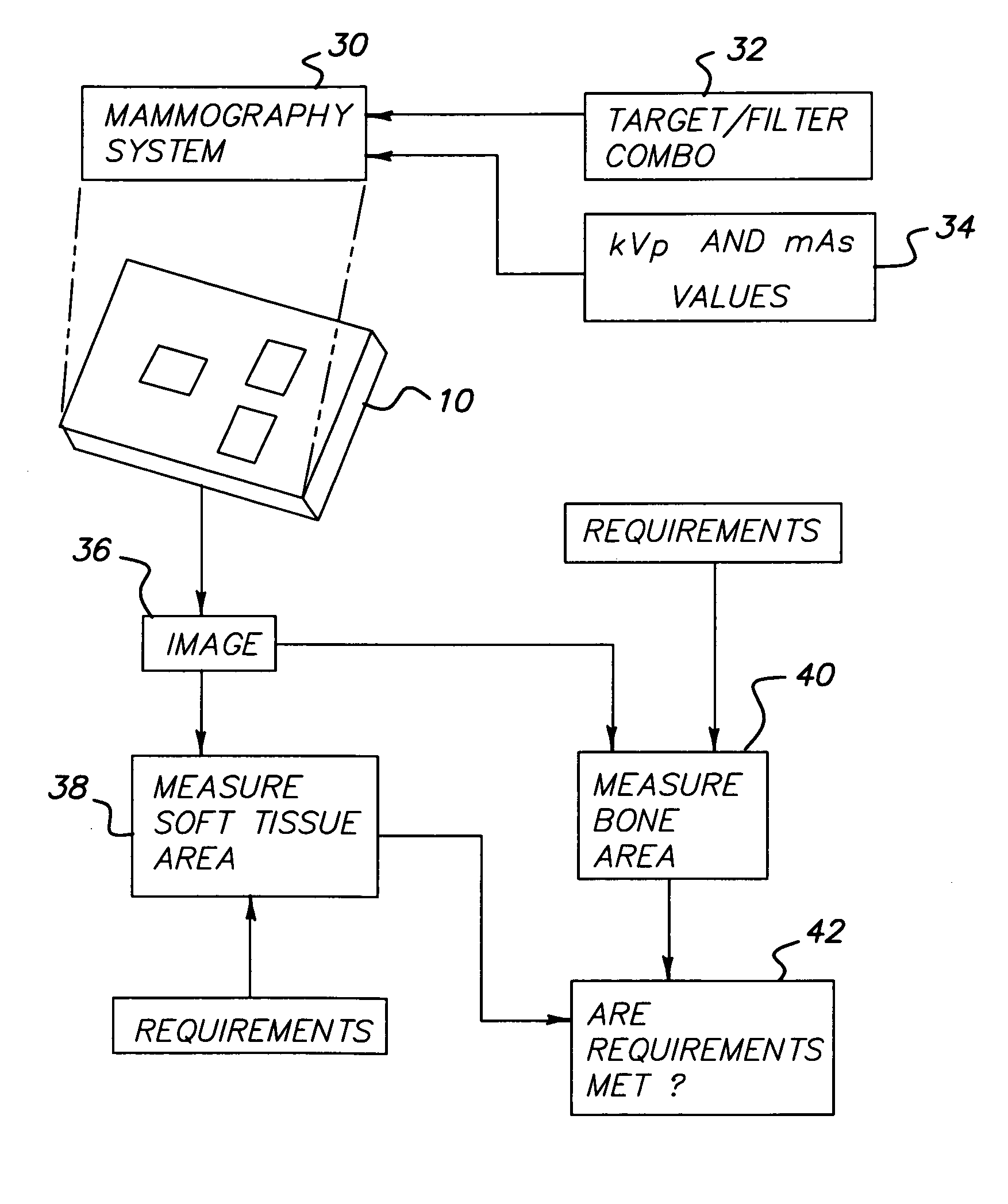
[0028] It is noted that the American Cancer Society recommends that women over the age of 40 years obtain annual mammograms. Millions of women have their annual screening mammograms each year at hospitals or breast imaging centers. Accordingly, Applicants have noted it would be desirable for women to have both their annual mammography screening and a bone mineral density screening done in one visit, at one location, and using one imaging system.

[0029] Conventionally, extremities (e.g., hands and feet) are imaged using conventional x-ray system, which generates an x-ray beam adapted to capture both low and high-density objects (i.e., bone and soft tissue) on a detector (film or digital) that are designed with a wide dynamic range. In contrast, mam...


PUM

PropertyMeasurementUnit
thicknessaaaaaaaaaa
thicknessaaaaaaaaaa
thicknessesaaaaaaaaaa

Abstract

An apparatus and method calibration method, particularly to determine an x-ray generation technique to acquire a hand x-ray image using a mammography x-ray system for assessing Radiographic Absorptiometry (RA) based BMD (Bone Mineral Density).

Description

FIELD OF THE INVENTION [0001] The invention relates generally to the field of mammography imaging. More specifically, the invention relates to x-ray beam calibration when assessing Radiographic Absorptiometry (RA) based BMD (Bone Mineral Density) using a mammography imaging system. BACKGROUND OF THE INVENTION [0002] Osteoporosis is a skeletal disorder characterized by reduced bone strength. It can result in increased risk to factures, height loss, hunched backs, and pain. Bone strength is a function of bone mineral density (BMD) and bone quality. It is believed that bone mineral density peaks about the age of 30 for both men and women, and then declines gradually. Some statistics have indicated that osteoporosis affects approximately 20 million people and is a cause of about 1.3 million fracture incidents in the United States each year. As such, screening for bone mineral density is often desired. [0003] Several common techniques have been used to measure bone mineral density, inclu...

Claims


Application Information

Patent Timeline
Patent Type & Authority Applications(United States)
IPC IPC(8): G01B15/02G01N23/06
CPCA61B6/502A61B6/583A61B6/505
Inventor YANG, CHANG-YING J.HUO, ZHIMIN
Owner CARESTREAM HEALTH INC
Why Eureka
Try Eureka