Use of roma for characterizing genomic rearrangements

a genomic rearrangement and roma technology, applied in the field of roma for characterizing genomic rearrangements, can solve the problems of insufficient predictors of treatment efficacy of other diseases and disorders, use of histopathological and clinical criteria, and significant and unmet need for accurate diagnostic methods to improve patient car

Inactive Publication Date: 2007-09-06
COLD SPRING HARBOR LAB INC +1
View PDF18 Cites 44 Cited by
  • Summary
  • Abstract
  • Description
  • Claims
  • Application Information

AI Technical Summary

Benefits of technology

[0014] Accordingly, relative copy numbers of regions (R) and / or (I), and (X) may be used to determine the likely response of the patient to a therapy that targets or treats an effect of the genetic rearrangements at the genetic locus X, such as misexpression (e.g., qualitative and / or quantitative changes in transcripts or transcript levels) of particular genes within the genetic locus X. Therapies directed to or especially effective in situations of over- or under-expression of the genetic locus X and / or its gene products may then be considered more likely to ameliorate or be effective in the patient's proposed treatment regimen, based on the relative copy number information that has been ascertained according to methods of the invention.

Problems solved by technology

Yet, the classical histopathological and clinical criteria used to assess the likelihood of response to the most commonly used modalities used to treat cancer and other diseases and disorders are inadequate predictors of treatment efficacy.
Consequently, there is a significant and unmet need for accurate diagnostic methods that improve patient care and disease outcome.
However, tools for high-resolution, comprehensive genome analysis have been lacking and consequently no cancer genome signatures that predict a patient's response to anti-cancer modalities have been discovered.
Despite these advances, there are few drugs or therapeutic regimens to date which have been successfully tailored for the individual patient or for a particular patient subpopulation (treatment stratification).
However, it is unclear whether the current technology has failed to detect certain genetic rearrangements at the HER2 locus, and therefore has excluded certain breast cancer patients who may respond to and benefit from Hercepting, alone or combined with another chemotherapy agent.
It remains unclear, however, whether all these patients can benefit from such combination therapy.

Method used


Image

  • Use of roma for characterizing genomic rearrangements
  • Use of roma for characterizing genomic rearrangements
  • Use of roma for characterizing genomic rearrangements

Examples


example 1

Materials and Methods

[0101] Formalin-fixed, paraffin-embedded (FFPE) tissue samples were obtained from the Pathology Department of Memorial Sloan-Kettering Cancer Center (MSKCC). The samples consist of unmounted 15 micron microtome slices taken from recently archived tumor tissue blocks. In most cases DNA was prepared between three and six months of original processing of the tissue for clinical histopathology. In all cases, samples were selected, prepared and coded at MSKCC before being sent to CSHL for ROMA analysis. Samples were given a score for amplification based on ROMA results and those scores were compared to the original pathology laboratory results at MSKCC. No additional patient identification or other clinical information was transferred to CSHL in this study.

[0102] ROMA and IHC / FISH results were compared between two different sample sets. The first set (Set A) was made up of 25 samples selected to contain a larger proportion of IHC+ / FISH+ cases than would be present ...

example 2

Probe Design for FISH

[0104] Hybridization probes for FISH were constructed in one of two methods. For the interdigitation analysis, probes were created from bacterial artificial chromosomes (BAC) selected using the UCSD Genome Browser. For the determination of copy number in the deletions and amplifications of the aneuploid tumors, probes were made with PCR amplification of primers identified through the PROBER algorithm designed in this laboratory (Navin et al. 2006). Genomic sequences of 100 kb containing target amplifications were tiled with 50 probes (800-1400 bp).

[0105] Oligonucleotide primers were ordered in 96-well plates from Sigma Genosys and resuspended to 25 μM. Probes were amplified with the PCR Mastermix kit from Eppendorf (Cat. 0,032,002.447) from EBV immortalized cell line DNA (Chp-Skn-1) DNA (100 ng) with 55° C. annealing, 72° C. extension, 2 min extension time, and 23 cycles. Probes were purified with Qiagen PCR purification columns (Cat. 28,104) and combined into...

example 3

Correlation of FISH and ROMA in Selected Cases (SET A)

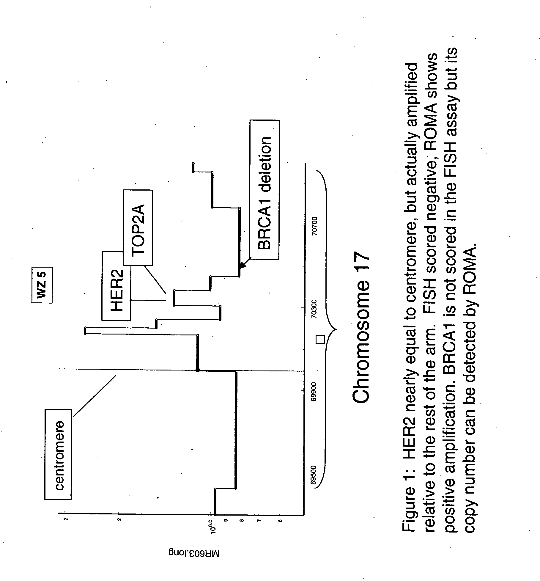
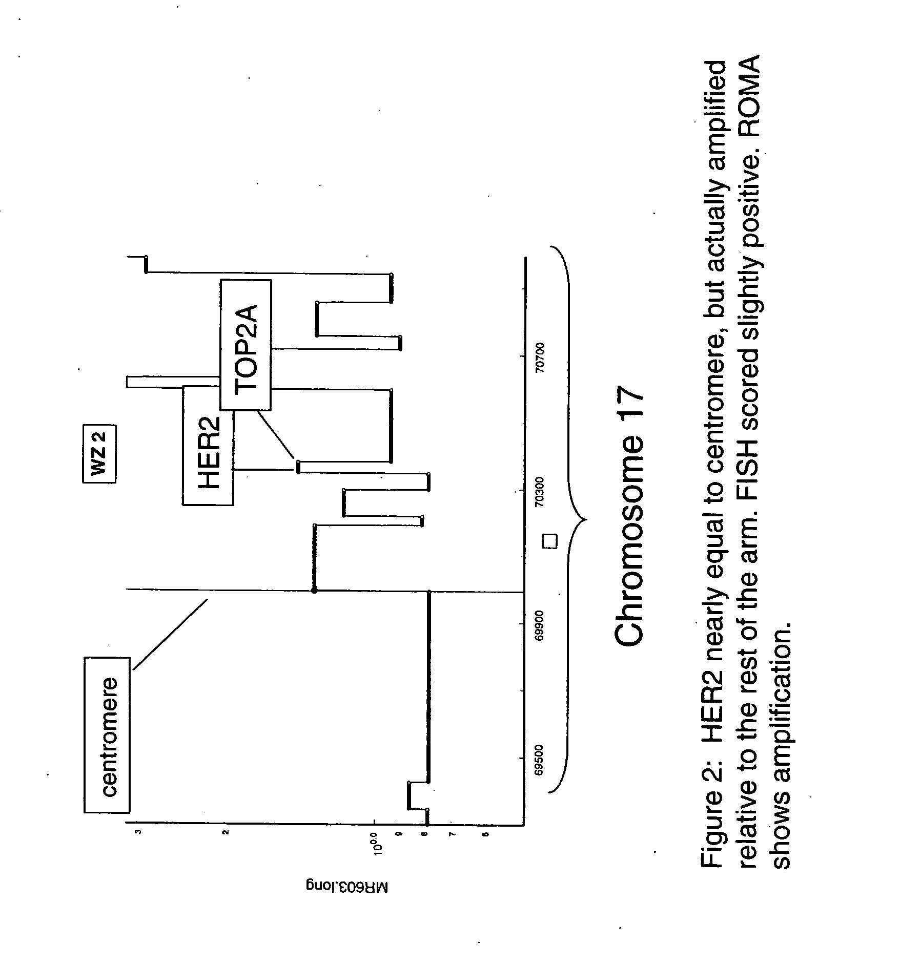
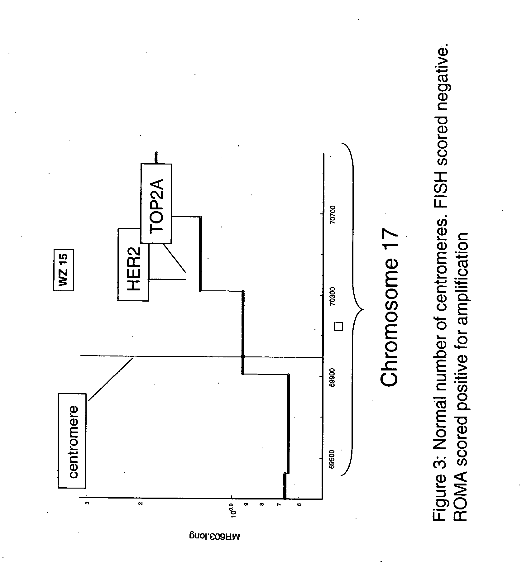
[0108] The data presented in Table 1 show a remarkably good agreement between FISH and ROMA data. The data for the predominantly Her-2+ cases in Set A in Table 1 show that all cases that scored negative by FISH were scored negative by ROMA (4 / 4). For the cases scored positive by FISH, all but one were detected by ROMA (20 / 21). Furthermore, with one exception (BTL19) the estimates for degree of amplification by the two techniques show a strong correlation. The single case that was scored as a false negative by ROMA (BTL16) had only 20% tumor cells and thus sets an apparent lower limit on the ratio of tumor / normal nuclei in the sample tissue for efficient detection by a mass technique such as ROMA.


PUM

PropertyMeasurementUnit
fluorescence in situ hybridizationaaaaaaaaaa
compositionsaaaaaaaaaa
sizeaaaaaaaaaa

Abstract

The present invention relates to methods and compositions for detecting genomic rearrangements (e.g., amplification) at one or more genetic loci and various applications of such methods and compositions. Examples of genetic loci include HER2, TOP2A and other loci on the human chromosome 17.

Description

CROSS-REFERENCE TO RELATED APPLICATIONS [0001] This application claims the benefit of and priority to U.S. Provisional Application No. 60 / 751,382, filed on Dec. 14, 2005, and No. 60 / 857,921, filed on Nov. 8, 2006, the contents of which are hereby incorporated by reference in their entirety.FIELD OF THE INVENTION [0002] The present invention relates to methods and compositions for detecting genomic rearrangements (e.g., amplification) at one or more genetic loci and various applications of such methods and compositions. BACKGROUND OF THE INVENTION [0003] Genomic rearrangements, including amplifications and deletions, account for the onset, development and progression of many diseases. Well-known examples include various cancers, and inherited disorders and predispositions. As each individual patient, as well as each individual tumor, has certain unique genetic traits, patients and tumors with similar phenotypic characteristics may not have the same underlying genotypes, and therefore...

Claims


Application Information

Patent Timeline
Patent Type & Authority Applications(United States)
IPC IPC(8): C12Q1/68G06F19/00C07H21/04
CPCC12Q2600/106C12Q1/6886
Inventor WIGLER, MICHAELHICKS, JAMESNORTON, LARRYZETTENBERG, ANDERS
Owner COLD SPRING HARBOR LAB INC
Why Eureka
Try Eureka