
Eureka
For R&D, Eureka makes reading and utilizing patents & technical documents easy.
Eureka AIR
Designed for self-driven R&D workflows. Generate viable solutions, solve complex R&D challenges, empower your innovation with AI.

Eureka Materials
Designed for material experts only. Revolutionize your material R&D, from search, analyze, to developing new materials.

TechResearch
Generate reliable direction feasibility study reports for your R&D in just a few steps.

TechSeek
Discover and master advanced knowledge NOW. Basics, ideas, possibilities, all at once.

TechMind
As an expert in R&D Theories, TechMind can generates customized viable solutions instantly.

TechRisk
Analyze your overall solution with one click, know your potential R&D risks in advance.

TechMonitor
Get weekly tech updates, stay abreast of the latest tech innovations and key insights.
Use of protein PSA3 as a marker for colorectal cancer
- Summary
- Abstract
- Description
- Claims
- Application Information
AI Technical Summary
Problems solved by technology
Method used
Image

Examples
specific embodiments
Example 1
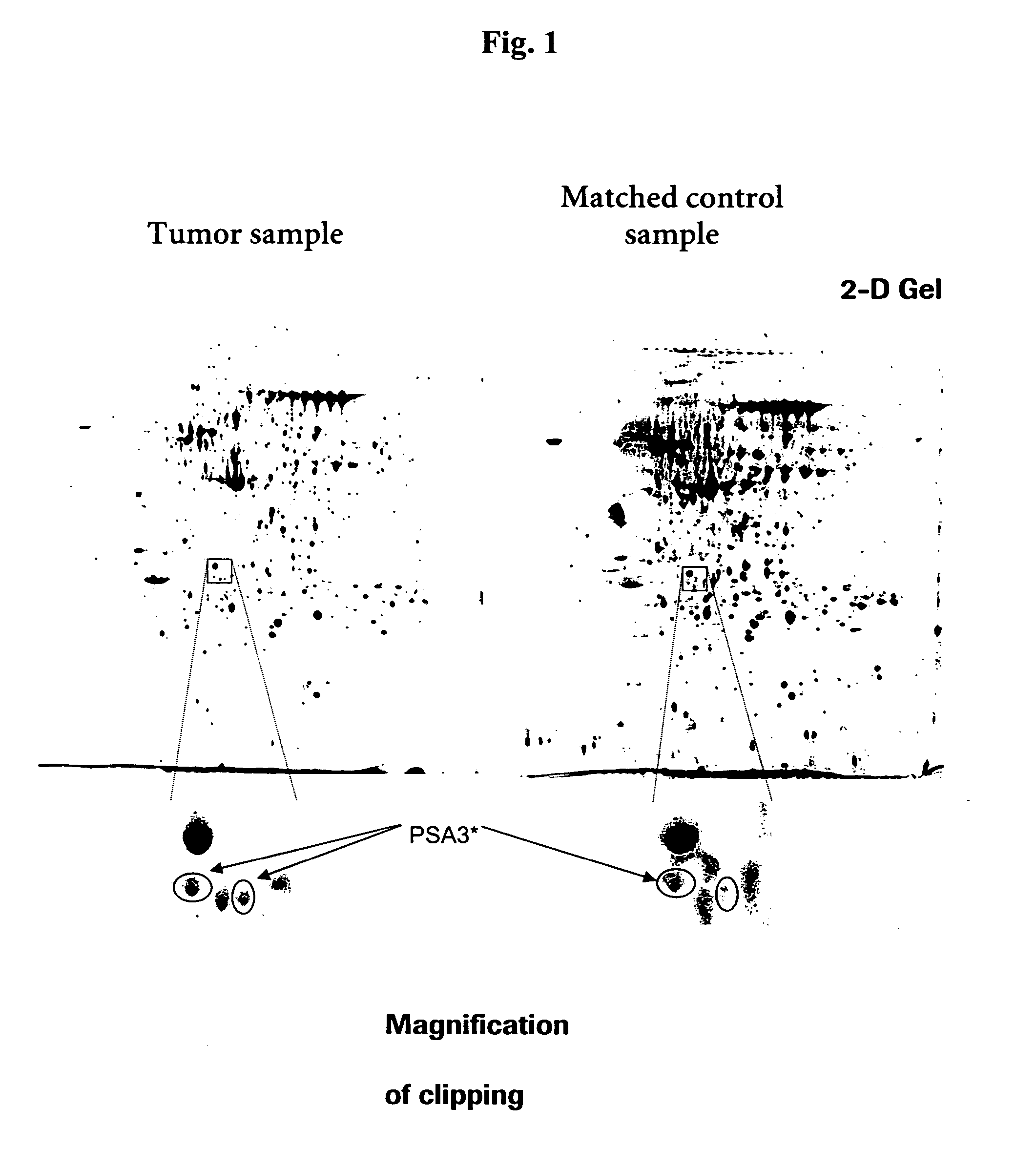
Identification of PSA3 as a Potential Colorectal Cancer Marker
[0086] Sources of Tissue
[0087] In order to identify tumor-specific proteins as potential diagnostic markers for colorectal cancer, analysis of three different kinds of tissue using proteomics methods is performed.
[0088] In total, tissue specimen from 10 patients suffering from colorectal cancer are analyzed. From each patient three different tissue types are collected from therapeutic resections: tumor tissue (>80% tumor) (T), adjacent healthy tissue (N) and stripped mucosa from adjacent healthy mucosa (M). The latter two tissue types serve as matched healthy control samples. Tissues are immediately snap frozen after resection and stored at −80° C. before processing. Tumors are diagnosed by histopathological criteria.
[0089] Tissue Preparation
[0090] 0.8-1.2 g of frozen tissue are put into a mortar and completely frozen by liquid nitrogen. The tissue is pulverized in the mortar, dissolved in the 10-fold volum...
example 2
Generation of Antibodies to the Colorectal Cancer Marker Protein PSA3
[0096] Polyclonal antibody to the colorectal cancer marker protein PSA3 is generated for further use of the antibody in the measurement of serum and plasma and blood levels of PSA3 by immunodetection assays, e.g. Western Blotting and ELISA.
[0097] Recombinant Protein Expression in E. coli
[0098] In order to generate antibodies to PSA3, recombinant expression of the protein is performed for obtaining immunogens. The expression is done applying a combination of the RTS 100 expression system and E. coli. In a first step, the DNA sequence is analyzed and recommendations for high yield cDNA silent mutational variants and respective PCR-primer sequences are obtained using the “ProteoExpert RTS E. coli HY” system. This is a commercial web based service (www.proteoexpert.com). Using the recommended primer pairs, the “RTS 100 E. coli Linear Template Generation Set, His-tag” (Roche Diagnostics GmbH, Mannheim, Germany, Cat. ...
example 3
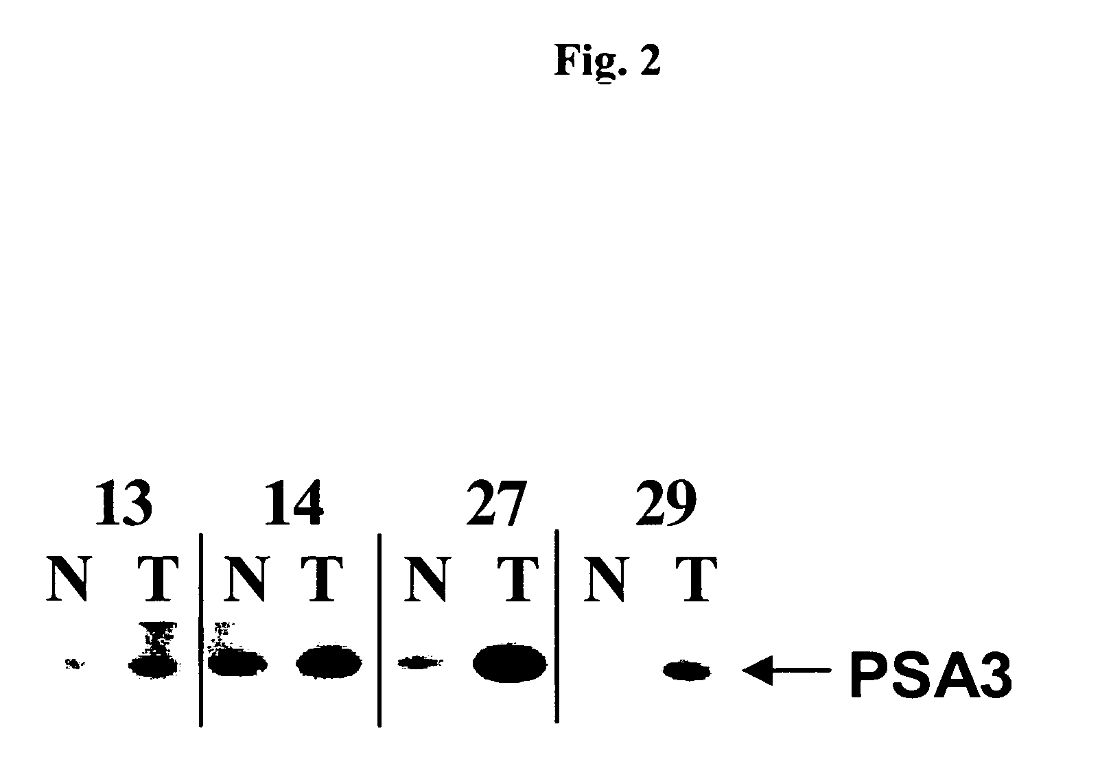
Western Blotting for the Detection of PSA3 in Human Colorectal Cancer Tissue Using Polyclonal Antibody as Generated in Example 2
[0119] Tissue lysates from tumor samples and healthy control samples are prepared as described in Example 1, “Tissue preparation”.
[0120] SDS-PAGE and Western-Blotting are carried out using reagents and equipment of Invitrogen, Karlsruhe, Germany. For each tissue sample tested, 10 μg of tissue lysate are diluted in reducing NuPAGE® (Invitrogen) SDS sample buffer and heated for 10 min at 95° C. Samples are run on 4-12% NuPAGE® gels (Tris-Glycine) in the MES running buffer system. The gel-separated protein mixture is blotted onto nitrocellulose membranes using the Invitrogen XCell II™ Blot Module (Invitrogen) and the NuPAGE® transfer buffer system. The membranes are washed 3 times in PBS / 0.05% Tween-20 and blocked with Roti®-Block blocking buffer (A151.1; Carl Roth GmbH, Karlsruhe, Germany) for 2 h. The primary antibody, polyclonal rabbit anti-PSA3 serum (ge...
PUM
 Abstract
Description
Claims
Application Information
 - R&D Engineer
- R&D Manager
- IP Professional
- Industry Leading Data Capabilities
- Powerful AI technology
- Patent DNA Extraction
Browse by: Latest US Patents, China's latest patents, Technical Efficacy Thesaurus, Application Domain, Technology Topic, Popular Technical Reports.
© 2024 PatSnap. All rights reserved.Legal|Privacy policy|Modern Slavery Act Transparency Statement|Sitemap|About US| Contact US: help@patsnap.com