Novel thrombospondin-1 polynucleotides encoding variant thrombospondin-1 polypeptides and methods using same
Inactive Publication Date: 2007-09-20
COJOCARU GAD +6
1 Cites 3 Cited by
AI-Extracted Technical Summary
Problems solved by technology
The background art also does not teach or suggest variants of TSP-1 and protein that are useful as therapeut...
Benefits of technology
[0013] In view of its critical role in angiogenesis and oncogenesis, there is an unmet need to develop therapies based on TSP-1. The background art does not teach or suggest variants of TSP-1 protein. The back...
Abstract
Novel polypeptides and polynucleotides encoding same are provided. Also provided methods and phamaceutical compositions which can be used to treat various disorders such as cancer and retinopathies, using the polypeptides and polynucleotides of the present invention.
Application Domain
Immunoglobulins against blood coagulation factorsSenses disorder +20
Technology Topic
DrugDisease +7
Image
Examples
- Experimental program(5)
Example
Example 1
[0313] Description of the methodology undertaken to uncover the biomolecular sequences of the present invention and uses therefor
[0314] Human ESTs and cDNAs were obtained from GenBank versions 136 (Jun. 15, 2003 ncbi “dot” nih “dot” gov/genbank/release “dot” notes/gb136 “dot” release “dot” notes) and NCBI genome assembly of April 2003. Novel splice variants were predicted using the LEADS clustering and assembly system as described in U.S. Pat. No: 6,625,545, U.S. patent application Ser. No. 10/426,002, both of which are hereby incorporated by reference as if fully set forth herein. Briefly, the software cleans the expressed sequences from repeats, vectors and immunoglobulins. It then aligns the expressed sequences to the genome taking alternatively splicing into account and clusters overlapping expressed sequences into “clusters” that represent genes or partial genes.
[0315] These were annotated using the GeneCarta (Compugen, Tel-Aviv, Israel) platform. The GeneCarta platform includes a rich pool of annotations, sequence information (particularly of spliced sequences), chromosomal information, alignments, and additional information such as SNPs, gene ontology terms, expression profiles, functional analyses, detailed domain structures, known and predicted proteins and detailed homology reports.
[0316] Brief description of the methodology used to obtain annotative sequence information is summarized infra (for detailed description see U.S. patent application Ser. No. 10/426,002, published as US20040101876 on May 27 2004).
[0317] The ontological annotation approach—An ontology refers to the body of knowledge in a specific knowledge domain or discipline such as molecular biology, microbiology, immunology, virology, plant sciences, pharmaceutical chemistry, medicine; neurology, endocrinology, genetics, ecology, genomics, proteomics, cheminformatics, pharmacogenomics, bioinformatics, computer sciences, statistics, mathematics, chemistry, physics and artificial intelligence.
[0318] An ontology includes domain-specific concepts—referred to, herein, as sub-ontologies. A sub-ontology may be classified into smaller and narrower categories. The ontological annotation approach is effected as follows.
[0319] First, biomolecular (i.e., polynucleotide or polypeptide) sequences are computationally clustered according to a progressive homology range, thereby generating a plurality of clusters each being of a predetermined homology of the homology range.
[0320] Progressive homology is used to identify meaningful homologies among biomolecular sequences and to thereby assign new ontological annotations to sequences, which share requisite levels of homologies. Essentially, a biomolecular sequence is assigned to a specific cluster if displays a predetermined homology to at least one member of the cluster (i.e., single linkage). A “progressive homology range” refers to a range of homology thresholds, which progress via predetermined increments from a low homology level (e.g. 35%) to a high homology level (e.g. 99%).
[0321] Following generation of clusters, one or more ontologies are assigned to each cluster. Ontologies are derived from an annotation preassociated with at least one biomolecular sequence of each cluster; and/or generated by analyzing (e.g., text-mining) at least one biomolecular sequence of each cluster thereby annotating biomolecular sequences.
[0322] The hierarchical annotation approach—“Hierarchical annotation” refers to any ontology and subontology, which can be hierarchically ordered, such as, a tissue expression hierarchy, a developmental expression hierarchy, a pathological expression hierarchy, a cellular expression hierarchy, an intracellular expression hierarchy, a taxonomical hierarchy, a functional hierarchy and so forth.
[0323] The hierarchical annotation approach is effected as follows. First, a dendrogram representing the hierarchy of interest is computationally constructed. A “dendrogram” refers to a branching diagram containing multiple nodes and representing a hierarchy of categories based on degree of similarity or number of shared characteristics.
[0324] Each of the multiple nodes of the dendrogram is annotated by at least one keyword describing the node, and enabling literature and database text mining, such as by using publicly available text mining software. A list of keywords can be obtained from the GO Consortium (www.geneontlogy.org). However, measures are taken to include as many keywords, and to include keywords which might be out of date. For example, for tissue annotation, a hierarchy is built using all available tissue/libraries sources available in the GenBank, while considering the following parameters: ignoring GenBank synonyms, building anatomical hierarchies, enabling flexible distinction between tissue types (normal versus pathology) and tissue classification levels (organs, systems, cell types, etc.).
[0325] In a second step, each of the biomolecular sequences is assigned to at least one specific node of the dendrogram.
[0326] The biomolecular sequences can be annotated biomolecular sequences, unannotated biomolecular sequences or partially annotated biomolecular sequences.
[0327] Annotated biomolecular sequences can be retrieved from pre-existing annotated databases as described hereinabove.
[0328] For example, in GenBank, relevant annotational information is provided in the definition and keyword fields. In this case, classification of the annotated biomolecular sequences to the dendrogram nodes is directly effected. A search for suitable annotated biomolecular sequences is performed using a set of keywords which are designed to classify the biomolecular sequences to the hierarchy (i.e., same keywords that populate the dendrogram).
[0329] In cases where the biomolecular sequences are unannotated or partially annotated, extraction of additional annotational information is effected prior to classification to dendrogram nodes. This can be effected by sequence alignment, as described hereinabove. Alternatively, annotational information can be predicted from structural studies. Where needed, nucleic acid sequences can be transformed to amino acid sequences to thereby enable more accurate annotational prediction.
[0330] Finally, each of the assigned biomolecular sequences is recursively classified to nodes hierarchically higher than the specific nodes, such that the root node of the dendrogram encompasses the full biomolecular sequence set, which can be classified according to a certain hierarchy, while the offspring of any node represent a partitioning of the parent set.
[0331] For example, a biomolecular sequence found to be specifically expressed in “rhabdomyosarcoma”, will be classified also to a higher hierarchy level, which is “sarcoma”, and then to “Mesenchymal cell tumors” and finally to a highest hierarchy level “Tumor”. In another example, a sequence found to be differentially expressed in endometrium cells, will be classified also to a higher hierarchy level, which is “uterus”, and then to “women genital system” and to “genital system” and finally to a highest hierarchy level “genitourinary system”. The retrieval can be performed according to each one of the requested levels.
[0332] Annotating gene expression according to relative abundance—Spatial and temporal gene annotations are also assigned by comparing relative abundance in libraries of different origins. This approach can be used to find genes, which are differentially expressed in tissues, pathologies and different developmental stages. In principal, the presentation of a contig in at least two tissues of interest is determined and significant over or under representation of the contig in one of the at least two tissues is assessed to identify differential expression. Significant over or under representation is analyzed by statistical pairing.
[0333] Annotating spatial and temporal expression can also be effected on splice variants. This is effected as follows. First, a contig which includes exonal sequence presentation of the at least two splice variants of the gene of interest is obtained. This contig is assembled from a plurality of expressed sequences. Then, at least one contig sequence region, unique to a portion (i.e., at least one and not all) of the at least two splice variants of the gene of interest, is identified. Identification of such unique sequence region is effected using computer alignment software. Finally, the number of the plurality of expressed sequences in the tissue having the at least one contig sequence region is compared with the number of the plurality of expressed sequences not-having the at least one contig sequence region, to thereby compare the expression level of the at least two splice variants of the gene of interest in the tissue.
[0334] Data concerning therapies, indications and possible pharmacological activities of the polypeptides of the present invention was obtained from PharmaProject (PJB Publications Ltd 2003 www “dot” pjbpubs “dot” com/cms “dot” asp?pageid=340) and public databases, including LocusLink (www “dot” genelynx “dot” org/cgi-bin/resource?res=locuslink) and Swissprot (www “dot” ebi “dot” ac “dot” uk/swissprot/index “dot” html). Functional structural analysis of the polypeptides of the present invention was effected using Interpro domain analysis software (Interpro default parameters, the analyses that were run are HMMPfam, HMMSmart, ProfileScan, FprintScan, and BlastProdom). Subecilular localization was analysed using ProLoc software (Einat Hazkani-Covo, Erez Y. Levanon, Galit Rotman, Dan Graur, Amit Novik. Evolution of multicellularity in metazoa: comparative analysis of the subcellular localization of proteins in Saccharomyces, Drosophila and Caenorhabditis. Cell Biology International (2004;28(3):171-8).
[0335] Identifying gene products by interspecies sequence comparison—The present inventors have designed and configured a method of predicting gene expression products based on interspecies sequence comparison. Specifically, the method is based on the identification of conserved alternatively spliced exons for which there might be no supportive expression data.
[0336] Alternatively spliced exons have unique characteristics differentiating them from constitutively spliced ones. Using machine-learning techniques a combination of such characteristics was elucidated that defines alternatively spliced exons with very high probability. Any human exon having this combination of characteristics is therefore predicted to be alternatively spliced. Using this method, the present inventors were able to detect putative splice variants that are not supported by human ESTs.
[0337] The method is effected as follows. First, alternatively spliced exons of a gene of interest are identified by scoring exon sequences of the gene of interest according to at least one sequence parameter as follows: (i) exon length—conserved alternatively spliced exons are relatively shorter than constitutively spliced ones; (ii) division by 3 —alternatively spliced exons are cassette exons that are sometimes inserted and sometimes skipped; Since alternatively spliced exons frequently contain sequences that regulate their splicing important parameters for scoring alternatively spliced exons include (iii) conservation level to a non-human ortholohgous sequence; (iv) length of conserved intron sequences upstream of each of the exon sequences; (v) length of conserved intron sequences downstream of each of the exon sequences; (vi) conservation level of the intron sequences upstream of each of the exon sequences; and (vii) conservation level of the intron sequences downstream of each of the exon sequences.
[0338] Exon sequences scoring above a predetermined threshold represent alternatively spliced exons of the gene of interest.
[0339] Once alternatively spliced exons are identified, the chromosomal location of each of the alternatively spliced exons is analyzed with respect to coding sequence of the gene of interest to thereby predict expression products of the gene of interest. When performed along with computerized means, mass prediction of gene products can be effected.
[0340] In addition, for identifying new gene products by interspecies sequence comparison, the expressed sequences derived from non-human species can be used for new human splice variants prediction.
Example
Example 2
Description for Cluster Humthrom
[0341] Cluster HUMTHROM features 5 transcripts the names for which are given in Table 1. The selected protein variants are given in Table 2. TABLE 1 Transcripts of interest Transcript Name HUMTHROM_1_T12 (SEQ ID NO:1) HUMTHROM_1_T14 (SEQ ID NO:2) HUMTHROM_1_T15 (SEQ ID NO:3) HUMTHROM_1_T17 (SEQ ID NO:4) HUMTHROM_1_T32 (SEQ ID NO:5)
[0342] TABLE 2 Proteins of interest Corresponding Protein Name Transcript(s) HUMTHROM_1_P8 HUMTHROM_1_T12 (SEQ ID NO:48) (SEQ ID NO:1) HUMTHROM_1_P10 HUMTHROM_1_T15 (SEQ ID NO:49) (SEQ ID NO:3) HUMTHROM_1_P12 HUMTHROM_1_T17 (SEQ ID NO:50) (SEQ ID NO:4) HUMTHROM_1_P22 HUMTHROM_1_T32 (SEQ ID NO:51) (SEQ ID NO:5) HUMTHROM_1_P27 HUMTHROM_1_T14 (SEQ ID NO:52) (SEQ ID NO:2)
[0343] These sequences are variants of the known protein Thrombospondin 1 precursor (SEQ ID NO:44) (SwissProt accession identifier TSP-1_HUMAN), referred to herein as the previously known protein.
[0344] Protein Thrombospondin 1 precursor (SEQ ID NO:44) is known or believed to have the following function(s): Adhesive glycoprotein that mediates cell-to-cell and cell-to-matrix interactions. Can bind to fibrinogen, fibronectin, laminin, type V collagen and integrins alpha-V/beta-1, alpha- V/beta-3 and alpha-Ilb/beta-3. Known polymorphisms for this sequence are as shown in Table 3. TABLE 3 Amino acid mutations for Known Protein SNP position(s) on amino acid sequence Comment 84 T -> A 523 T -> A
[0345] The previously known protein also has the following indication(s) and/or potential therapeutic use(s): Cancer, general. It has been investigated for clinical/therapeutic use in humans, for example as a target for an antibody or small molecule, and/or as a direct therapeutic; available information related to these investigations is as follows. Potential pharmaceutically related or therapeutically related activity or activities of the previously known protein are as follows: Angiogenesis inhibitor; Thrombospondin antagonist. A therapeutic role for a protein represented by the cluster has been predicted. The cluster was assigned this field because there was information in the drug database or the public databases (e.g., described herein above) that this protein, or part thereof, is used or can be used for a potential therapeutic indication: Anticancer, other; Imaging agent; Recombinant, other.
[0346] The following GO Annotation(s) apply to the previously known protein. The following annotation(s) were found: development, which are annotation(s) related to Biological Process; endopeptidase inhibitor activity; signal transducer activity, which are annotation(s) related to Molecular Function; and extracellular region, which are annotation(s) related to Cellular Component.
[0347] The GO assignment relies on information from one or more of the SwissProt/TremBl Protein knowledgebase, available from; or Locuslink, available from.
[0348] Acording to the present invention, TSP variants can be used for treatment of primary and metastatic cancer, targeting a broad spectrum of cancers, including but not limited to prostate cancer, renal cancer, cervical carcinomas, breast cancer, colon and colorectal cancer, pancreatic cancer, ovarian cancer, bladder cancer, lung cancer, melanoma, brain cancer, soft tissue sarcomas, lymphomas, head-and-neck, glioblastomas, and other tumors and metastatic cancers. This includes the use of the TSP-1 variants in this invention as monotherapy for cancer, or in combination therapy with any of various other cytotoxic agents, or anti-angiogenic and/or anti-tumor agents.
[0349] TSP variants of the present invention can be used for treatment of retinal angiogenesis in a number of human ocular diseases, such as diabetic retinopathy, retinopathy of prematurity, and age-related macular degeneration.
[0350] As noted above, cluster HUMTHROM features 5 transcript(s), which were listed in Table 1 above. These transcript(s) encode for protein(s) which are variant(s) of protein Thrombospondin 1 precursor (SEQ ID NO:44). A description of each variant protein according to the present invention is now provided.
[0351] Variant protein HUMTHROM—1_P8 (SEQ ID NO:48) according to the present it is encoded by transcript(s) HUMTHROM—1_T12 (SEQ ID NO:1).
[0352] The localization of the variant protein was determined according to results from a number of different software programs and analyses, including analyses from SignalP and other specialized programs. The variant protein is believed to be secreted.
[0353] Variant protein HUMTHROM—1_P8 (SEQ ID NO:48) also has the following non-silent SNPs (Single Nucleotide Polymorphisms) as listed in Table 4 (given according to their position(s) on the amino acid sequence, with the alternative amino acid)(s) listed;). TABLE 4 Amino acid mutations SNP position(s) on amino Alternative acid sequence amino acid(s) 42 K -> 79 V -> M 163 D -> G 181 V -> 237 S -> N 329 E -> G 478 A ->
[0354] The glycosylation sites of variant protein HUMTHROM—1_P8 (SEQ ID NO:48), as compared to the known protein Thrombospondin 1 precursor (SEQ ID NO:44), are described in Table 5 (given according to their position(s) on the amino acid sequence in the first column; the second column indicates whether the glycosylation site is present in the variant protein; and the last column indicates whether the position is different on the variant protein). TABLE 5 Glycosylation site(s) Position(s) on known Present in Position(s) on variant amino acid sequence variant protein? protein 248 Yes 248 360 Yes 360 385 Yes 385 394 Yes 394 438 Yes 438 441 Yes 441 450 Yes 450 498 No 507 No 708 No 1067 No
[0355] The variant protein has the following domains, as determined by using InterPro. The domains are described in Table 6. TABLE 6 InterPro domain(s) Analysis Domain description type Position(s) on protein Thrombospondin, subtype 1 FPrintScan 436-449, 454-465, 473-484 Thrombospondin, type I HMMPfam 383-428, 439-489 von Willebrand factor, type C HMMPfam 318-372 Thrombospondin, type I HMMSmart 382-429, 438-490 Thrombospondin, N-terminal HMMSmart 24-221 von Willebrand factor, type C HMMSmart 318-372 Thrombospondin, type I ProfileScan 379-429, 435-490 von Willebrand factor, type C ProfileScan 316-373 von Willebrand factor, type C ScanRegExp 336-372
[0356] Variant protein HUMTHROM—1_P8 (SEQ ID NO:48) is encoded by the transcript HUMTHROM—1_T12 (SEQ ID NO:1). The coding portion of transcript HUMTHROM—1_T12 (SEQ ID NO:1) starts at position 326 and ends at position 2059. The transcript also has the following SNPs as listed in Table 7 (given according to their position on the nucleotide sequence, with the alternative nucleic acid listed). TABLE 7 Nucleic acid SNPs SNP position(s) on Alternative nucleotide sequence nucleic acid(s) 21 G -> C 151 G -> A 188 T -> C 451 G -> 560 G -> A 813 A -> G 868 C -> 1035 G -> A 1311 A -> G 1615 G -> A 1735 C -> T 1757 G -> 2199 A -> G 2204 C -> T 2431 T -> C 2519 C -> 2528 G -> 2599 A -> G 2675 C -> 2727 C -> 2731 A -> G 3230 C -> G 3230 C -> 3500 T -> C 3505 G -> 3536 A -> 3550 A -> 3603 A -> G 3661 G -> C 3892 C -> 3932 A -> G 3982 A -> 4105 T -> C 4183 A -> 4224 C -> T 4423 G -> A 4450 T -> A 4490 A -> T 4559 C -> A 4643 A -> T 4730 G -> A 4808 C -> G 4821 C -> T 4856 A -> C 5033 T -> G 5121 T -> 5135 T -> 5251 T -> G 5251 T -> 5275 T -> 5420 C -> A 5420 C -> 5489 T -> C 5489 T -> 5605 T -> C 5606 C -> T 5674 T -> 5777 A -> G 5852 T -> 5974 C -> G 6052 T -> G 6057 T -> G 6125 A -> G
[0357] Variant protein HUMTHROM—1_P10 (SEQ ID NO:49) according to the present invention is encoded by transcript HUMTHROM—1_T15 (SEQ ID NO:3). One or more alignments to one or more previously published protein sequences are shown in FIG. 7. A brief description of the relationship of the variant protein according to the present invention to each such aligned protein is as follows:
1. Comparison report between HUMTHROM—1_P10 (SEQ ID NO:49) and TSP-1_HUMAN_V1 (SEQ ID NO:47):
[0358] A. An isolated chimeric polypeptide encoding for HUMTHROM—1_P10 (SEQ ID NO:49), comprising a first amino acid sequence being at least 90% homologous to MGLAWGLGVLFLMHVCGTNRIPESGGDNSVFDIFELTGAARKGSGRRLVKG PDPSSPAFRIEDANLIPPVPDDKFQDLVDAVRAEKGFLLLASLRQMKKTRGTL LALERKDHSGQVFSVVSNGKAGTLDLSLTVQGKQHVVSVEEALLATGQWKS ITLFVQEDRAQLYIDCEKMENAELDVPIQSVFTRDLASIARLRIAKGGVNDNF QGVLQNVRFVFGTTPEDILRNKGCSSSTSVLLTLDNNVVNGSSPAIRTNYIGH KTKDLQAICGISCDELSSMVLELRGLRTIVTTLQDSfRKVTEENKELANELRRP PLCYHNGVQYRNNEEWTVDSCTECHCQNSVTICKKVSCPIMPCSNATVPDGE CCPRCWPSDSADDGWSPWSEWTSCSTSCGNGIQQRGRSCDSLNNRCEGSSVQ TRTCHIQECDKRFKQDGGWSHWSPWSSCSVTCGDGVITRIRLCNSPSPQMNG KPCEGEARETKACKKDACPINGGWGPWSPWDICSVTCGGGVQKRSRLCNNP TPQFGGKDCVGDVTENQICNKQDCPIDGCLSNPCFAGVKCTSYPDGSWKCGA CPPGYSGNGIIQCTDVDECKEVPDACFNHNGEHRCENTDPGYNCLPCPPRFTG SQPFGQGVEHATANKQVCKPRNPCTDGTHDCNKNAKCNYLGHYSDPMYRC ECKPGYAGNGIICGEDTDLDGWPNENLVCVANATYHCKKDNCPNLPNSGQE DYDKDGIGDACDDDDDNDKIPDDR corresponding to amino acids 1-751 of TSP-1_HUMAN_V1 (SEQ ID NO:47), which also corresponds to amino acids 1-751 of HUMTHROM—1_P10 (SEQ ID NO:49), and a second amino acid sequence being at least 70%, optionally at least 80%, preferably at least 85%, more preferably at least 90% and most preferably at least 95% homologous to a polypeptide having the sequence VKTVFYPFFIFSVQQQPETLWDSRKLHGYSKKYTKSIHRIIRNYSLCSSSLRM corresponding to amino acids 752-804 of HUMTHROM—1_P10 (SEQ ID NO:49), wherein said first amino acid sequence and second amino acid sequence are contiguous and in a sequential order.
[0359] B. An isolated polypeptide encoding for an edge portion of HUMTHROM—1_P10 (SEQ ID NO:49), comprising an amino acid sequence being at least 70%, optionally at least about 80%, preferably at least about 85%, more preferably at least about 90% and most preferably at least about 95% homologous to the sequence VKTVFYPFFIFSVQQQPETLWDSRKLHGYSKKYTKSIHRIIRNYSLCSSSLRM of HUMTHROM—1_P10 (SEQ ID NO:49).
[0360] It should be noted that the known protein sequence (TSP-1_HUMAN) has one or more changes than the sequence given at the end of the application and named as being the amino acid sequence for TSP-1_HUMAN_V1 (SEQ ID NO:47). These changes were previously known to occur and are listed in the table below. TABLE 8 Changes to TSP-1_HUMAN_V1 (SEQ ID NO: 47) SNP position on amino acid sequence Type of change 84 conflict
[0361] The localization of the variant protein was determined according to results from a number of different software programs and analyses, including analyses from SignalP and other specialized programs. The variant protein is believed to be secreted.
[0362] Variant protein HUMTHROM—1_P10 (SEQ ID NO:49) also has the following non-silent SNPs (Single Nucleotide Polymorphisms) as listed in Table 9, (given according to their position(s) on the amino acid sequence, with the alternative amino acid(s) listed). TABLE 9 Amino acid mutations SNP position(s) on amino Alternative acid sequence amino acid(s) 42 K -> 79 V -> M 163 D -> G 181 V -> 237 S -> N 329 E -> G 478 A -> 523 T -> A 600 F -> S 629 P -> 632 Q -> 656 D -> G 681 G -> 699 P -> 700 N -> S
[0363] The glycosylation sites of variant protein HUMTHROM—1_P10 (SEQ ID NO:49), as compared to the known protein Thrombospondin 1 precursor (SEQ ID NO:44), are described in Table 10 (given according to their position(s) on the amino acid sequence in the first column; the second column indicates whether the glycosylation site is present in the variant protein; and the last column indicates whether the position is different on the variant protein). TABLE 10 Glycosylation site(s) Position(s) on known Present in Position(s) on variant amino acid sequence variant protein? protein 248 Yes 248 360 Yes 360 385 Yes 385 394 Yes 394 438 Yes 438 441 Yes 441 450 Yes 450 498 Yes 498 507 Yes 507 708 Yes 708 1067 No
[0364] The variant protein has the following domains, as determined by using InterPro. The domains are described in Table 11. TABLE 11 InterPro domain(s) Analysis Domain description type Position(s) on protein Thrombospondin, subtype 1 FPrintScan 436-449, 454-465, 473-484 EGF-like HMMPfam 650-689 Thrombospondin, type I HMMPfam 383-428, 439-489, 496-546 von Willebrand factor, type C HMMPfam 318-372 Thrombospondin type 3 repeat HMMPfam 691-706, 727-739 EGF-like calcium-binding HMMSmart 542-587, 588-645 Type I EGF HMMSmart 550-587, 591-645, 649-690 Thrombospondin, type I HMMSmart 382-429, 438-490, 495-547 Thrombospondin, N-terminal HMMSmart 24-221 von Willebrand factor, type C HMMSmart 318-372 Thrombospondin, type I ProfileScan 379-429, 435-490, 492-547 von Willebrand factor, type C ProfileScan 316-373 EGF-like ScanRegExp 676-689 von Willebrand factor, type C ScanRegExp 336-372
[0365] Variant protein HUMTHROM—1_P10 (SEQ ID NO:49) is encoded by the transcript HUMTHROM—1_T15 (SEQ ID NO:3). The coding portion of transcript HUMTHROM—1_T15 (SEQ ID NO:3) starts at position 326 and ends at position 2737. The transcript also has the following SNPs as listed in Table 12 (given according to their position on the nucleotide sequence, with the alternative nucleic acid listed). TABLE 12 Nucleic acid SNPs SNP position(s) on Alternative nucleotide sequence nucleic acid(s) 21 G -> C 151 G -> A 188 T -> C 451 G -> 560 G -> A 813 A -> G 868 C -> 1035 G -> A 1311 A -> G 1615 G -> A 1735 C -> T 1757 G -> 1892 A -> G 1897 C -> T 2124 T -> C 2212 C -> 2221 G -> 2292 A -> G 2368 C -> 2420 C -> 2424 A -> G 3490 C -> G 3490 C -> 3760 T -> C 3765 G -> 3796 A -> 3810 A -> 3863 A -> G 3921 G -> C 4152 C -> 4192 A -> G 4242 A -> 4365 T -> C 4443 A -> 4484 C -> T 4683 G -> A 4710 T -> A 4750 A -> T 4819 C -> A 4903 A -> T 4990 G -> A 5068 C -> G 5081 C -> T 5116 A -> C 5293 T -> G 5381 T -> 5395 T -> 5511 T -> G 5511 T -> 5535 T -> 5680 C -> A 5680 C -> 5749 T -> C 5749 T -> 5865 T -> C 5866 C -> T 5934 T -> 6037 A -> G 6112 T -> 6234 C -> G 6312 T -> G 6317 T -> G 6385 A -> G
[0366] Variant protein HUMTHROM—1_P12 (SEQ ID NO:50) according to the present invention is encoded by transcript HUMTHROM—1_T17 (SEQ ID NO:4). One or more alignments to one or more previously published protein sequences are shown in FIG. 7. A brief description of the relationship of the variant protein according to the present invention to each such aligned protein is as follows:
1. Comparison report between HUMTHROM—1_P12 (SEQ ID NO:50) and TSP-1_HUMAN_V1 (SEQ ID NO:47):
[0367] A. An isolated chimeric polypeptide encoding for HUMTHROM—1_P12 (SEQ ID NO:50), comprising a first amino acid sequence being at least 90% homologous to MGLAWGLGVLFLMHVCGTNRIPESGGDNSVFDIFELTGAARKGSGRRLVKG PDPSSPAFRIEDANLIPPVPDDKFQDLVDAVRAEKGFLLLASLRQMKKTRGTL LALERKDHSGQVFSVVSNGKAGTLDLSLTVQGKQHVVSVEEALLATGQWKS ITLFVQEDRAQLYIDCEKMENAELDVPIQSVFTRDLASIARLRIAKGGVNDNF QGVLQNVRFVFGTTPEDILRNKGCSSSTSVLLTLDNNVVNGSSPAIRTNYIGH KTKDLQAICGISCDELSSMVLELRGLRTIVTTLQDSIRKVTEENKELANELRRP PLCYHNGVQYRNNEEWTVDSCTECHCQNSVTICKKVSCPIMPCSNATVPDGE CCPRCWPSDSADDGWSPWSEWTSCSTSCGNGIQQRGRSCDSLNNRCEGSSVQ TRTCHIQECDKRFKQDGGWSHWSPWSSCSVTCGDGVITRIRLCNSPSPQMNG KPCEGEARETKACKKDACPINGGWGPWSPWDICSVTCGGGVQKRSRLCNNP TPQFGGKDCVGDVTENQICNKQDCPIDGCLSNPCFAGVKCTSYPDGS WKCGA CPPGYSGNGIQCTDVDECKEVPDACFNHNGEHRCENTDPGYNCLPCPPRFTG SQPFGQGVEHATANKQV corresponding to amino acids 1-643 of TSP-1_HUMAN_V1 (SEQ ID NO:47), which also corresponds to amino acids 1-643 of HUMTHROM—1_P12 (SEQ ID NO:50), and a second amino acid sequence being at least 70%, optionally at least 80%, preferably at least 85%, more preferably at least 90% and most preferably at least 95% homologous to a polypeptide having the sequence QSTRRVNQRTGELSLTKITGSGRNVISYPSPKKKGRGDECTV corresponding to amino acids 644-685 of HUMTHROM—1_P12 (SEQ ID NO:50), wherein said first amino acid sequence and second amino acid sequence are contiguous and in a sequential order.
[0368] B. An isolated polypeptide encoding for an edge portion of HUMTHROM—1_P12 (SEQ ID NO:50), comprising an amino acid sequence being at least 70%, optionally at least about 80%, preferably at least about 85%, more preferably at least about 90% and most preferably at least about 95% homologous to the sequence QSTRRVNQRTGELSLTKITGSGRNVISYPSPKKKGRGDECTV of HUMTHROM—1_P12 (SEQ ID NO:50).
[0369] The localization of the variant protein was determined according to results from a number of different software programs and analyses, including analyses from SignalP and other specialized programs. The variant protein is believed to be secreted.
[0370] Variant protein HUMTHROM—1_P12 (SEQ ID NO:50) also has the following non-silent SNPs (Single Nucleotide Polymorphisms) as listed in Table 14, (given according to their position(s) on the amino acid sequence, with the alternative amino acid(s) listed; the last column indicates whether the SNP is known or not; the presence of known SNPs in variant protein HUMTHROM—1_P12 (SEQ ID NO:50) sequence des support for the deduced sequence of this variant protein according to the present invention). TABLE 14 Amino acid mutations SNP position(s) on amino acid sequence Alternative amino acid(s) 42 K -> 79 V -> M 163 D -> G 181 V -> 237 S -> N 329 E -> G 478 A -> 523 T -> A 600 F -> S 629 P -> 632 Q ->
[0371] The glycosylation sites of variant protein HUMTHROM—1_P12 (SEQ ID NO:50), as compared to the known protein Thrombospondin 1 precursor (SEQ ID NO:44), are described in Table 15 (given according to their position(s) on the amino acid sequence in the first column; the second column indicates whether the glycosylation site is present in the variant protein; and the last column indicates whether the position is different on the variant protein). TABLE 15 Glycosylation site(s) Position(s) on known Present in variant Position(s) on variant amino acid sequence protein? protein 248 Yes 248 360 Yes 360 385 Yes 385 394 Yes 394 438 Yes 438 441 Yes 441 450 Yes 450 498 Yes 498 507 Yes 507 708 No 1067 No
[0372] The variant protein has the following domains, as determined by using InterPro. The domains are described in Table 16. TABLE 16 InterPro domain(s) Analysis Domain description type Position(s) on protein Thrombospondin, subtype 1 FPrintScan 436-449, 454-465, 473-484 Thrombospondin, type I HMMPfam 383-428, 439-489, 496-546 von Willebrand factor, type C HMMPfam 318-372 EGF-like calcium-binding HMMSmart 542-587, 588-632 Type I EGF HMMSmart 550-587, 591-631 Thrombospondin, type I HMMSmart 382-429, 438-490, 495-547 Thrombospondin, N-terminal HMMSmart 24-221 von Willebrand factor, type C HMMSmart 318-372 Thrombospondin, type I ProfileScan 379-429, 435-490, 492-547 von Willebrand factor, type C ProfileScan 316-373 von Willebrand factor, type C ScanRegExp 336-372
[0373] Variant protein HUMTHROM—1_P12 (SEQ ID NO:50) is encoded by the transcript HUMTHROM—1_T17 (SEQ ID NO:4). The coding portion of transcript HUMTHROM—1_T17 (SEQ ID NO:4 portion starts at position 326 and ends at position 2380. The transcript also has the following SNPs as listed in Table 17 (given according to their position on the nucleotide sequence, with the alternative nucleic acid listed). TABLE 17 Nucleic acid SNPs SNP position(s) on Alternative nucleic nucleotide sequence acid(s) 21 G -> C 151 G -> A 188 T -> C 451 G -> 560 G -> A 813 A -> G 868 C -> 1035 G -> A 1311 A -> G 1615 G -> A 1735 C -> T 1757 G -> 1892 A -> G 1897 C -> T 2124 T -> C 2212 C -> 2221 G -> 2742 A -> G 2818 C -> 2870 C -> 2874 A -> G 3373 C -> G 3373 C -> 3643 T -> C 3648 G -> 3679 A -> 3693 A -> 3746 A -> G 3804 G -> C 4035 C -> 4075 A -> G 4125 A -> 4248 T -> C 4326 A -> 4367 C -> T 4566 G -> A 4593 T -> A 4633 A -> T 4702 C -> A 4786 A -> T 4873 G -> A 4951 C -> G 4964 C -> T 4999 A -> C 5176 T -> G 5264 T -> 5278 T -> 5394 T -> G 5394 T -> 5418 T -> 5563 C -> A 5563 C -> 5632 T -> C 5632 T -> 5748 T -> C 5749 C -> T 5817 T -> 5920 A -> G 5995 T -> 6117 C -> G 6195 T -> G 6200 T -> G 6268 A -> G
[0374] Variant protein HUMTHROM—1_P22 (SEQ ID NO:51) according to the present is encoded by transcript HUMTHROM—1_T32 (SEQ ID NO:5). One or more alignments to one or more previously published protein sequences are shown in FIG. 7. A brief description of the relationship of the variant protein according to the present invention to each such aligned protein is as follows:
1. Comparison report between HUMTHROM—1_P22 (SEQ ID NO:51) and TSP-1_HUMAN_V1 (SEQ ID NO:47):
[0375] A. An isolated chimeric polypeptide encoding for HUMTHROM—1_P22 (SEQ ID NO:51), comprising a first amino acid sequence being at least 90% homologous to WGLGVLFLMHVCGTNRIPESGGDNSVFDIFELTGAARKGSGRRLVKG SPAFRIEDANLIPPVPDDKFQDLVDAVRAEKGFLLLASLRQMKKTRGTL RKDHSGQVFSVVSNGKAGTLDLSLTVQGKQHVVSVEEALLATGQWKS VQEDRAQLYIDCEKMENAELDVPIQSVFTRDLASIARLRIAKGGVNDNF QNVRFVFGTTPEDILRNKGCSSSTSVLLTLDNNVVNGSSPAIRTNYIGH LQAICGISCDELSSMVLELRGLRTIVTTLQDSIRKVTEENKELANELRRP HNGVQYRNNEEWTVDSCTECHCQNSVTICKKVSCPIMPCSNATVPDGE CWPSDSADDGWSPWSEWTSCSTSCGNGIQQRGRSCDSLNNRCEGSSVQ HIQECDKRFKQDGGWSHWSPWSSCSVTCGDGVITRIRLCNSPSPQMNG KPCEGEARETKACKKDACP corresponding to amino acids 1-490 of TSP-1_HUMAN_V1 (SEQ ID NO:47), which also corresponds to amino acids 1-490 of HUMTHROM—1_P22 (SEQ ID NO:51), a second bridging amino acid sequence comprising of N, and a third amino acid sequence being at least 90% homologous to GCLSNPCFAGVKCTSYPDGSWKCGACPPGYSGNGIQCTDVDECKEVPDACFN HNGEHRCENTDPGYNCLPCPPRFTGSQPFGQGVEHATANKQVCKPRNPCTDG THDCNKNAKCNYLGHYSDPMYRCECKPGYAGNGIICGEDTDLDGWPNENLV CVANATYHCKKDNCPNLPNSGQEDYDKDGIGDACDDDDDNDKIPDDRDNCP FHYNPAQYDYDRDDVGDRCDNCPYNHNPDQADTDNNGEGDACAADIDGDG ILNERDNCQYVYNVDQRDTDMDGVGDQCDNCPLEHNPDQLDSDSDRIGDTC DNNQDIDEDGHQNNLDNCPYVPNANQADHDKDGKGDACDHDDDNDGIPDD KDNCRLVPNPDQKDSDGDGRGDACKDDFDHDSVPDIDDICPENVDISETDFR RFQMIPLDPKGTSQNDPNWVVRHQGKELVQTVNCDPGLAVGYDEFNAVDFS GTFFINTERDDDYAGFVFGYQSSSRFYVVMWKQVTQSYWDTNPTRAQGYSG LSVKVVNSTTGPGEHLRNALWHTGNTPGQVRTLWHDPRHIGWKDFTAYRW RLSHRPKTGFIRVVMYEGKKIMADSGPIYDKTYAGGRLGLFVFSQEMVFFSD LKYECRDP corresponding to amino acids 550-1170 of TSP-1_HUMAN_V1 (SEQ ID NO:47), which also corresponds to amino acids 492-1112 of HUMTHROM—1_P22 (SEQ ID NO:51), wherein said first amino acid sequence, second amino acid sequence and third amino acid sequence are contiguous and in a sequential order.
[0376] B. An isolated polypeptide encoding for an edge portion of HUMTHROM—1_P22 (SEQ ID NO:51), comprising a polypeptide having a length “n”, wherein n is at least about 10 amino acids in length, optionally at least about 20 amino acids in length, preferably at least about 30 amino acids in length, more preferably at least about 40 amino acids in length and most preferably at least about 50 amino acids in length, wherein at least three amino acids comprise PNG having a structure as follows (numbering according to HUMTHROM—1_P22 (SEQ ID NO:51)): a sequence starting from any of amino acid numbers 490-x to 490; and ending at any of amino acid numbers 492+((n−2)−x), in which x varies from 0 to n−2.
[0377] The localization of the variant protein was determined according to results from a number of different software programs and analyses, including analyses from SignalP and other specialized programs. The variant protein is believed to be secreted.
[0378] Variant protein HUMTHROM—1_P22 (SEQ ID NO:51) also has the following non-silent SNPs (Single Nucleotide Polymorphisms) as listed in Table 19, (given according to their position(s) on the amino acid sequence, with the alternative amino acid(s) listed). TABLE 19 Amino acid mutations SNP position(s) on amino acid sequence Alternative amino acid(s) 42 K -> 79 V -> M 163 D -> G 181 V -> 237 S -> N 329 E -> G 478 A -> 542 F -> S 571 P -> 574 Q -> 598 D -> G 623 G -> 641 P -> 642 N -> S 808 G -> 900 R -> 910 K -> 915 N -> 933 N -> D 952 G -> A 1029 P -> 1042 I -> M 1059 K -> 1100 V -> A
[0379] The glycosylation sites of variant protein HUMTHROM—1_P22 (SEQ ID NO:51), as compared to the known protein Thrombospondin 1 precursor (SEQ ID NO:44), are described in Table 20 (given according to their position(s) on the amino acid sequence in the first column; the second column indicates whether the glycosylation site is present in the variant protein; and the last column indicates whether the position is different on the variant protein). TABLE 20 Glycosylation site(s) Position(s) on known Present in variant Position(s) on variant amino acid sequence protein? protein 248 Yes 248 360 Yes 360 385 Yes 385 394 Yes 394 438 Yes 438 441 Yes 441 450 Yes 450 498 No 507 No 650 Yes 650 1009 Yes 1009
[0380] The variant protein has the following domains, as determined by using InterPro. The domains are described in Table 21. TABLE 21 InterPro domain(s) Analysis Domain description type Position(s) on protein Thrombospondin, subtype 1 FPrintScan 436-449, 454-465, 473-484 EGF-like HMMPfam 592-631 Thrombospondin, type I HMMPfam 383-428, 439-489 von Willebrand factor, type C HMMPfam 318-372 Thrombospondin type 3 repeat HMMPfam 633-648, 669-681, 682-697, 705-717, 728-740, 741-756, 764-776, 787-799, 802-817, 825-837, 838-853, 861-873, 874-889 Thrombospondin, C-terminal HMMPfam 914-1112 EGF-like calcium-binding HMMSmart 485-529, 530-587 Type I EGF HMMSmart 492-529, 533-587, 591-632 Thrombospondin, type I HMMSmart 382-429, 438-490 Thrombospondin, N-terminal HMMSmart 24-221 von Willebrand factor, type C HMMSmart 318-372 Thrombospondin, type I ProfileScan 379-429, 435-490 von Willebrand factor, type C ProfileScan 316-373 EGF-like ScanRegExp 618-631 von Willebrand factor, type C ScanRegExp 336-372
[0381] Variant protein HUMTHROM—1_P22 (SEQ ID NO:51) is encoded by the transcript HUMTHROM—1_T32 (SEQ ID NO:5). The coding portion of transcript HUMTHROM—1_T32 (SEQ ID NO:5) portion starts at position 326 and ends at position 3661. The transcript also has the following SNPs as listed in Table 22 (given according to their position on the nucleotide sequence, with the alternative nucleic acid listed). TABLE 22 Nucleic acid SNPs SNP position(s) on Alternative nucleic nucleotide sequence acid(s) 21 G -> C 151 G -> A 188 T -> C 451 G -> 560 G -> A 813 A -> G 868 C -> 1035 G -> A 1311 A -> G 1615 G -> A 1735 C -> T 1757 G -> 1950 T -> C 2038 C -> 2047 G -> 2118 A -> G 2194 C -> 2246 C -> 2250 A -> G 2749 C -> G 2749 C -> 3019 T -> C 3024 G -> 3055 A -> 3069 A -> 3122 A -> G 3180 G -> C 3411 C -> 3451 A -> G 3501 A -> 3624 T -> C 3702 A -> 3743 C -> T 3942 G -> A 3969 T -> A 4009 A -> T 4078 C -> A 4162 A -> T 4249 G -> A 4327 C -> G 4340 C -> T 4375 A -> C 4552 T -> G 4640 T -> 4654 T -> 4770 T -> G 4770 T -> 4794 T -> 4939 C -> A 4939 C -> 5008 T -> C 5008 T -> 5124 T -> C 5125 C -> T 5193 T -> 5296 A -> G 5371 T -> 5493 C -> G 5571 T -> G 5576 T -> G 5644 A -> G
[0382] Variant protein HUMTHROM—1_P27 (SEQ ID NO:52) according to the present invention is encoded by transcript HUMTHROM—1_T14 (SEQ ID NO:2).
[0383] The localization of the variant protein was determined according to results from a number of different software programs and analyses, including analyses from SignalP and other specialized programs. The variant protein is believed to be secreted.
[0384] Variant protein HUMTHROM—1_P27 (SEQ ID NO:52) also has the following non-silent SNPs (Single Nucleotide Polymorphisms) as listed in Table 23, (given according to their position(s) on the amino acid sequence, with the alternative amino acid(s) listed). TABLE 23 Amino acid mutations SNP position(s) on amino acid sequence Alternative amino acid(s) 42 K -> 79 V -> M 163 D -> G 181 V -> 237 S -> N 329 E -> G 478 A -> 523 T -> A
[0385] The glycosylation sites of variant protein HUMTHROM—1_P27 (SEQ ID NO:52), as compared to the known protein Thrombospondin 1 precursor (SEQ ID NO:44), are described in Table 24 (given according to their position(s) on the amino acid sequence in the first column; the second column indicates whether the glycosylation site is present in the variant protein; and the last column indicates whether the position is different on the variant protein). TABLE 24 Glycosylation site(s) Position(s) on known Present in variant Position(s) on variant amino acid sequence protein? protein 248 Yes 248 360 Yes 360 385 Yes 385 394 Yes 394 438 Yes 438 441 Yes 441 450 Yes 450 498 Yes 498 507 Yes 507 708 No 1067 No
[0386] The variant protein has the following domains, as determined by using InterPro. The domains are described in Table 25. TABLE 25 InterPro domain(s) Analysis Domain description type Position(s) on protein Thrombospondin, subtype 1 FPrintScan 436-449, 454-465, 473-484 Thrombospondin, type I HMMPfam 383-428, 439-489, 496-546 von Willebrand factor, type C HMMPfam 318-372 Thrombospondin, type I HMMSmart 382-429, 438-490, 495-547 Thrombospondin, N-terminal HMMSmart 24-221 von Willebrand factor, type C HMMSmart 318-372 Thrombospondin, type I ProfileScan 379-429, 435-490, 492-547 von Willebrand factor, type C ProfileScan 316-373 von Willebrand factor, type C ScanRegExp 336-372
[0387] Variant protein HUMTHROM—1_P27 (SEQ ID NO:52) is encoded by the transcript HUMTHROM—1_T14 (SEQ ID NO:2). The coding portion of transcript HUMTHROM—1_T14 (SEQ ID NO:2) portion starts at position 326 and ends at position 1990. The transcript also has the following SNPs as listed in Table 26 (given according to their position on the nucleotide sequence, with the alternative nucleic acid listed). TABLE 26 Nucleic acid SNPs SNP position(s) on Alternative nucleic nucleotide sequence acid(s) 21 G -> C 151 G -> A 188 T -> C 451 G -> 560 G -> A 813 A -> G 868 C -> 1035 G -> A 1311 A -> G 1615 G -> A 1735 C -> T 1757 G -> 1892 A -> G 1897 C -> T 2011 G -> T 2383 T -> C 2471 C -> 2480 G -> 2551 A -> G 2627 C -> 2679 C -> 2683 A -> G 3182 C -> G 3182 C -> 3452 T -> C 3457 G -> 3488 A -> 3502 A -> 3555 A -> G 3613 G -> C 3844 C -> 3884 A -> G 3934 A -> 4057 T -> C 4135 A -> 4176 C -> T 4375 G -> A 4402 T -> A 4442 A -> T 4511 C -> A 4595 A -> T 4682 G -> A 4760 C -> G 4773 C -> T 4808 A -> C 4985 T -> G 5073 T -> 5087 T -> 5203 T -> G 5203 T -> 5227 T -> 5372 C -> A 5372 C -> 5441 T -> C 5441 T -> 5557 T -> C 5558 C -> T 5626 T -> 5729 A -> G 5804 T -> 5926 C -> G 6004 T -> G 6009 T -> G 6077 A -> G
[0388] The function of TSP-1 and its splice variants can be examined according to a variety of in vitro and in vivo models. These models examine a variety of different TSP-1 related functions, and examine whether a particular splice variant possesses anti-angiogenic activity.
Example
Example 3
Validation, Cloning and Expression of TSP-1 Variants
[0389] This example relates to the validation, cloning and expression of TSP-1 variants according to the present invention. The following TSP-1 variants were selected: TSP-1—1170 (wt) (SEQ ID NO:54); TSP-1—1112 (SEQ ID NO:56); TSP-1—685 (SEQ ID NO:58); TSP-1—555 (SEQ ID NO:60); TSP-1—173 (positive control) (SEQ ID NO:62).
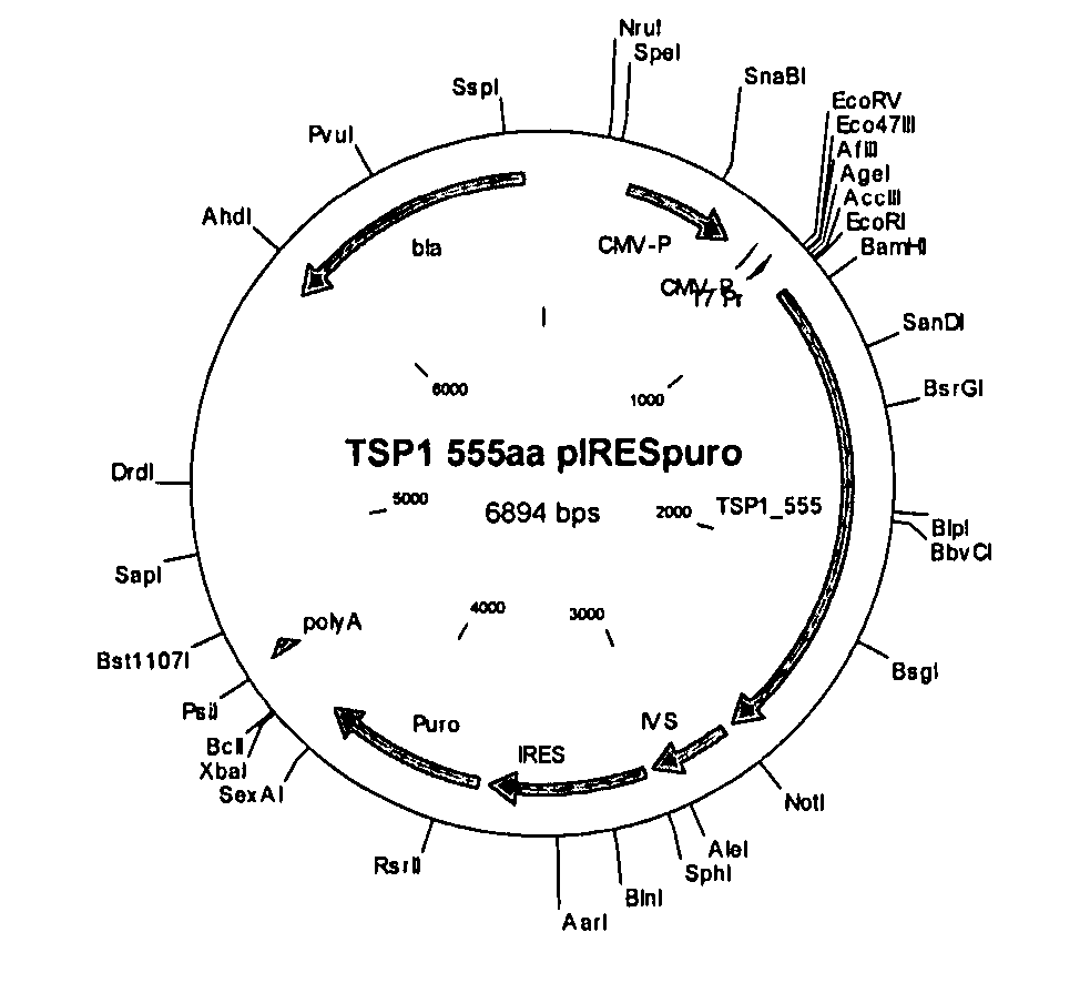
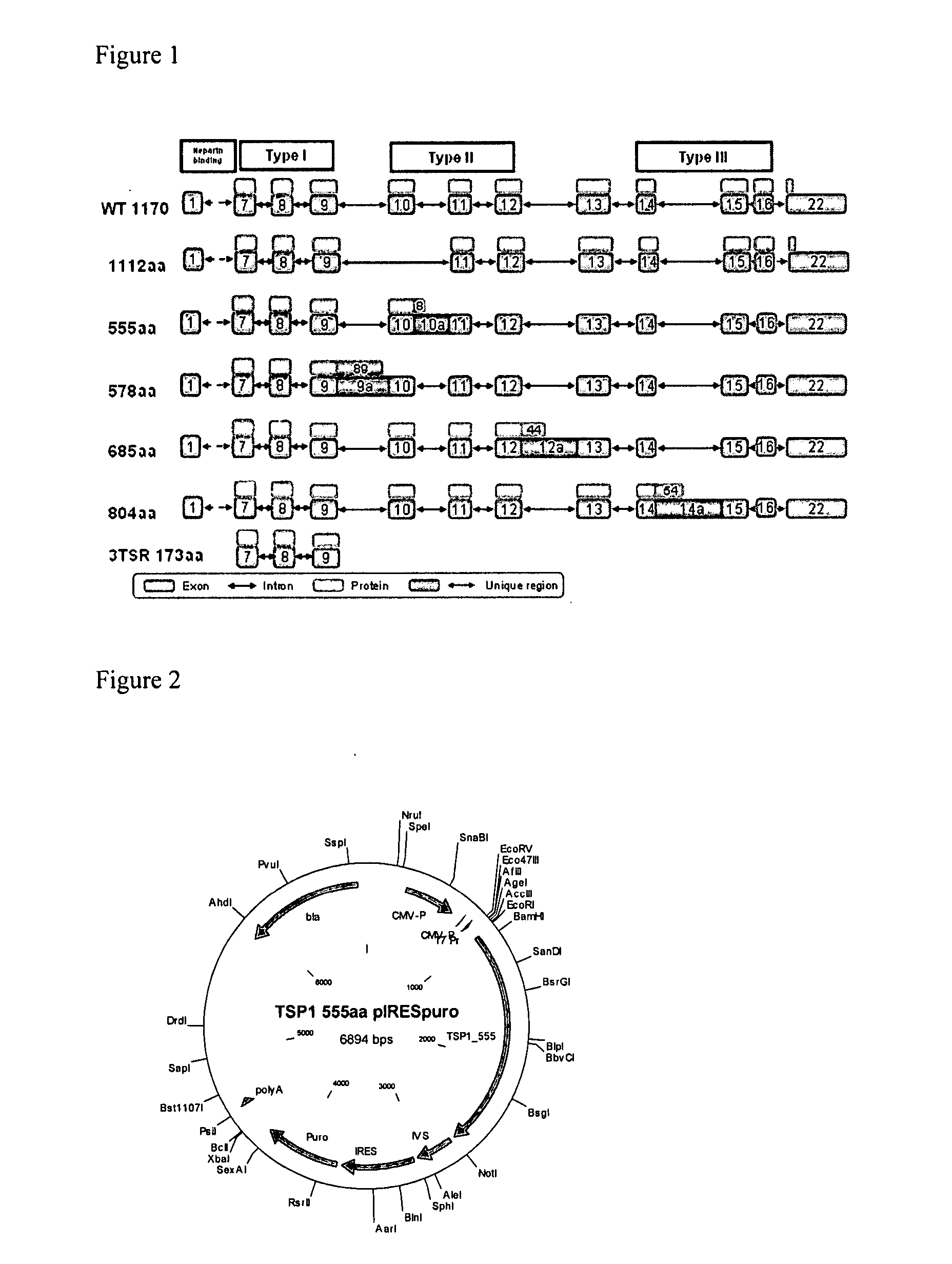
[0390]FIG. 1 provides a schematic drawing of TSP-1 variants of the present invention as well as a known TSP-1 and a previously described P173 anti-angiogenic TSP-1 fragment, also known as the 3TSR fragment (Miao et al. (2001), Cancer Research 61, 7830-7839; Short et al. (2005), J. Cell Biology 168, 643-653). TSP-1 variants of the present invention, depicted in FIG. 1, are TSP-1—1112 (SEQ ID NO:5, 51); TSP-1—685 (SEQ ID NO:4, 50); TSP-1—555 (SEQ ID NO:2, 52), TSP-1—578 (SEQ ID NO:1, 48) and TSP-1—804 (SEQ ID NO:3, 49). All variants include the 3TSR domains that are necessary for activity. Of the five variants, four variants were caused by intron retention and therefore have unique tails. One variant, P1112, is caused by the skipping of the 10th exon. The 3TSR fragment (P173) that was previously shown by Prof. J. Lawler (Miao et al. (2001), Cancer Research 61, 7830-7839; Short et al. (2005), J. Cell Biology 168, 643-653) to exhibit anti-angiogenic activity, and the known WT 1170 variants are shown as well. Exons are represented by orange boxes, while introns are represented by two headed arrows. Proteins are shown in yellow boxes. The unique regions are colored green.The heparin binding domain and the TSR domains are indicated.
[0391] Validation of TSP-1—555 variant of the present invention (SEQ ID NO:2):
[0392] The expression of TSP-1—555 and TSP—578 variants was validated at the mRNA level. The TSP-1—555 transcript was validated using cDNA prepared from RNA mix extracted from heart and brain tissues (Ichilov); bone cell line (SaOs-2-#ATCC HTB-85) and fibroblasts cell line (BJ # ATCC CRL2522). The experimental method used was as follows.
[0393] RT PCR—Purified RNA (1 μg) was mixed with 150 ng Random Hexamer primers (Invitrogen) and 500 μM dNTP in a total volume of 15.6 μl. The mixture was incubated for 5 min at 65° C. and then quickly chilled on ice. Thereafter, 5 μl of 5× SuperscriptII first strand buffer (Invitrogen), 2.4 μl 0.1M DTT and 40 units RNasin (Promega) were added, and the mixture was incubated for 10 min at 25° C., followed by further incubation at 42° C. for 2 min. Then, 1 μl (200 units) of SuperscriptII (Invitrogen) was added and the reaction (final volume of 25 μl) was incubated for 50 min at 42° C. and then inactivated at 70° C. for 15 min. The resulting cDNA was diluted 1:20 in TE buffer (10 mM Tris pH=8, 1 mM EDTA pH=8). The table 66 below shows the sequences of the primers used for the PCR reaction of TSP 555 (SEQ ID NO: 2), while table 67 shows the sequences of PCR pprimers used for the PCR reaction of TSP578 (SEQ ID NO: 1). Orientation for the primers is given as F (forward) or R (reverse). TABLE 66 Nucleotide coordinates on target sequence Oligonucleotide sequence (SEQ (ID) Orientation ID NO: 2): 5′ GCTCCTGCGATAGCCTCAAC-3′ F 1536 (100-350_F_TSP-1_T17_N23) SEQ ID No: 63 5′-CAAATCGCTCAGGACTAACC-3′ R 2077 (100-353_R_TSP-1_T14_N30) SEQ ID NO: 64
[0394] TABLE 67 Nucleotide coordinates on target sequence Oligonucleotide sequence (SEQ (ID) Orientation ID NO: 1): 5′ TGATAGCTGCACTGAGTGTC-3′ F 1324 (100-346_F_TSP-1_T12_N16) (SEQ ID NO: 120) 5′-CTCTATGACCCACTGAACTG-3′ R 1892 (100-347_R_TSP-1_T12_N28) (SEQ ID NO: 121)
PCR amplification and analysis
[0395] cDNA (5ul), prepared as described above (RT PCR), was used as a template in PCR reactions. The amplification was done using AccuPower PCR PreMix (Bioneer, Korea, Cat# K2016), under the following conditions: lul—of each primer (10 uM) plus 13 ul—H2O were added into AccuPower PCR PreMix tube with a reaction program of 5 minutes at 94° C.; 35 cycles of: [30 seconds at 94° C., 30 seconds at 55° C. 60 seconds at 72° C] and 10 minutes at 72° C. At the end of the PCR amplification, products were analyzed on agarose gels stained with ethidium bromide and visualized with UV light. The PCR products were extracted from the gel using QiaQuickTM gel extraction kit (Qiagen, Cat #28706). The extracted DNA products were sequenced by direct sequencing using the gene specific primers described above (Hy-Labs, Israel).
[0396] The PCR products sequence for TSP 555 (SEQ ID NO:122) and TSP 578 (SEQ ID NO:123) are given below. The primers sequence is underlined. PCR product for TSP 555 GCTCCTGCGATAGCCTCAACAACCGATGTGAGGGCTCCTCGGTCCAGACA CGGACCTGCCACATTCAGGAGTGTGACAAGAGATTTAAACAGGATGGTGG CTGGAGCCACTGGTCCCCGTGGTCATCTTGTTCTGTGACATGTGGTGATG GTGTGATCACAAGGATCCGGCTCTGCAACTCTCCCAGCCCCCAGATGAAC GGGAAACCCTGTGAAGGCGAAGCGCGGGAGACCAAAGCCTGCAAGAAAGA CGCCTGCCCCATCAATGGAGGCTGGGGTCCTTGGTCACCATGGGACATCT GTTCTGTCACCTGTGGAGGAGGGGTACAGAAACGTAGTCGTCTCTGCAAC AACCCCACACCCCAGTTTGGAGGCAAGGACTGCGTTGGTGATGTAACAGA AAACCAGATCTGCAACAAGCAGGACTGTCCAATTGGTGAGCCACGCAGCC CAGGATGAAACGACCCAGGAGCTTTGCTCTTTTACTGAATGCTGCAGTCA GCATTCGAGGAGATTCCAGCTTGGTTAGTCCTGAGCGATTTG
[0397] PCR product for TSP 578 TGATAGCTGCACTGAGTGTCACTGTCAGAACTCAGTTACCATCTGCAAAA AGGTGTCCTGCCCCATCATGCCCTGCTCCAATGCCACAGTTCCTGATGGA GAATGCTGTCCTCGCTGTTGGCCCAGCGACTCTGCGGACGATGGCTGGTC TCCATGGTCCGAGTGGACCTCCTGTTCTACGAGCTGTGGCAATGGAATTC AGCAGCGCGGCCGCTCCTGCGATAGCCTCAACAACCGATGTGAGGGCTCC TCGGTCCAGACACGGACCTGCCACATTCAGGAGTGTGACAAGAGATTTAA ACAGGATGGTGGCTGGAGCCACTGGTCCCCGTGGTCATCTTGTTCTGTGA CATGTGGTGATGGTGTGATCACAAGGATCCGGCTCTGCAACTCTCCCAGC CCCCAGATGAACGGGAAACCCTGTGAAGGCGAAGCGCGGGAGACCAAAGC CTGCAAGAAAGACGCCTGCCCCAGTAAGTGTGAGGTCCGCTGCAAGGGTG AGCATGGGCAGCAGCTCTGCCCAGCTGGTTGCCTGGCATCTGCAGCCTGC
Cloning
[0398] The nucleotide sequences of all of the TSP-1 variants were codon optimized to boost protein expression in a mammalian system. The optimized sequences were synthesized by BlueHeron (USA) by using their proprietary gene synthesis technology with the addition of sequence encoding the StrepII and His tags at the 3′.
[0399] The optimized sequences were cloned into EcoRI-Notl sites of plRESpuro3 expression vector. An exemplary, illustrative non-limiting plasmid, suitable for use with the present invention, is the pIRESpuro3 vector. FIG. 2 shows a schematic map of TSP-1—555 in the pIRESpuro3 vector.
[0400] The optimized cloned sequences of all the TSP-1 variants, containing the Strep-His tag, are given in FIG. 3. The relevant ORFs (open reading frames) including the tag sequences are shown in bold; StrepHis tag sequences are underlined. FIG. 3A demonstrates the nucleic acid (SEQ ID NO: 53) and the amino acid sequence (SEQ ID NO:54) of TSP-1-1170; FIG. 3B demonstrates the nucleic acid (SEQ ID NO:55) and the amino acid (SEQ ID NO:56) sequence of TSP-1-1112; FIG. 3C demonstrates the nucleic acid (SEQ ID NO:57) and the amino acid (SEQ ID NO:58) sequence of TSP-1-685; FIG. 3D demonstrates the nucleic acid (SEQ ID NO:59) and the amino acid (SEQ ID NO:60) sequence of TSP-1-555; FIG. 3E demonstrates the nucleic acid (SEQ ID NO:61) and the amino acid (SEQ ID NO:62) sequence of TSP-1-173.
Transfection of TSP-1 Constructs
[0401] The TSP-1 constructs were transfected into HEK-293T cells (ATCC # CRL-11268) as follows. One day prior to transfection, one well from a 6 well plate was plated with 500,000 cells in 2 ml DMEM. On the day of transfection, the FuGENE 6 Transfection Reagent (Roche, Cat#: 1-814-443) was warmed to ambient temperature and mixed prior to use. 6 μl of FuGENE Reagent were diluted into 100 μl DMEM (Dulbecco's modified Eagle's medium; Biological Industries, Cat#: 01-055-1A). Next, 2 micrograms of construct DNA were added. The contents were gently mixed and incubated at room temperature (RT) for 15 minutes. 100 μl of the complex mixture was added dropwise to the cells and swirled. The cells were incubated overnight at 37° C. with 5% CO2. Following about 48 h, transfected cells were split and subjected to antibiotic selection with 5 microgram/ml puromycin. The surviving cells were propagated for about three weeks.
Expression Analysis
[0402] The supernatants of the TSP-1 puromycin resistant cells were bound to NiNTA beads as follows: for each sample, 50ul Ni-NTA agarose (Qiagen #1018244) were washed twice with water and ×2 with ×1 IMIDAZOLE buffer (Biologicals industries #01-914-5A) and then centrifuged for 5 min at 950× g. 1 ml of cell supernatant was added to the beads and the samples were gently shaken for 45 min. at RT. Then, the samples were spun down and washed with ×1 IMIDAZOLE buffer, and centrifuged again at 950× g for 5 min. The samples were eluted with 50 ul SDS sample buffer, incubated for 5 min. at 100° C. and loaded on a 12% SDS-PAGE gel.
[0403] Following electrophoresis, proteins on the gel were transferred to nitrocellulose membranes for 60 min at 35 V using Invitrogen's transfer buffer and X-Cell II blot module. Following transfer, the blots were blocked with 5% skim milk in wash buffer (0.05% Tween-20 in PBS) for at least 60 min. at room temperature with shaking. Following blocking, the blots were incubated for 60 min at room temperature with a commercially available mouse anti Histidine Tag (Serotec, Cat# MCA1396) and diluted in 1/5 blocking buffer, followed by washing with wash buffer and incubation with the secondary antibody Goat anti Mouse HRP (Jackson, Cat# 115-035-146) and diluted 1:25,000 in 1/5 blocking buffer. Next, ECL (Enhanced Chemiluminescence) detection was performed according to the manufacturer's instructions (Amersham; Cat # RPN2209).
[0404] The results, demonstrating stable TSP-1 expression, are shown in FIG. 4. FIG. 4A lane 5 represents the expression of TSP-1—173 (3TSR) (SEQ ID NO:62); lane 7 represents TSP-1—555 (SEQ ID NO:60); lane 1 represents the molecular weight marker (Rainbow Amersham RPN800); lane 2 represents mock plRESpuro3 (also referred to herein as “mock”, or cells that were transfected with the vector alone, without any variant or known TSP-1 sequence); and lane 8 represents Strep-His control (˜100 ng). FIG. 4B lane 2 represents the expression of TSP-1—685 (SEQ ID NO:58); lane 1 represents molecular weight marker (Rainbow Amersham RPN800); and lane 8 represents Strep-His control (˜100 ng). FIG. 4C lane 13 represents the expression of TSP-1—1170 (SEQ ID NO:54); lane 12 represents molecular weight marker (Rainbow Amersham RPN800); lane 22 represents Strep-His control (˜100 ng). FIG. 4D lane 10 represents the expression of TSP-1—1112 (SEQ ID NO:56); lane 1 represents molecular weight marker (Rainbow Amersham RPN800); and lane 12 represents Strep-His control (˜100 ng).
PUM
Property | Measurement | Unit |
Fraction | 0.95 | fraction |
Fraction | 0.883 | fraction |
Fraction | 0.909 | fraction |
Description & Claims & Application Information
We can also present the details of the Description, Claims and Application information to help users get a comprehensive understanding of the technical details of the patent, such as background art, summary of invention, brief description of drawings, description of embodiments, and other original content. On the other hand, users can also determine the specific scope of protection of the technology through the list of claims; as well as understand the changes in the life cycle of the technology with the presentation of the patent timeline. Login to view more.