Membrane for use with implantable devices

a technology of membranes and implants, applied in the field of biointerface membranes, can solve the problems of unable to provide data safely and reliably for long periods of time, unable to obtain continuous and accurate glucose levels, and unable to meet the needs of patients,

Active Publication Date: 2010-04-08
DEXCOM INC
View PDF100 Cites 649 Cited by
  • Summary
  • Abstract
  • Description
  • Claims
  • Application Information

AI Technical Summary

Benefits of technology

[0025]The biointerface membranes, devices including these membranes and methods of use of these membranes provided by the invention allow for long term protection of implanted cells or drugs, as well as continuous information regarding, for example, glucose levels of a host over extended periods of time. Because of these abilities, the biointerface membranes of the present invention can be extremely important in the management of transplant patients, diabetic patients and patients requiring frequent drug treatment.Definitions

Problems solved by technology

Despite the increasing number of individuals diagnosed with diabetes and recent advances in the field of implantable glucose monitoring devices, currently used devices are unable to provide data safely and reliably for long periods of time (e.g., months or years) [See, e.g., Moatti-Sirat et al., Diabetologia 35:224-30 (1992)].
With reference to devices that may be implanted in tissue, a disadvantage of these devices has been that they tend to lose their function after the first few days to weeks following implantation.
Because of these limitations, it has previously been difficult to obtain continuous and accurate glucose levels.
A disadvantage of cell-impermeable membranes is that they often stimulate a local inflammatory response, called the foreign body response (FBR) that has long been recognized as limiting the function of implanted devices that require solute transport.
Previous efforts to overcome this problem have been aimed at increasing local vascularization at the device-tissue interface with limited success.
However, the present studies show that once the monolayer of cells (barrier cell layer) is established adjacent to the membrane, increasing angiogenesis is not sufficient to increase transport of molecules such as glucose and oxygen across the device-tissue interface.

Method used


Image

  • Membrane for use with implantable devices
  • Membrane for use with implantable devices
  • Membrane for use with implantable devices

Examples


example 1

Example 1

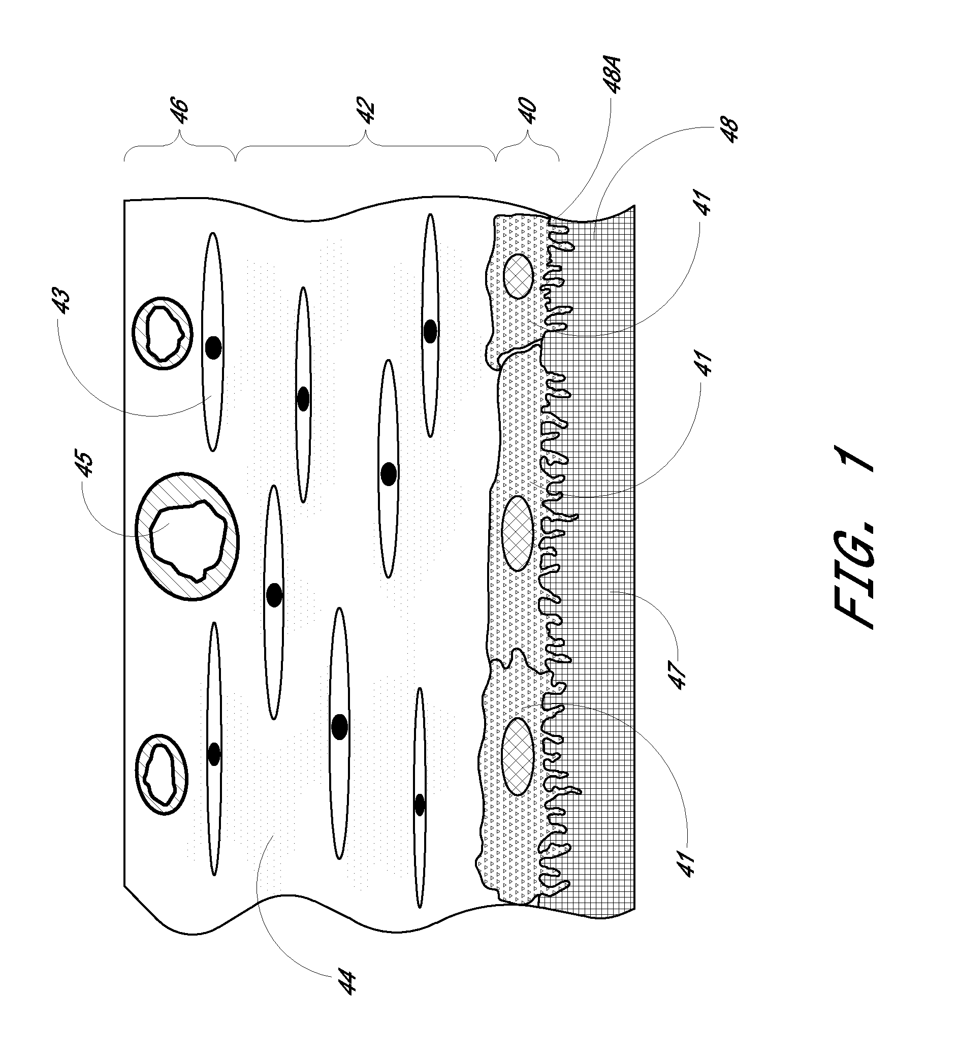
[0086]Preparation of Biointerface Membrane with Non-Woven Fibers

[0087]The barrier-cell disruptive domain may be prepared from a non-woven polyester fiber filtration membrane. The cell-impermeable domain may then be coated on this domain layer. The cell-impermeable domain was prepared by placing approximately 706 gm of dimethylacetamide (DMAC) into a 3 L stainless steel bowl to which a polycarbonateurethane solution (1325 g, Chronoflex AR 25% solids in DMAC and a viscosity of 5100 cp) and polyvinylpyrrolidone (125 g, Plasdone K-90D) were added. The bowl was then fitted to a planetary mixer with a paddle type blade and the contents were stirred for 1 hour at room temperature. This solution was then coated on the barrier-cell disruptive domain by knife-edge drawn at a gap of 0.006″ and dried at 60.degree. C. for 24 hours. The membrane is then mechanically secured to the sensing device by an O-ring.

example 2

Example 2

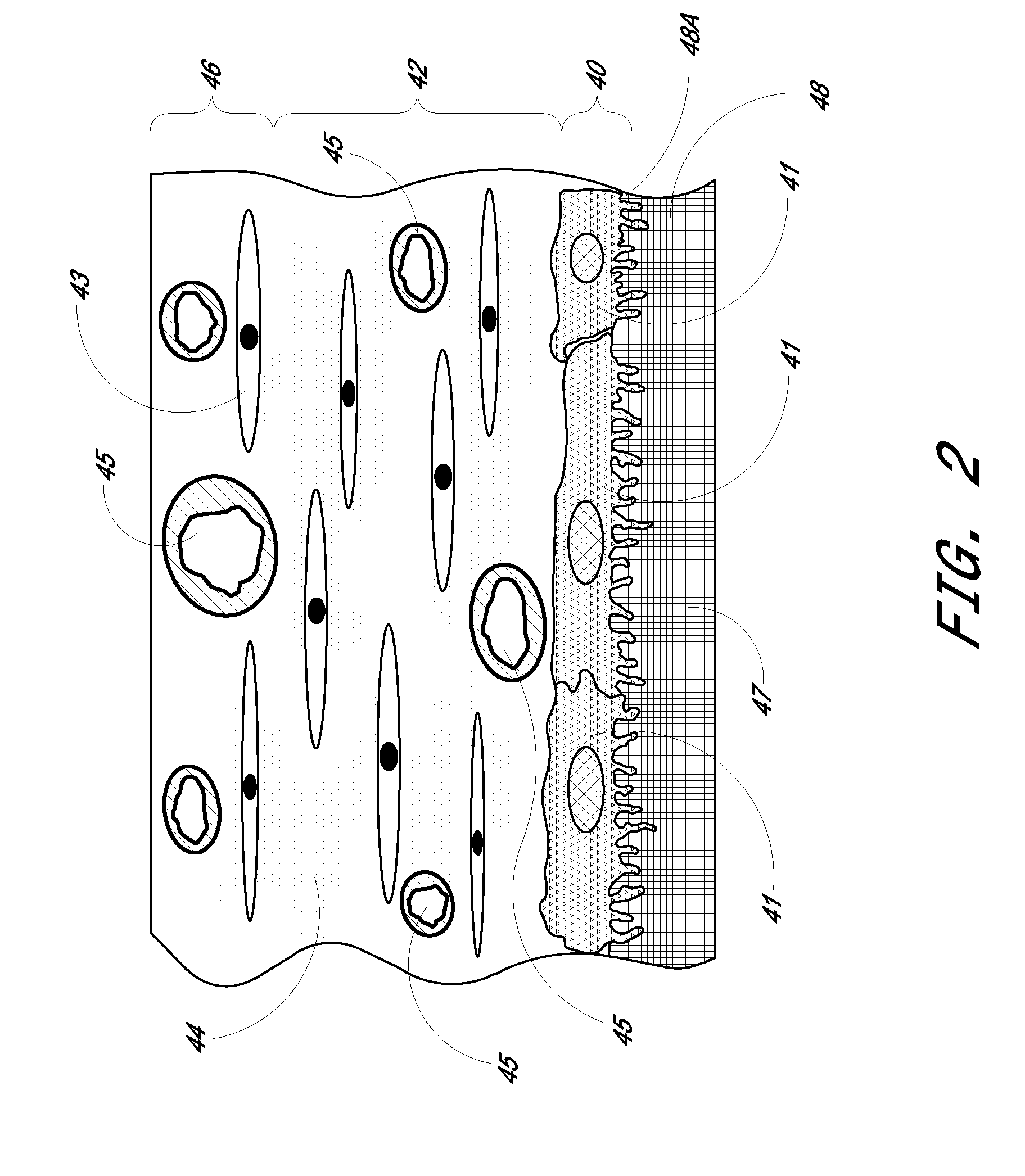
[0088]Preparation of Biointerface Membrane with Porous Silicone

[0089]The barrier-cell disruptive domain can be comprised of a porous silicone sheet. The porous silicone was purchased from Seare Biomatrix Systems, Inc. The cell-impermeable domain was prepared by placing approximately 706 gm of dimethylacetamide (DMAC) into a 3 L stainless steel bowl to which a polycarbonateurethane solution (1,325 gm, Chronoflex AR 25% solids in DMAC and a viscosity of 5100 cp) and polyvinylpyrrolidone (125 gm, Plasdone K-90D) were added. The bowl was then fitted to a planetary mixer with a paddle type blade and the contents were stirred for 1 hour at room temperature. The cell-impermeable domain coating solution was then coated onto a PET release liner (Douglas Hansen Co.) using a knife over roll set at a 0.012″ gap. This film was then dried at 305.degree. F. The final film was approximately 0.0015″ thick. The biointerface membrane was prepared by pressing the porous silicone onto the cast...

example 3

Example 3

[0090]Test Method for Glucose Measuring Device Function

[0091]In vivo sensor function was determined by correlating the sensor output to blood glucose values derived from an external blood glucose meter. We have found that non-diabetic dogs do not experience rapid blood glucose changes, even after ingestion of a high sugar meal. Thus, a 10% dextrose solution was infused into the sensor-implanted dog. A second catheter is placed in the opposite leg for the purpose of blood collection. The implanted sensor was programmed to transmit at 30-second intervals using a pulsed electromagnet. A dextrose solution was infused at a rate of 9.3 ml / minute for the first 25 minutes, 3.5 ml / minute for the next 20 minutes, 1.5 ml / minute for the next 20 minutes, and then the infusion pump was powered off Blood glucose values were measured in duplicate every five minutes on a blood glucose meter (LXN Inc., San Diego, Calif.) for the duration of the study. A computer collected the sensor output....


PUM

PropertyMeasurementUnit
thicknessaaaaaaaaaa
thicknessaaaaaaaaaa
thicknessaaaaaaaaaa

Abstract

The present invention provides a biointerface membrane for use with an implantable device that interferes with the formation of a barrier cell layer including; a first domain distal to the implantable device wherein the first domain supports tissue attachment and interferes with barrier cell layer formation and a second domain proximal to the implantable device wherein the second domain is resistant to cellular attachment and is impermeable to cells. In addition, the present invention provides sensors including the biointerface membrane, implantable devices including these sensors or biointerface membranes, and methods of monitoring glucose levels in a host utilizing the analyte detection implantable device of the invention. Other implantable devices which include the biointerface membrane of the present invention, such as devices for cell transplantation, drug delivery devices, and electrical signal delivery or measuring devices are also provided.

Description

CROSS-REFERENCE TO RELATED APPLICATIONS[0001]This application is a continuation of U.S. application Ser. No. 10 / 768,889 filed Jan. 29, 2004, now U.S. Pat. No. 7,632,228, which is a continuation of U.S. application Ser. No. 09 / 916,386, filed Jul. 27, 2001, now U.S. Pat. No. 6,702,857, the disclosures of which are hereby expressly incorporated by reference in their entirety and are hereby expressly made a portion of this application.FIELD OF THE INVENTION[0002]The present invention relates generally to biointerface membranes that may be utilized with implantable devices such as devices for the detection of analyte concentrations in a biological sample, cell transplantation devices, drug delivery devices and electrical signal delivering or measuring devices. The present invention further relates to methods for determining analyte levels using implantable devices including these membranes. More particularly, the invention relates to novel biointerface membranes, to sensors and implantab...

Claims


Application Information

Patent Timeline
Patent Type & Authority Applications(United States)
IPC IPC(8): A61B5/1486A61B5/145G01N27/327A61B5/1473A61L31/10A61L31/14C12M1/34C12M1/40C12Q1/26C12Q1/54G01N27/416
CPCA61B5/076A61B5/14865A61L31/14A61L31/10A61B2562/02Y10T428/249921A61B5/14532A61B5/14546A61B5/14735
Inventor BRAUKER, JAMES H.SHULTS, MARKTAPSAK, MARK A.
Owner DEXCOM INC
Why Eureka
Try Eureka