Imaging arrangement and method

Inactive Publication Date: 2007-09-27
XCOUNTER
View PDF23 Cites 15 Cited by
  • Summary
  • Abstract
  • Description
  • Claims
  • Application Information

AI Technical Summary

Benefits of technology

[0008]It is an object of the invention to provide improvements in medical imaging techniques, such as an

Problems solved by technology

Fluoroscopy involves the use of ionizing radiation and all fluorosco

Method used


Image

  • Imaging arrangement and method
  • Imaging arrangement and method
  • Imaging arrangement and method

Examples


Embodiment Construction

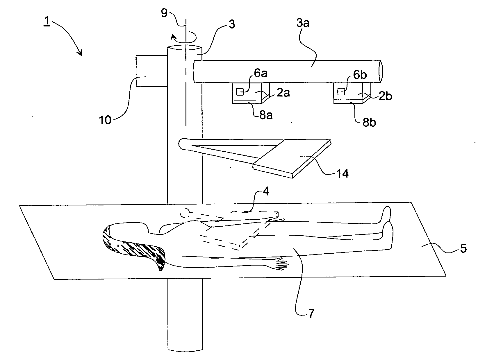
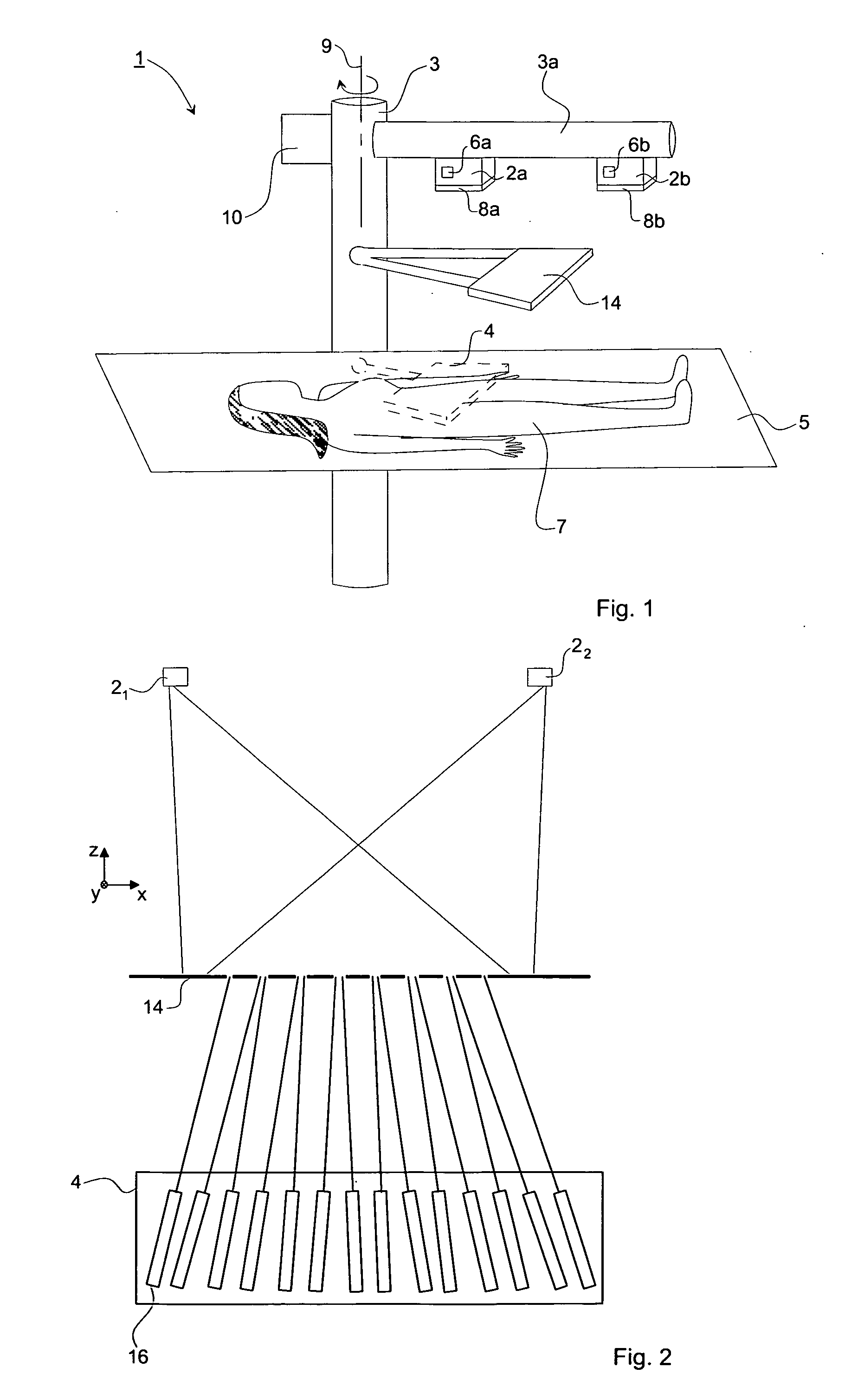
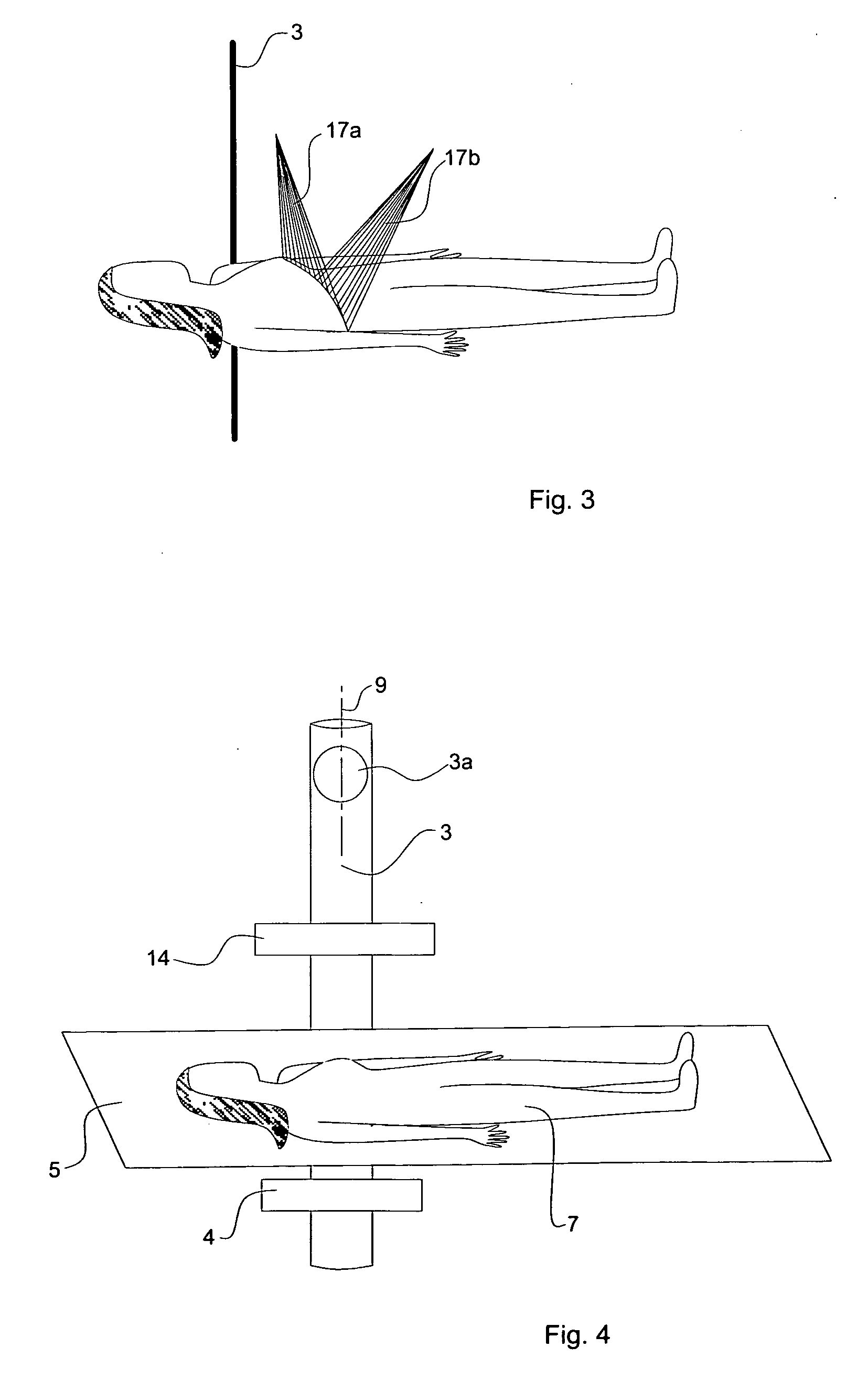
[0026]FIG. 1 illustrates schematically the basic components of the present invention. An imaging arrangement 1 in accordance with the invention comprises one or more pairs of radiation modules 2a, 2b arranged on a support arm 3a of a support structure 3. The imaging arrangement 1 further comprises a radiation detector 4 arranged on the support structure 3 beneath a patient positioning table, in the following denoted object table 5. The radiation detector 4 and the radiation modules 2a, 2b are arranged on opposite sides of the patient positioning table 5 so as to enable the radiation detector 4 to detect radiation emitted from the radiation modules 2a, 2b and passed through a patient 7 or other object that is being imaged. The radiation modules 2a, 2b, the collimator 14 and the radiation detector 4 are rigidly connected to each other by means of the support structure 3, 3a.

[0027]The radiation modules 2a, 2b comprise a radiation source, preferably X-ray tubes and in the following den...


PUM


Abstract

The invention relates to an imaging arrangement 1 for obtaining imaging data of an object 7. The imaging arrangement 1 comprises a support structure 3 having an axis of rotation 9 and comprising means 10 for rotating the support structure 3; a pair of first and second radiation modules 2a, 2b fixedly arranged on the support structure 3 along a radius perpendicular to the axis of rotation 9, wherein each of the radiation modules 2a, 2b comprises a radiation source 6a, 6b simultaneously emitting radiation; a radiation detector 4 for detecting radiation as emitted from the radiation modules 2a, 2b and passed through the object 7 being imaged; and a collimator 14 arranged between, as seen in a direction parallel to the axis of rotation 9, the pair of radiation modules 2a, 2b and the radiation detector 4. The invention also relates to a corresponding method.

Description

PRIORITY STATEMENT[0001]This application claims benefit of priority under 35 U.S.C. §119 from Swedish Patent Publication No. SE 0600636-5, filed on Mar. 21, 2006, and Patent Application No. SE 0602518-3, filed on Nov. 27, 2006, in the Swedish Patent Office, the disclosures of which are incorporated herein by reference in their entirety.FIELD OF THE INVENTION [0002]The present invention relates generally to apparatuses and methods for obtaining time resolved imaging data of an object.BACKGROUND OF THE INVENTION[0003]Medical X-ray imaging of patients, for example angiography, is most important and a frequently used examination method. Angiography is a radiological method for examination of blood or lymph vessels. Blood has the same radiodensity as the surrounding tissues and a radiocontrast agent (which absorbs X-rays) is therefore added to the blood to make angiography visualization possible. A catheter is inserted into the vessel, e.g. by Seldinger technique, and a water soluble con...

Claims


Application Information

Patent Timeline
IPC IPC(8): H05G1/60A61B6/00G01N23/00G21K1/12
CPCA61B6/025A61B6/4007A61B6/4035A61B6/504A61B6/481A61B6/482A61B6/4476
Inventor FRANCKE, TOM
Owner XCOUNTER
Why Eureka
Try Eureka