High frame rate quantitative doppler flow imaging using unfocused transmit beams

a quantitative doppler and transmit beam technology, applied in tomography, instruments, applications, etc., can solve the problems of high equipment cost, more extensive processing capabilities, and high equipment cost for significant adoption, so as to improve the contrast resolution of the final image and enhance the acoustic information

Active Publication Date: 2009-12-31
VERASONICS
View PDF27 Cites 104 Cited by
  • Summary
  • Abstract
  • Description
  • Claims
  • Application Information

AI Technical Summary

Benefits of technology

[0029]In accordance with another aspect of the foregoing embodiment, the method includes combining results of multiple cycles of generating acoustic signals, receiving echoes, and obtaining echo signals from the received echoes to derive enhanced acoustic information.
[0030]In accordance with another aspect of the foregoing embodiment, the enhanced acoustic information includes spatial compounding that improves contrast resolution of a final image generated therefrom.

Problems solved by technology

Unfortunately, there are still many applications for ultrasound systems where the equipment costs are too high for significant adoption.
These performance capabilities are found only on the more expensive ultrasound systems, which have more extensive processing capabilities.
Unfortunately, the low manufacturing volumes of ultrasound systems results in substantial overhead and fixed costs for these unique circuits, and thus the transition to digital signal processing has not significantly reduced system cost.
Color Flow Doppler can provide approximate mean flow velocities in the region of interest, but accuracy is limited due to the short acquisition sequences needed to maintain reasonable frame rates.
Using a small ROI can improve frame rates, but may limit the assessment of flow abnormalities.
In general, high quality Doppler imaging is more technically difficult than echographic imaging in great part because backscattering from blood is very weak compared to tissue.
Well known fundamental challenges to producing uncluttered and artifact-free Color Flow images include:The requirement for highly repeatable transmit pulses, and very low noise and phase jitter in the acquisition hardware.Flow signals are often of the same order of magnitude as various sources of noise, but averaging has an adverse impact on frame rate and other motion artifacts.The large contrast between the scattering amplitudes of tissue and blood leads to difficulty in discriminating between vessel walls (strong echo) and moving blood (weak echo), even when the velocity contrast is high.
This is particularly challenging for small blood vessels since the sample volume for Doppler pulses can be larger than the vessel diameter.

Method used


Image

  • High frame rate quantitative doppler flow imaging using unfocused transmit beams
  • High frame rate quantitative doppler flow imaging using unfocused transmit beams
  • High frame rate quantitative doppler flow imaging using unfocused transmit beams

Examples


Embodiment Construction

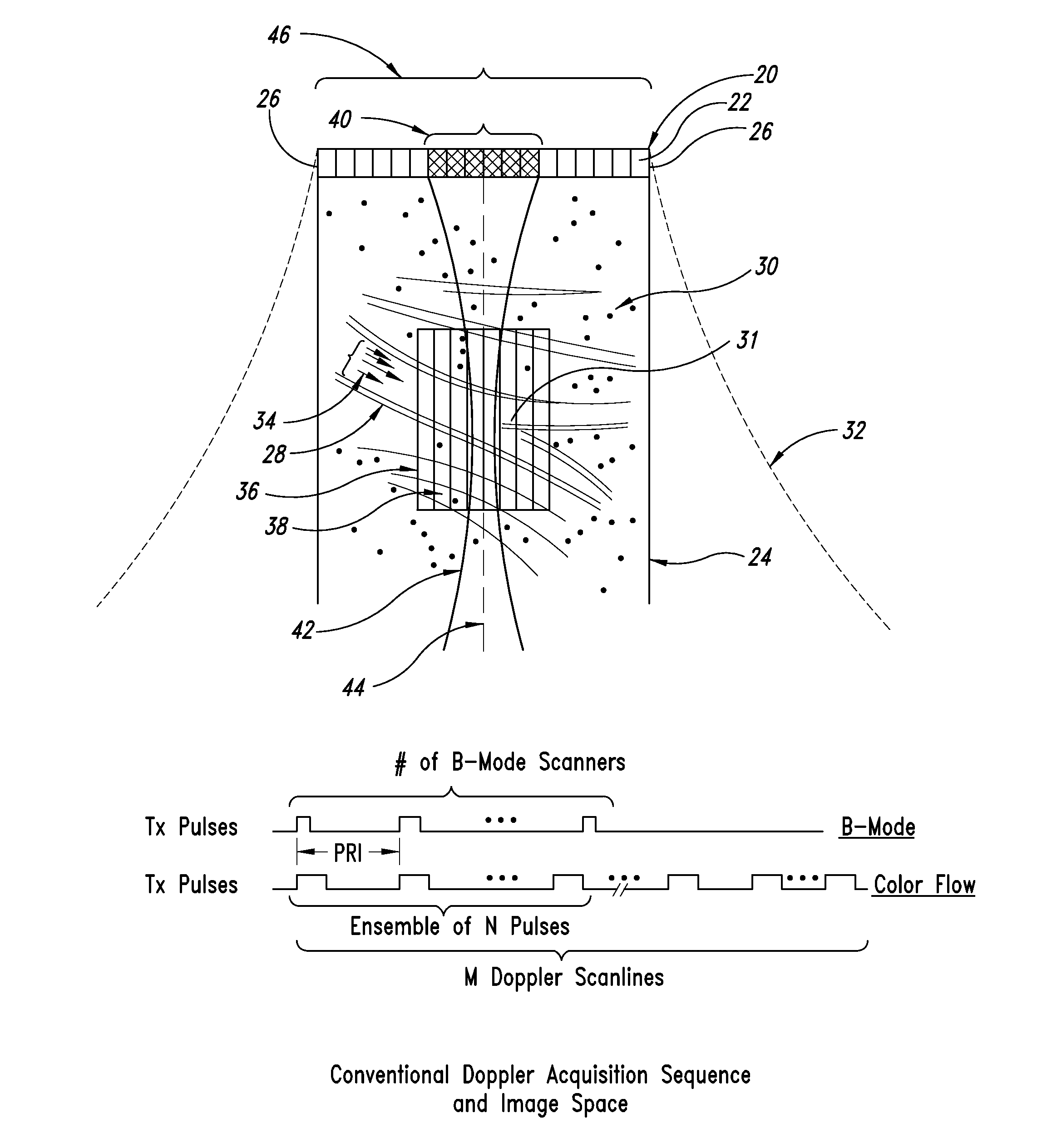
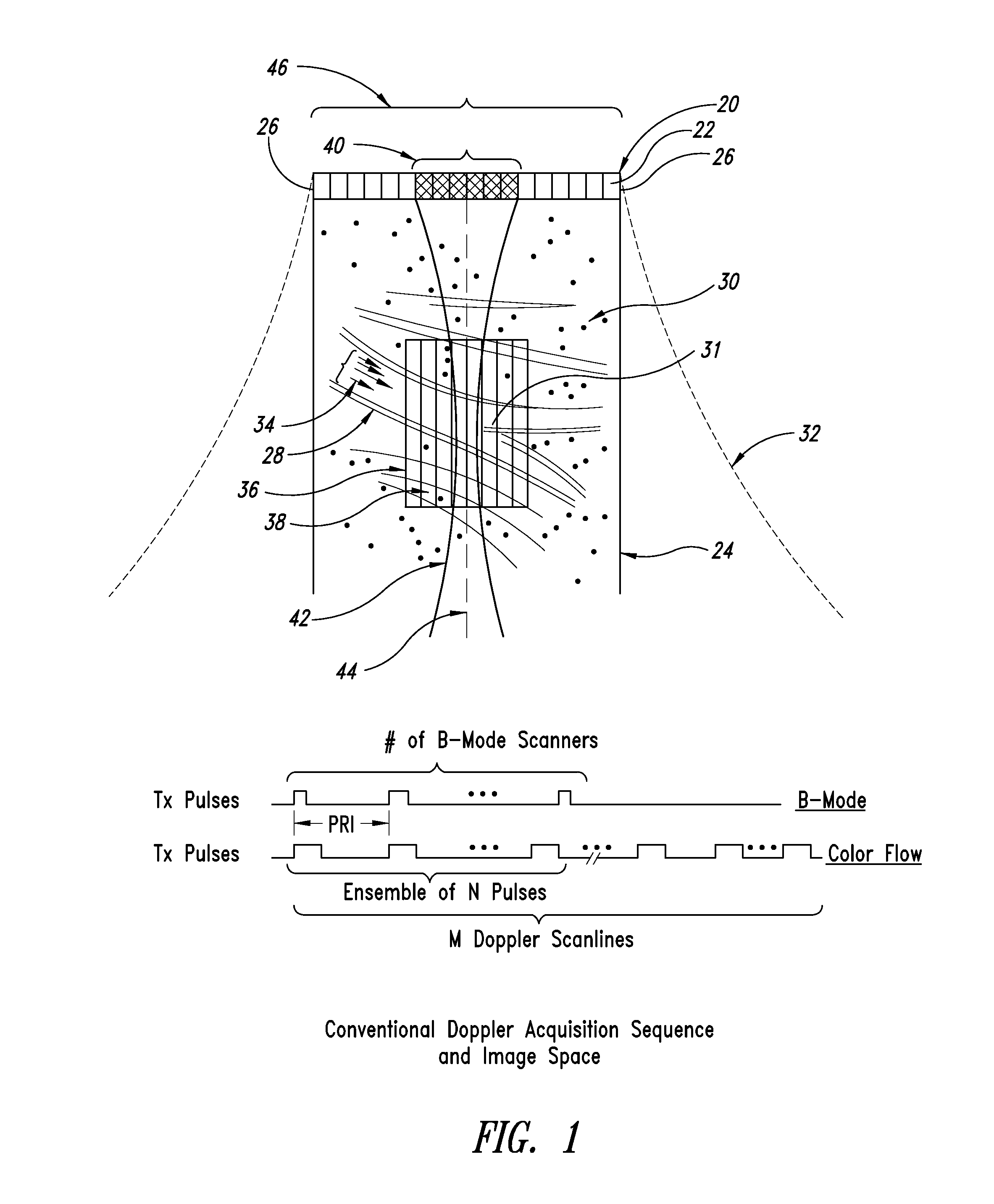
[0058]A conventional Doppler acquisition sequence and image scene are shown in FIG. 1 for a single beam direction that consists of an ensemble of N transmit-receive events (with 8≦N≦16). Conventional ultrasound systems form focused transmit beams and focus the received data dynamically, using delay-and-sum beam forming and several other processing steps required to form the image. In applicants' U.S. patent application Ser. No. 11 / 911,633, entitled “Ultrasound Imaging System with Pixel Oriented Processing,” a method of image reconstruction is described below in conjunction with FIGS. 7-10 that greatly reduces the processing load compared to conventional beam forming and permits using a wide variety of non-conventional transmit fields.

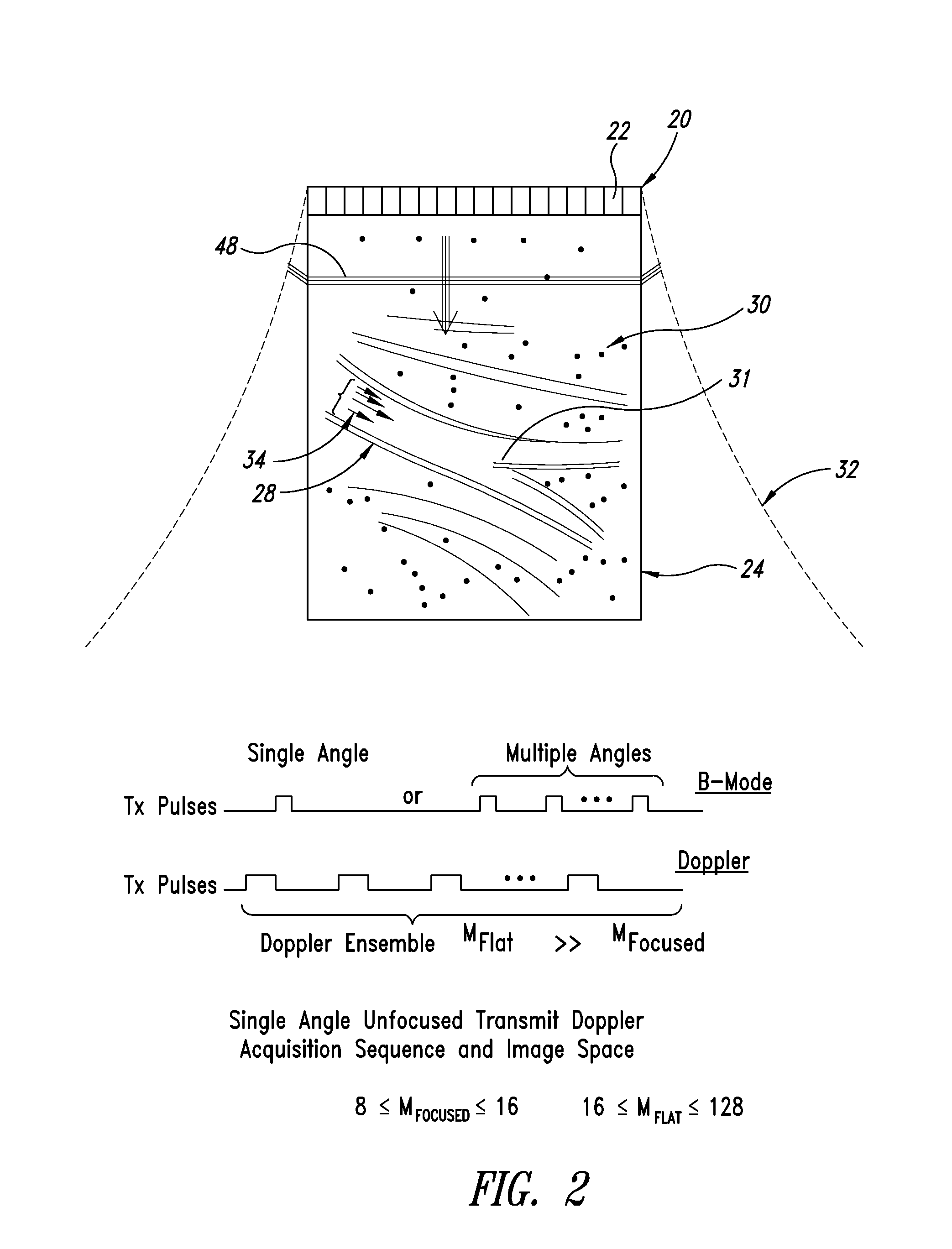
[0059]For example, one non-conventional transmit field is the flat-focus transmit mode in which all transducer elements are fired in phase to produce a segment of a plane wave (for a linear array) that can be used to ensonify the entire field of view wi...


PUM


Abstract

An ultrasound imaging system with pixel oriented processing is provided in which a method of producing a Doppler velocity image is accomplished by emitting unfocused acoustic signals into a medium over substantially an entire field; receiving scattered and reflected ultrasonic signals on a transducer array in response to the emission; processing the received ultrasonic signals to extract information to construct a Doppler velocity signal corresponding to at least one point in the medium; and generating on a display device the Doppler velocity image from the processed Doppler velocity signal. Acquisition sequences and signal processing algorithms are described that provide improved quantification of fluid flow parameters, including improved discrimination between regions of blood flow and tissue. Very high frame rate Spectral Doppler and Vector Doppler acquisition modes for real-time and post-acquisition visualization over a large field of view are described.

Description

BACKGROUND OF THE DISCLOSURE[0001]1. Technical Field[0002]The present disclosure is directed to a system and method of capturing and processing ultrasound data and generating images therefrom that represent fluid flow.[0003]2. Description of the Related Art[0004]Ultrasound Imaging has developed into an effective tool for diagnosing a wide variety of disease states and conditions. The market for ultrasound equipment has seen steady growth over the years, fueled by improvements in image quality and the capability to differentiate various types of tissue. Unfortunately, there are still many applications for ultrasound systems where the equipment costs are too high for significant adoption. Examples are application areas such as breast cancer detection, prostate imaging, musculoskeletal imaging, and interventional radiology. In these areas and others, the diagnostic efficacy of ultrasound imaging depends on excellent spatial and contrast resolution for differentiation and identification...

Claims


Application Information

Patent Timeline
Patent Type & Authority Applications(United States)
IPC IPC(8): A61B8/00
CPCA61B8/06A61B8/13G01S7/52028G01S7/52034G01S7/52047G01S15/8977G01S15/8979G01S15/8984G01S15/8995G01S7/52085
Inventor DAIGLE, RONALD ELVINKACZDOWSKI, PETER JOHN
Owner VERASONICS
Why Eureka
Try Eureka