Image diagnostic processing device and image diagnostic processing program

a technology of image diagnostic and processing program, which is applied in the field of image diagnostic processing device and image diagnostic processing program, can solve the problems of not accurately discriminating nodules and lung blood vessels by feature quantities, and achieve the effect of accurate discrimination

Active Publication Date: 2007-10-04
KOBE UNIV +1
View PDF5 Cites 30 Cited by
  • Summary
  • Abstract
  • Description
  • Claims
  • Application Information

AI Technical Summary

Benefits of technology

[0009] With respect to the automatic detection of the CT lung nodule, an approach for extracting a region which becomes a nodule candidate (hereinafter, referred to as a nodule candidate region), obtaining a plurality of feature quantities for characterizing the nodule candidate region and judging whether the nodule candidate region is a nodule or not based on the feature quantities may be used. However, since the nodule is similar to a portion of the lung blood vessel in the feature, the nodule and the lung blood vessel may not be accurately discriminated by the feature quantities for characterizing the nodule candidate region.

Problems solved by technology

However, since the nodule is similar to a portion of the lung blood vessel in the feature, the nodule and the lung blood vessel may not be accurately discriminated by the feature quantities for characterizing the nodule candidate region.

Method used


Image

  • Image diagnostic processing device and image diagnostic processing program
  • Image diagnostic processing device and image diagnostic processing program
  • Image diagnostic processing device and image diagnostic processing program

Examples


first embodiment

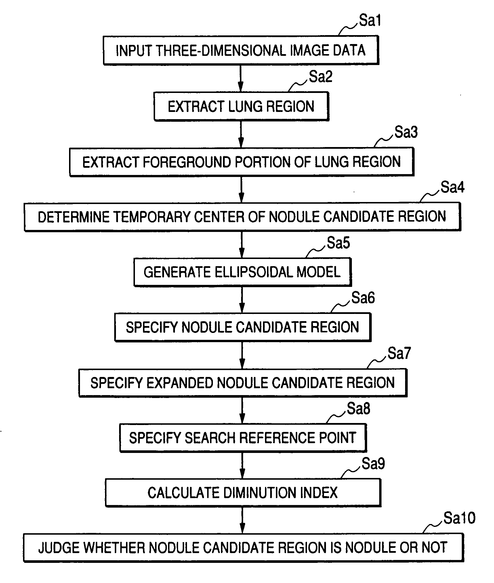
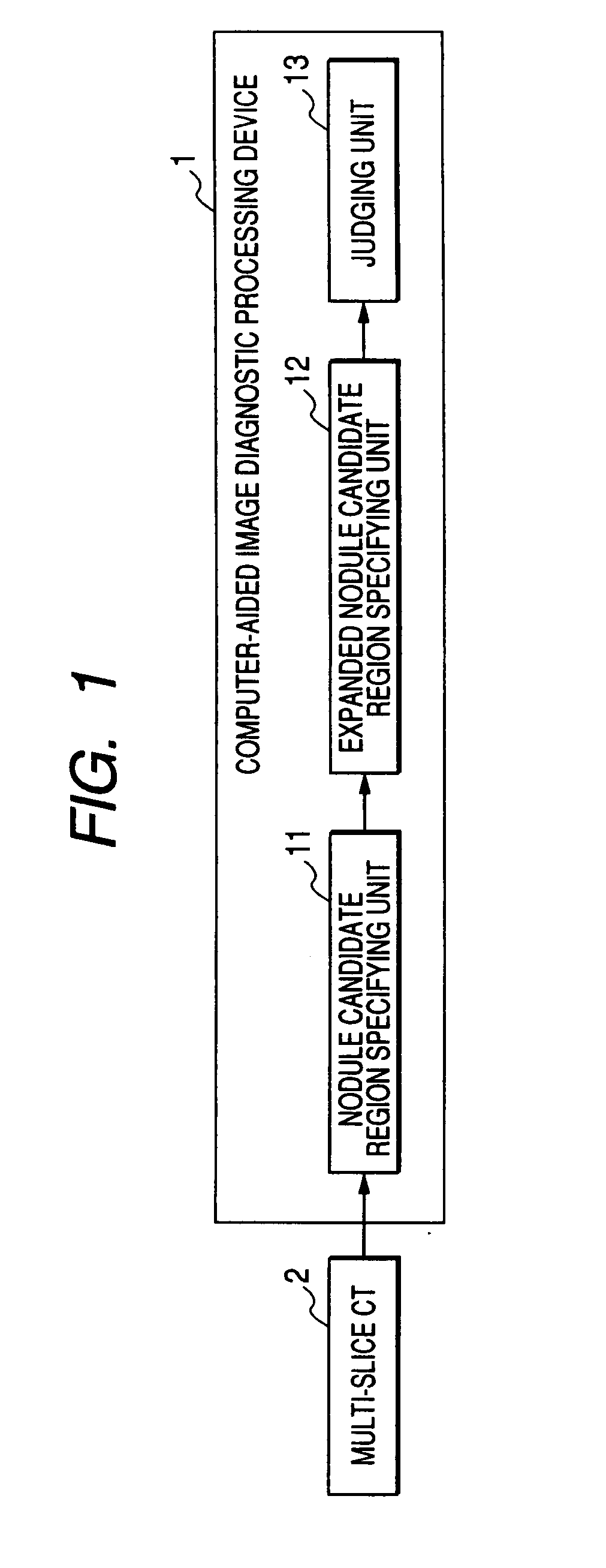
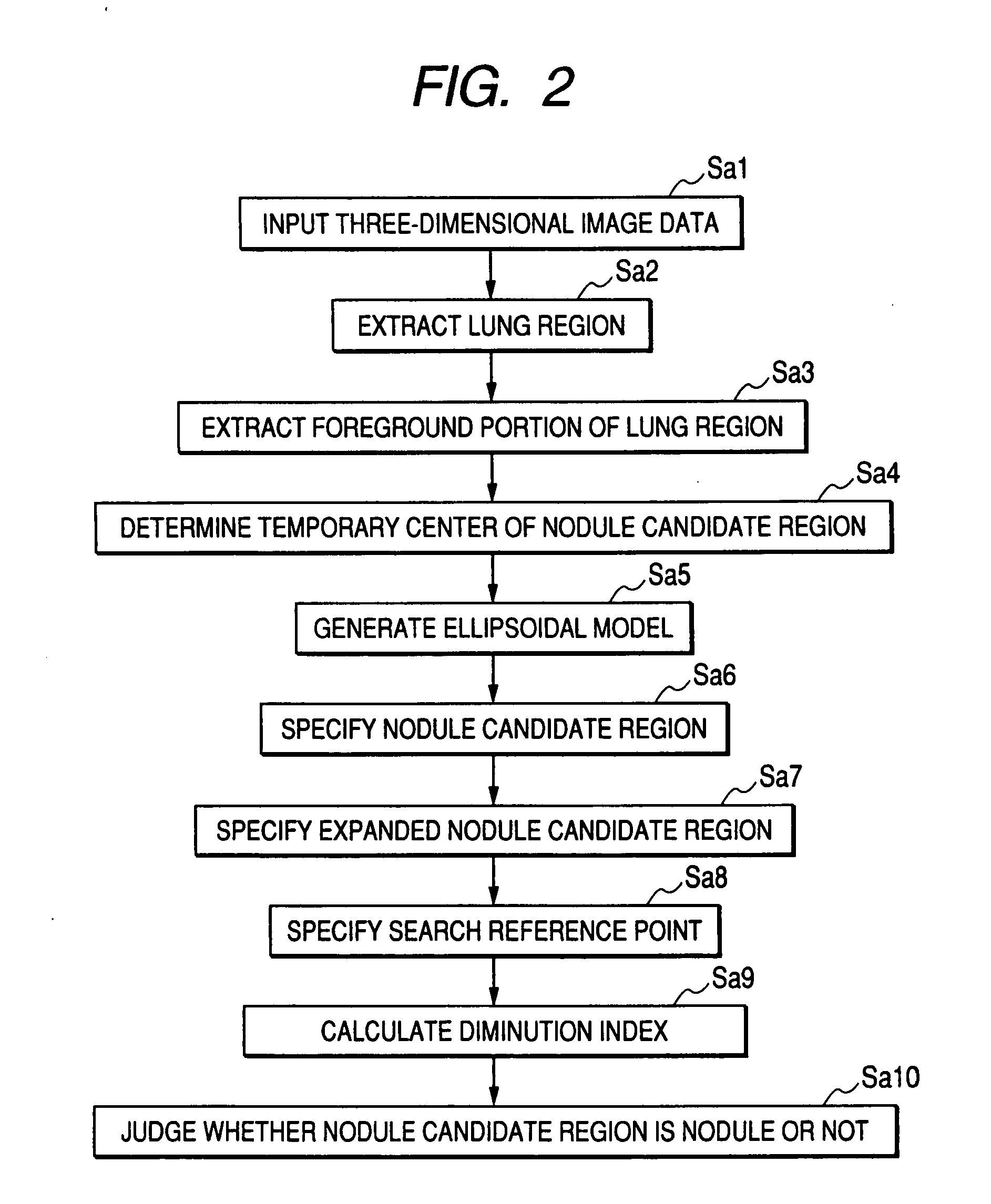
[0055]FIG. 1 is a view showing the configuration of a computer-aided image diagnostic processing device 1 according to a first embodiment using an image diagnostic processing device according to the present invention.

[0056] The computer-aided image diagnostic processing device 1 shown in FIG. 1 processes three-dimensional image data acquired by a multi-slice CT 2. As shown in FIG. 1, the computer-aided image diagnostic processing device 1 includes a nodule candidate region specifying unit 11, an expanded nodule candidate region specifying unit 12 and a judging unit 13.

[0057] The computer-aided image diagnostic processing device 1 can, for example, use a general-purpose computer device as basic hardware. The nodule candidate region specifying unit 11, the expanded nodule candidate region specifying unit 12 and the judging unit 13 can be embodied by executing an image diagnostic processing program on a processor mounted in the computer device. At this time, the computer-aided image ...

second embodiment

[0101]FIG. 17 is a view showing the configuration of a computer-aided image diagnostic processing device 3 using the image diagnostic processing device according to the present invention. In FIG. 17, the same portions as FIG. 1 are denoted by the same reference numerals and their detailed description will be omitted.

[0102] The computer-aided image diagnostic processing device 3 shown in FIG. 17 processes three-dimensional image data acquired by a multi-slice CT 2. As shown in FIG. 17, the computer-aided image diagnostic processing device 3 includes a nodule candidate region specifying unit 11 and a judging unit 31.

[0103] The computer-aided image diagnostic processing device 3 can, for example, use a general-purpose computer device as basic hardware, similar to the computer-aided image diagnostic processing device 1 according to the first embodiment.

[0104] The judging unit 31 judges whether a nodule candidate region is a nodule or not, based on a foreground occupancy ratio of an e...

third embodiment

[0114] The first or second embodiment is efficient for the detection of solid nodules. However, with respect to a ground glass opacity (GGO) type nodule, it is difficult to apply an ellipsoidal model to the GGO type nodules.

[0115] In the GGO type nodule, the existing lung blood vessel or a component having a higher intensity is observed. That is, an object having relatively high contrast, which is surrounded by an object having relatively low contrast such as the GGO, may exist. Accordingly, in the first or second embodiment, the following problems may be caused.

[0116] (1) When the lung region is divided into the foreground portion and the background portion by an adaptive thresholding process, only a portion of the nodule may enter the foreground portion in the GGO type nodule. In such a case, the value for transforming the distance of the foreground portion is set to the initial radius of the ellipsoidal model, the actual radius of the nodule is significantly small.

[0117] (2) S...


PUM


Abstract

An image diagnostic processing device includes peripheral region specifying means which specifies a peripheral region connecting to an abnormal candidate region included in an image representing the inside of a subject, and judging means which judges whether the abnormal candidate region is an anatomic abnormal region or not, based on a first feature quantity of the abnormal candidate region and a second feature quantity of the peripheral region.

Description

CROSS-REFERENCE TO RELATED APPLICATIONS [0001] This is a Continuation Application of PCT Application No. PCT / JP2006 / 303597, filed Feb. 27, 2006, which was published under PCT Article 21(2) in Japanese. [0002] This application is based upon and claims the benefit of priority from prior Japanese Patent Application No. 2005-056098, filed Mar. 1, 2005, the entire contents of which are incorporated herein by reference.BACKGROUND OF THE INVENTION [0003] 1. Field of the Invention [0004] The present invention relates to an image diagnostic processing device and an image diagnostic processing program which performs a diagnostic process on anatomic abnormality such as nodule abnormality or wen abnormality in a three-dimensional image collected using a medical image diagnostic modality such as an X-ray computed tomographic imaging device, an X-ray diagnostic device, a magnetic resonance imaging device or an ultrasonic diagnostic device. [0005] 2. Description of the Related Art [0006] Now, as l...

Claims


Application Information

Patent Timeline
Patent Type & Authority Applications(United States)
IPC IPC(8): G06K9/00
CPCG06T2207/30061G06T7/0012
Inventor MATSUMOTO, SUMIAKIYAMAGATA, HITOSHI
Owner KOBE UNIV
Why Eureka
Try Eureka