
Intraocular lens with distortion free valve

a technology of distortion-free valve and intraocular lens, which is applied in the field of implantable intraocular lens, can solve the problems of increasing intraocular pressure, iris bombe, and patients who undergo ocular surgery with an intraocular lens (iol) are at risk so as to prevent or reduce the occurrence of pseudophakic pupillary block and maintain distortion-free optics

Inactive Publication Date: 2007-10-18
STAAR SURGICAL COMPANY INC
View PDF14 Cites 59 Cited by
  • Summary
  • Abstract
  • Description
  • Claims
  • Application Information

AI Technical Summary

Benefits of technology

[0018] Briefly and in general terms, the present invention provides a new and improved intraocular lens having a valve that allows for the flow of aqueous humor through an intraocular lens while maintaining distortion-free optics. The invention is configured for fluid flow through the lens, between the posterior and anterior surfaces of the lens, thus assisting to prevent or reduce the occurrence of pseudophakic pupillary block.
[0019] The present invention is advantageous because it prevents pseudophakic pupillary block from occurring in the first place, thereby avoiding the need for additional surgeries in a patient who has had implantation of an IOL. Preventing pseudophakic pupillary block prevents both the medical complications and loss of vision that result from a block, as well as those complications that may result from additional surgeries required to open up a pseudophakic pupillary block.
[0020] Furthermore, another aspect of the invention is that it provides a distortion-free lens that is configured to permit flow of aqueous humor from the posterior surface of the lens through and out the anterior surface of the lens. In one aspect of the present invention, a soft lens including valves is configured to create pulsatile micro flow of fluid between two wedge-like surfaces during accommodation of an eye. In one embodiment, the valves and openings in the lens are self cleaning and are configured wherein clogging with internal eye debris and scar tissue is minimized.

Problems solved by technology

Patients who undergo ocular surgery with an implantation of an intraocular lens (IOL) are at risk of developing pseudophakic pupillary block.
If the condition is not recognized and treated early, it can lead to iris bombe, iridocorneal adhesion formation (starting at the periphery and extending toward the center), increasing intraocular pressure, progressive damage to the optic nerve head, and loss of vision.
Sulcus-supported lenses have a tendency to erode the ciliary processes and the ciliary body resulting in a breakdown of the blood-aqueous barrier.
The fibrous reactions in the capsular bag also can push the optic out of the bag, a process that may lead to pupil capture.
Pediatric patients are particularly vulnerable to pseudophakic pupillary block.
Furthermore, there is a greater tendency for the optic to come out of the small capsular bag and become captured by the pupil.
In neonates and young infants, there is a tendency for the iridectomy opening to shrink and ultimately close.
Pupillary block is particularly prone to occur with use of an artificial intraocular lens implanted in the anterior chamber, an angle-supported lens, or an iris claw (Artisan) lens.
Failure to relieve the pupillary block can lead to the development of chronic angle closure glaucoma and glaucomatous optic neuropathy with loss of vision.
These procedures may carry a risk of further complications.
However, known valve mechanisms do not provide flow completely through the lens from the posterior surface of the lens out through the anterior surface of the lens.
For this reason, implanting such a lens would not prevent pseudophakic pupillary block.
The patient's aqueous humor does not flow through the lens, and therefore this lens would not prevent pseudophakic pupillary block.
The device does not have a valve configured to control the rate of flow through the lens.
Furthermore, the central openings in the optic lead to optical distortion in the region where light passes through the central openings.
However, these are discrete implants that are not a component of known implantable intraocular lens devices.
Because known glaucoma shunts are discrete devices that are not part of a lens optic, distortion of vision is not a consideration in the design of those devices.
There is no known implantable intraocular lens device that is configured for the prevention of pseudophakic pupillary block.

Method used


Image

  • Intraocular lens with distortion free valve
  • Intraocular lens with distortion free valve
  • Intraocular lens with distortion free valve

Examples


Embodiment Construction

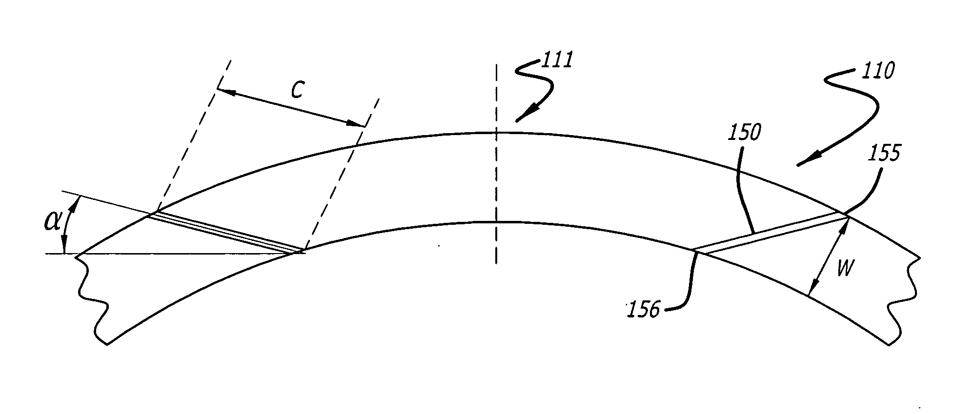
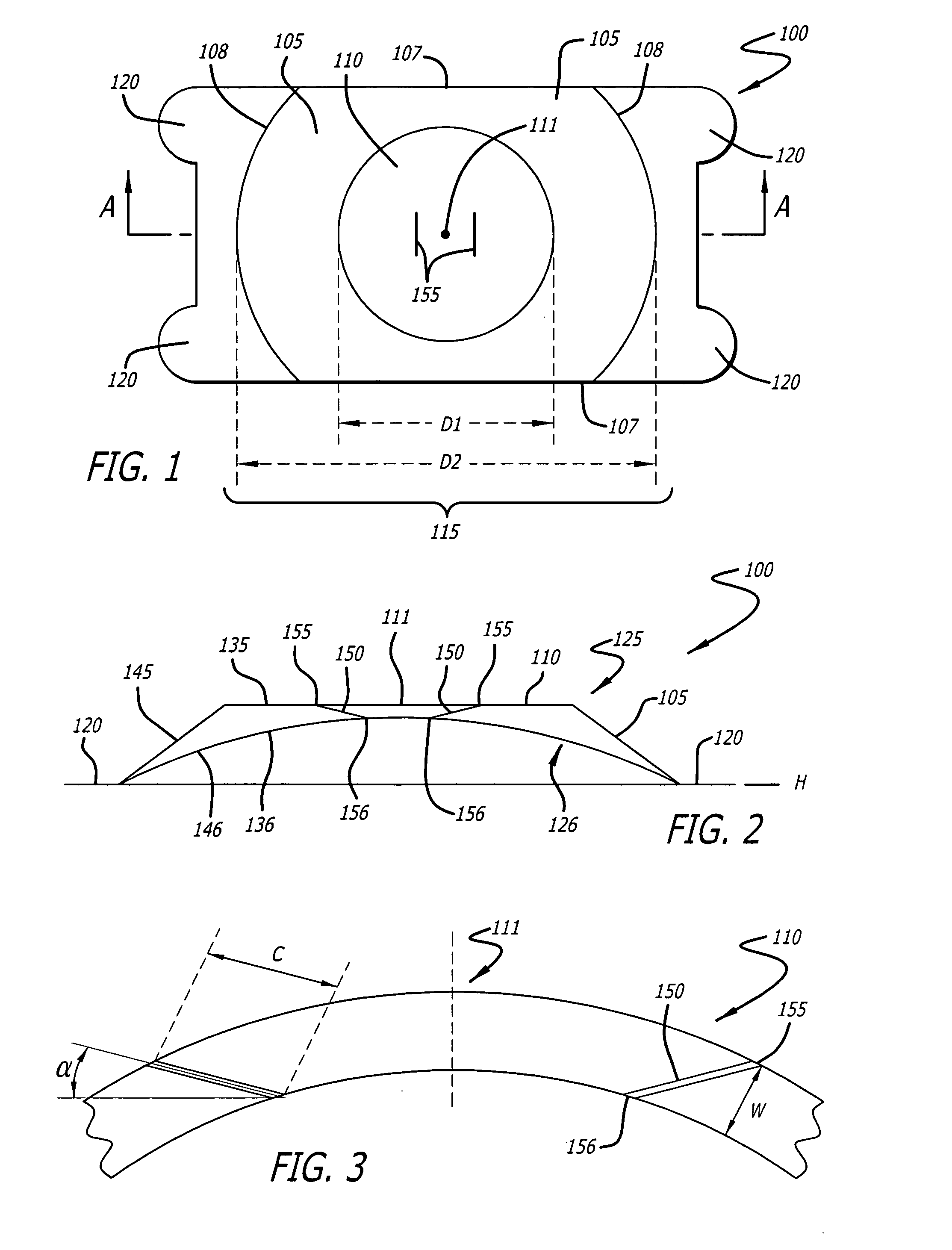
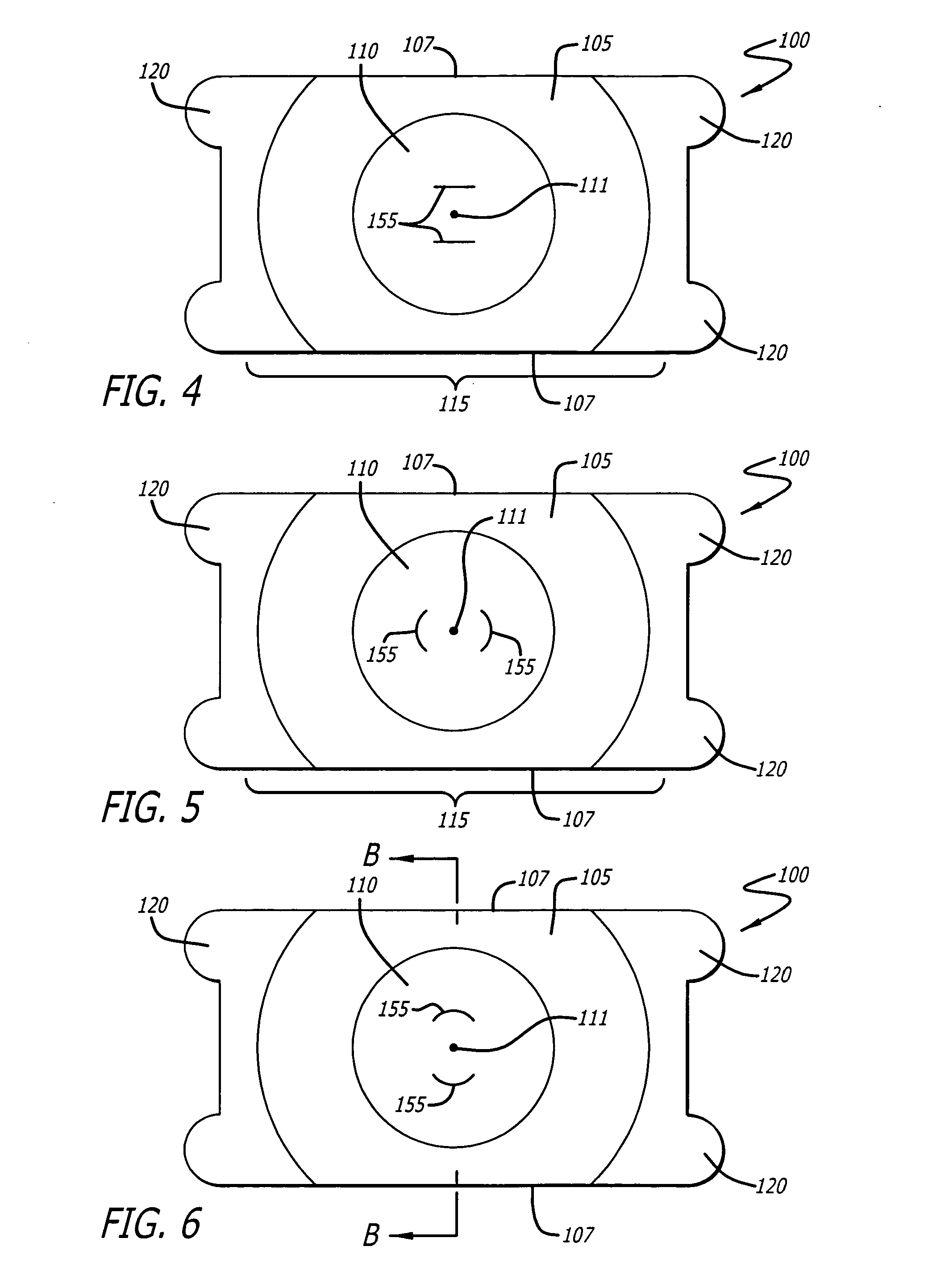
[0039] Referring to the drawings, which are provided for purposes of illustration and by way of example, one or more embodiments of the present invention of an intraocular lens (IOL) are illustrated in FIGS. 1-8.

[0040] Referring first to FIGS. 1-3, in general terms, there is provided in the present invention an intraocular lens (IOL) 100. The IOL includes an optic 115, having a peripheral zone 105 circumscribing an optical zone 110. In at least one embodiment, the IOL further includes one or more haptics or fixation members 120 that are coupled to the peripheral zone and extend outwardly from the peripheral zone to assist in retaining the optic in position in an eye of a patient.

[0041] In one embodiment the fixation member 120 or members are preferably located so that the optical zone 110 is free of such member or members. The peripheral zone 105 preferably forms, in effect, a frame which assists in strengthening the optic 115 against unwanted deformation after implantation in the...


PUM


Abstract

The invention provides a medical device and method to reduce the occurrence of pseudophakic pupillary block following implantation of an intraocular lens. The device includes an intraocular lens configured with channels traversing through the surfaces of the lens optic. The invention includes special edge angulations and locations in the optic zone that eliminate optical distortion from channels. The channels perform as valves configured to allow flow of a patient's aqueous humor from one surface of the lens through to the other surface of the lens, thereby preventing fluid pressure from building up in a patient's eye.

Description

FIELD OF THE INVENTION [0001] The present invention relates generally to implantable intraocular lenses. More specifically, the present invention relates to a distortion-free implantable intraocular lens including a valve configured to permit fluid flow through the lens, from the posterior surface of the lens to the anterior surface of the lens, whereby pseudophakic pupillary block may be prevented. BACKGROUND OF THE INVENTION [0002] Patients who undergo ocular surgery with an implantation of an intraocular lens (IOL) are at risk of developing pseudophakic pupillary block. Pupillary block refers to a failure of communication of aqueous humor between the anterior and posterior chambers of an eye caused by obstruction of the pupil and surgical openings in the iris. In pseudophakic pupillary block, the implanted intraocular lens is partly or wholly involved in an obstruction of aqueous flow through the pupil. Pseudophakic pupillary block can develop at any time after the lens implant s...

Claims


Application Information

Patent Timeline
IPC IPC(8): A61F2/16
CPCA61F2/1613A61F2002/1697A61F2/24A61F2/1601A61F2002/1699A61F2/2476A61F2/14A61F2/16
Inventor OSSIPOV, ALEXEIPAUL, THOMAS R.
Owner STAAR SURGICAL COMPANY INC
Learn More