Ultrasound Imaging Method of Extracting a Flow Signal

a flow signal and ultrasonic imaging technology, applied in ultrasonic/sonic/infrasonic diagnostics, instruments, and using reradiation, can solve the problems that component analysis does not lead to a flow components, and achieve the effect of reliable separation of doppler clutter

Inactive Publication Date: 2007-12-13
KONINKLIJKE PHILIPS ELECTRONICS NV
View PDF4 Cites 11 Cited by
  • Summary
  • Abstract
  • Description
  • Claims
  • Application Information

AI Technical Summary

Benefits of technology

[0011] It is therefore an object of the invention to provide a solution for reliably separating the Doppl

Problems solved by technology

A problem is that when a limited number of transmissions is performed, the obtained Doppler clutter and flow components have a large spectrum comprising more th

Method used


Image

  • Ultrasound Imaging Method of Extracting a Flow Signal
  • Ultrasound Imaging Method of Extracting a Flow Signal
  • Ultrasound Imaging Method of Extracting a Flow Signal

Examples


Embodiment Construction

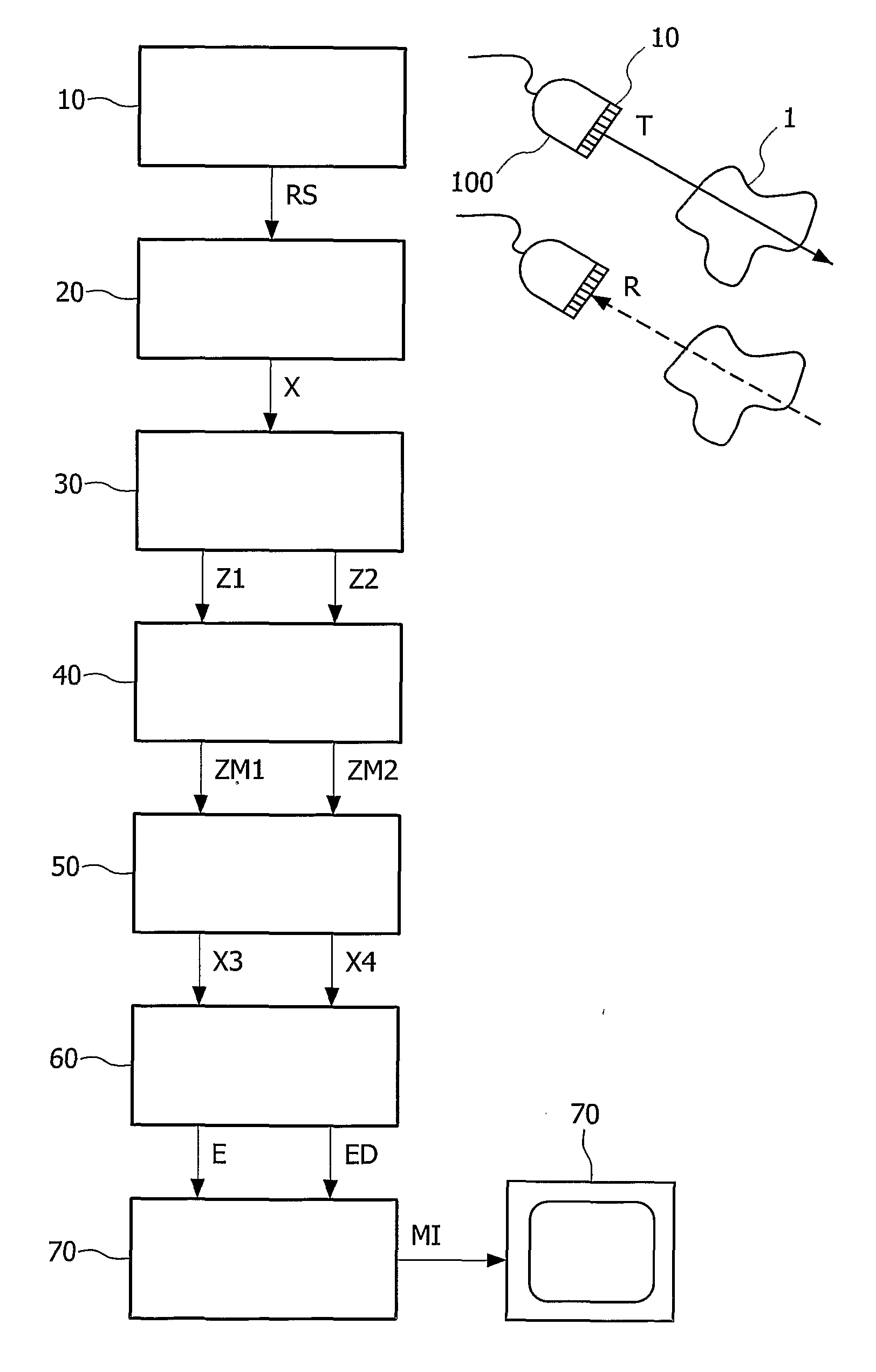
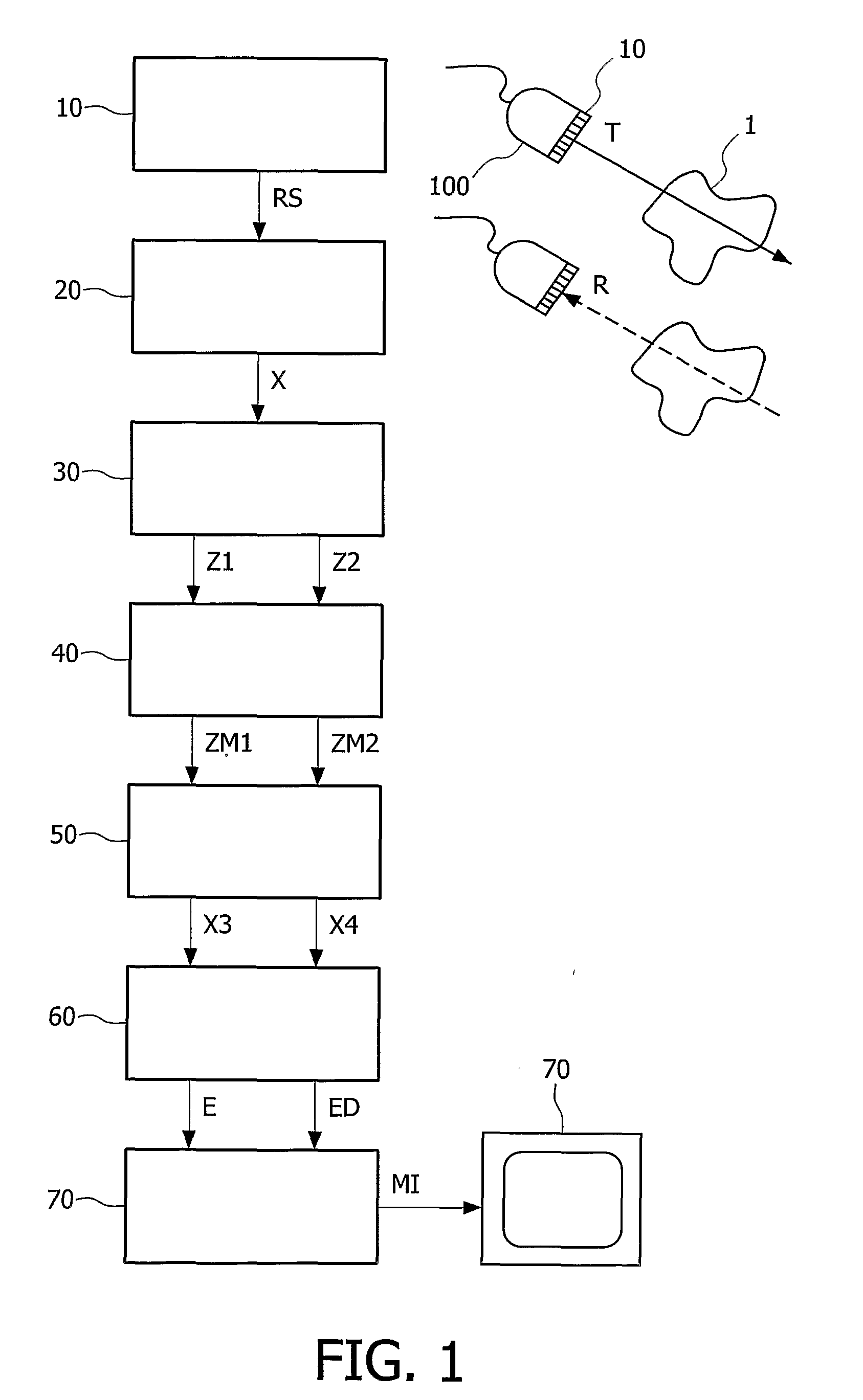
[0029] The invention relates to an ultrasound imaging method of extracting a flow component from echographic signals received from a region of interest comprising moving tissues and flowing fluids and of forming a motion image of said flow component. In the following, the particular domain of medical ultrasound imaging is considered and the moving tissues and flowing fluids are typically arterial or cardiac walls and blood flows. In this domain both the acquisition of 3D echographic data sets and the imaging of the blood flows offer a real added value for early diagnosis of arterial or cardiac diseases.

[0030] Referring to FIG. 1, the method in accordance with the invention comprises a step 10 of forming a set of beams of ultrasound data signals in order to receive echographic signals RS with a small number EL of time samples from a region of interest comprising moving objects, a step 20 of calculating Doppler signals X from said received echographic signals RS within said small num...


PUM


Abstract

The present invention relates to a method of extracting a flow signal from echographic signals received from a region of interest comprising moving tissues and flowing fluids. The method comprises a step of calculating Doppler signals from said echographic signals within a small number of time samples, a step of separating first and second estimated Doppler signals from said calculated Doppler signals, a step of calculating a linear combination of said first and second estimated Doppler signals which locally maximizes a temporal coherence, a step of deriving a third and fourth estimated Doppler signals from first and second maxima of said temporal coherence, a step of classifying said third and fourth estimated Doppler signals into an estimated Doppler clutter and flow components. The method finally comprises a step of forming a motion image of the flowing fluids from said estimated Doppler flow component.

Description

FIELD OF THE INVENTION [0001] The present invention relates to an ultrasound imaging method of extracting a flow signal from echographic signals received from a region of interest comprising moving tissues and flowing fluids. The present invention also concerns an ultrasound imaging system which is operated to use such a method. [0002] The present invention finds in particular its application in the domain of medical ultrasound imaging where the moving tissues are typically arterial or cardiac walls and the flowing fluids are blood flows. BACKGROUND OF THE INVENTION [0003] When transmitting a beam of ultrasound signals to a region of interest of the human body comprising moving tissues and / or flowing fluids, echographic signals are received, which comprise both a clutter component and a flow component. Prior art techniques have been developed for removing the clutter component and extracting some characteristics of the flow component. [0004] In the international patent application p...

Claims


Application Information

Patent Timeline
IPC IPC(8): G01F1/66F15B7/06F15B15/02F15B15/20F16D23/12F16D29/00F16D48/02
CPCF15B7/06F16D23/12F16D25/088F16D25/14A61B8/483F16D48/02F16D48/066G01S15/8981F16D29/005F16D2048/0212F16D2500/1026F16D2500/10412F16D2500/3024F16D2500/31413F16D2500/70404F16D2500/7041
Inventor BONNEFOUS, ODILE
Owner KONINKLIJKE PHILIPS ELECTRONICS NV
Why Eureka
Try Eureka