Devices and methods for atrial appendage exclusion

a technology for exclusion of atrial appendages and devices, applied in the field of appendix and methods for performing minimally invasive surgery, can solve the problems of increasing the risk of stroke, embolism and/or other circulatory complications, and the closure force of staples, and the inability to adapt to the tissu

Inactive Publication Date: 2008-02-14
MAQUET CARDIOVASCULAR LLC
View PDF99 Cites 166 Cited by
  • Summary
  • Abstract
  • Description
  • Claims
  • Application Information

AI Technical Summary

Benefits of technology

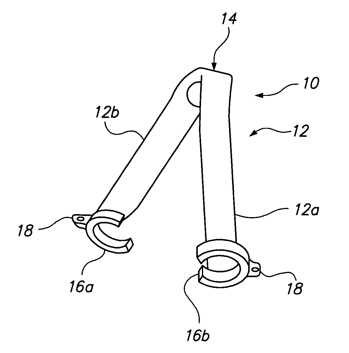
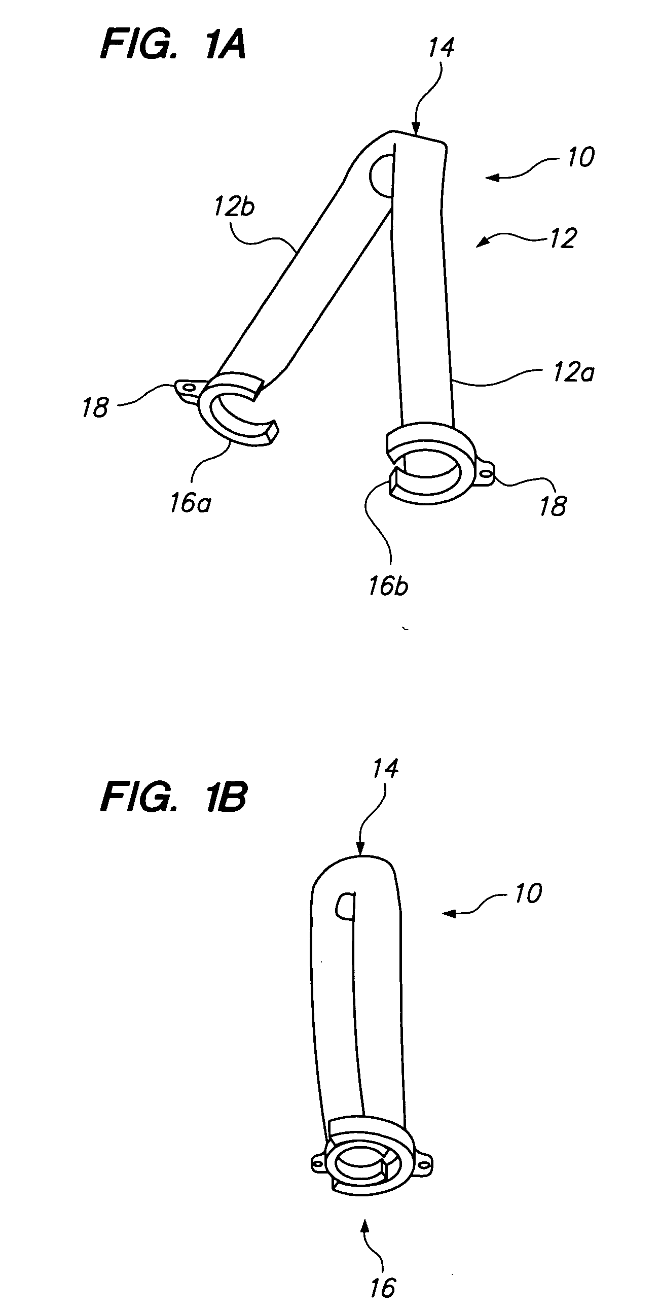
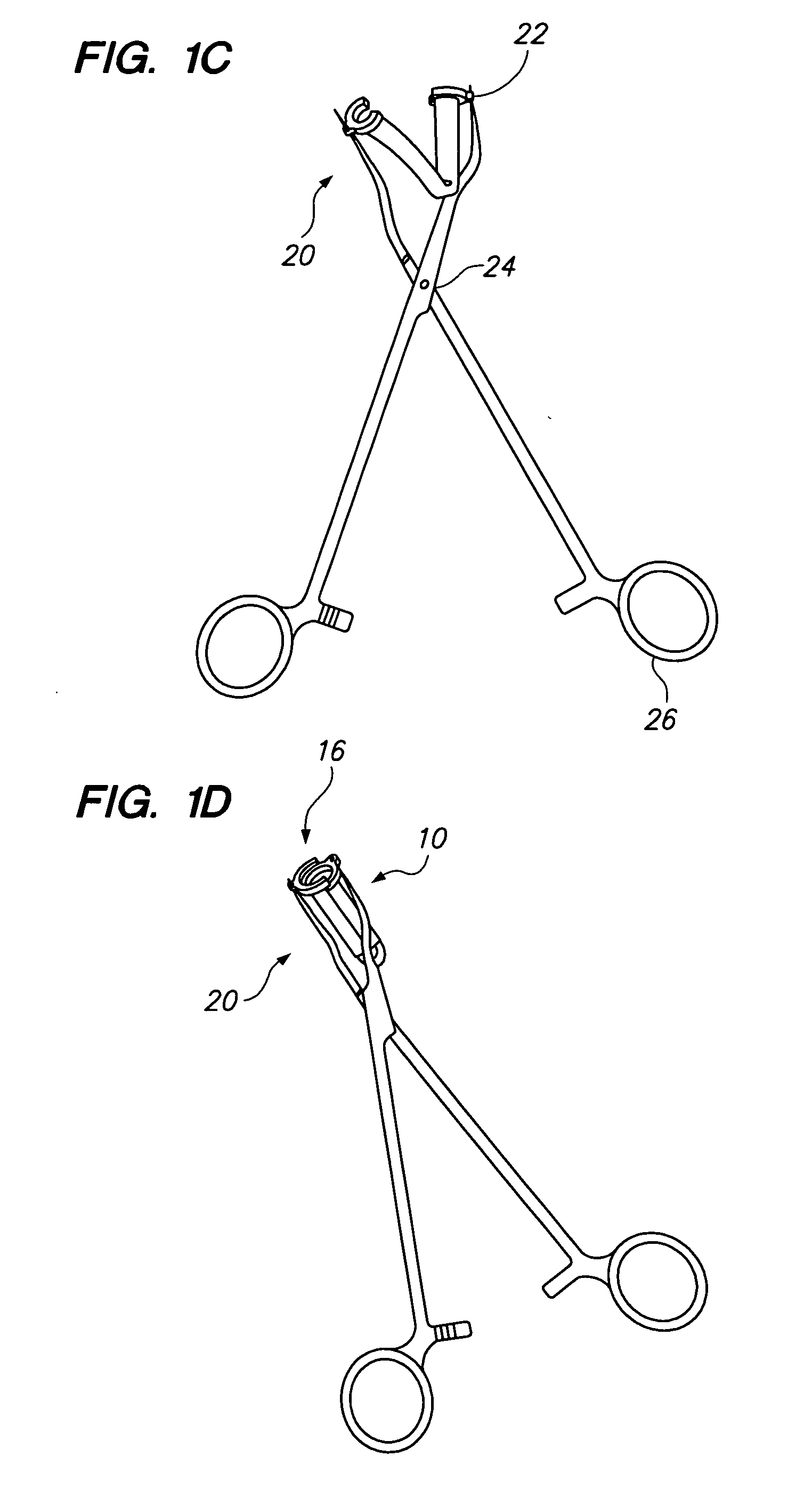
[0008]Devices, tools and methods for occluding fluid flow between two walls of tissue in a patient are provided. Two walls of tissue are compressed together with sufficient compressive force to prevent fluid flow between the two walls, while ensuring that the compressive force is not so great as to cause tissue necrosis.
[0016]A tissue wall coating device for minimizing bleeding caused by an incision or puncture of the tissue wall is provided, including a main body in the shape of an elastomeric sack being closed at one end and having an opening at a base portion end; and a ligature extending around the base portion end and configured to constrict the opening at the base portion end.
[0019]A method of reducing bleeding during a surgical procedure on an atrial appendix is provided, including: placing an elastomeric sack over the atrial appendix to form a slightly compressive interface between the sack and the atrial appendix; constricting a base opening of the sack by drawing on a ligature passing through the base opening, and thereby compressing the walls of tissue at the location contacted by the base opening; and fixing the ligature relative to the base opening to maintain the constriction.

Problems solved by technology

Once formed, these clots have the potential of detaching from the left atrial appendage and travel elsewhere in the body via the circulatory system, thereby increasing the risk of stroke, embolism and / or other circulatory complications.
While effective, this technique generally requires an open chest procedure, i.e., open heart surgery, as suturing an appendage closed is very difficult to perform in a closed-chest environment and is generally not attempted.
However, difficulties present with use of this technique, as there is a tendency for the staples to tear into the friable tissue of the appendage and cause bleeding, requiring the chest to be opened to repair the damage to the torn appendage.
Further, since staplers that are presently used for these procedures are not designed for use on an atrial appendage, but rather for gastrointestinal use, the closure force on the staples, as the staples are placed in the appendage, may not be suitable for the tissue to which the force is applied.
Further, the tissue thickness of the walls of the appendage may differ significantly from tissue thicknesses that the stapler is designed to close, resulting either in tissue damage to the appendage by the applied staple drawing the tissue walls too close together and thus crushing them, or incomplete closure, resulting in a failure to completely close off the appendage to the flow of blood.
Space occupying devices that are currently used also tend to leave areas of the appendage exposed to the blood path (circulation), with potential thrombus formation, and are particularly susceptible to this when delivered under closed chest conditions, such as via catheter, for example.

Method used


Image

  • Devices and methods for atrial appendage exclusion
  • Devices and methods for atrial appendage exclusion
  • Devices and methods for atrial appendage exclusion

Examples


Embodiment Construction

[0095]Before the present devices and methods are described, it is to be understood that this invention is not limited to particular embodiments described, as such may, of course, vary. It is also to be understood that the terminology used herein is for the purpose of describing particular embodiments only, and is not intended to be limiting, since the scope of the present invention will be limited only by the appended claims.

[0096]Where a range of values is provided, it is understood that each intervening value, to the tenth of the unit of the lower limit unless the context clearly dictates otherwise, between the upper and lower limits of that range is also specifically disclosed. Each smaller range between any stated value or intervening value in a stated range and any other stated or intervening value in that stated range is encompassed within the invention. The upper and lower limits of these smaller ranges may independently be included or excluded in the range, and each range wh...


PUM


Abstract

Devices, tools and methods for occluding fluid flow between two walls of tissue in a patient. Two walls of tissue are compressed together with sufficient compressive force to prevent fluid flow between the two walls, while ensuring that the compressive force is not so great as to cause tissue necrosis. The devices, tools and methods may be carried out using minimally invasive surgical techniques, such as in reduced-access surgical sites. Devices, tools and methods are provided for occluding an atrial appendix.

Description

FIELD OF THE INVENTION[0001]The field of the present invention is apparatus and methods for performing minimally invasive surgery, more particularly to cardiac procedures performed with minimally invasive surgical techniques and apparatus.BACKGROUND OF THE INVENTION[0002]In patients undergoing therapy for atrial fibrillation, for example atrial ablation therapy, it is desirable to exclude the left atrial appendage from the circulatory path, such as by sealing off the appendage from the remainder of the atrial cavity, or removing the appendage from the atrium. Blood clots tend to form in the atrial appendage of a patient experiencing atrial fibrillation. Once formed, these clots have the potential of detaching from the left atrial appendage and travel elsewhere in the body via the circulatory system, thereby increasing the risk of stroke, embolism and / or other circulatory complications.[0003]Even when atrial ablation is performed in an attempt to cure atrial ablation, atrial appendag...

Claims


Application Information

Patent Timeline
Patent Type & Authority Applications(United States)
IPC IPC(8): A61B17/08
CPCA61B17/064A61B17/0643A61B17/068A61B17/122A61B2017/1225A61B2017/00243A61B2017/0647A61B2017/0649A61B17/1227
Inventor CHIN, ALBERT K.DAVIS, JOHN W.SHROFF, KETANAGARWAL, AMITUEMURA, SHUJICANTU, ALFREDO R.WILLIS, GEOFFREY H.CALLAS, PETER L.GRESL, CHARLES
Owner MAQUET CARDIOVASCULAR LLC
Why Eureka
Try Eureka