
Molecular imaging of epithelial cells in lymph

Inactive Publication Date: 2008-02-28
BAYLOR COLLEGE OF MEDICINE
View PDF7 Cites 2 Cited by
  • Summary
  • Abstract
  • Description
  • Claims
  • Application Information

AI Technical Summary

Benefits of technology

[0015] The molecular specificity of antibodies reduces the probability of a false positive, while concurrently increasing signal to noise, and signal to background. The use of antibodies as the means for locating the dispersed cells increases the accuracy of staging the cancer during medical diagnosis. These advantages exceed current protocols because of the decreased likelihood of other non-specific interactions within the patient's lymph. Furthermore, the imaging can be conducted quickly, without prolonged periods of immobilization required in imaging machinery, or further discomfort to the patient.

Problems solved by technology

Despite recent advances combined with new extra-, and intracellular contrast agents, these techniques represent only non-specific means primarily useful in identifying enlarged lymphatic nodes.
Administered intravenously, this technique results in high-background signals reducing sensitivity in the detection of occult, micro, and difficult to detect, nodes.
Furthermore, macrophage uptake in the lymph rather than cancer cell specific uptake impedes accurate diagnosis of cancer.

Method used


Image

  • Molecular imaging of epithelial cells in lymph
  • Molecular imaging of epithelial cells in lymph
  • Molecular imaging of epithelial cells in lymph

Examples


example

Demonstration of Binding of NIR Dye Labeled, Anti-Human EpCAM to Cancer Epithelial Cells

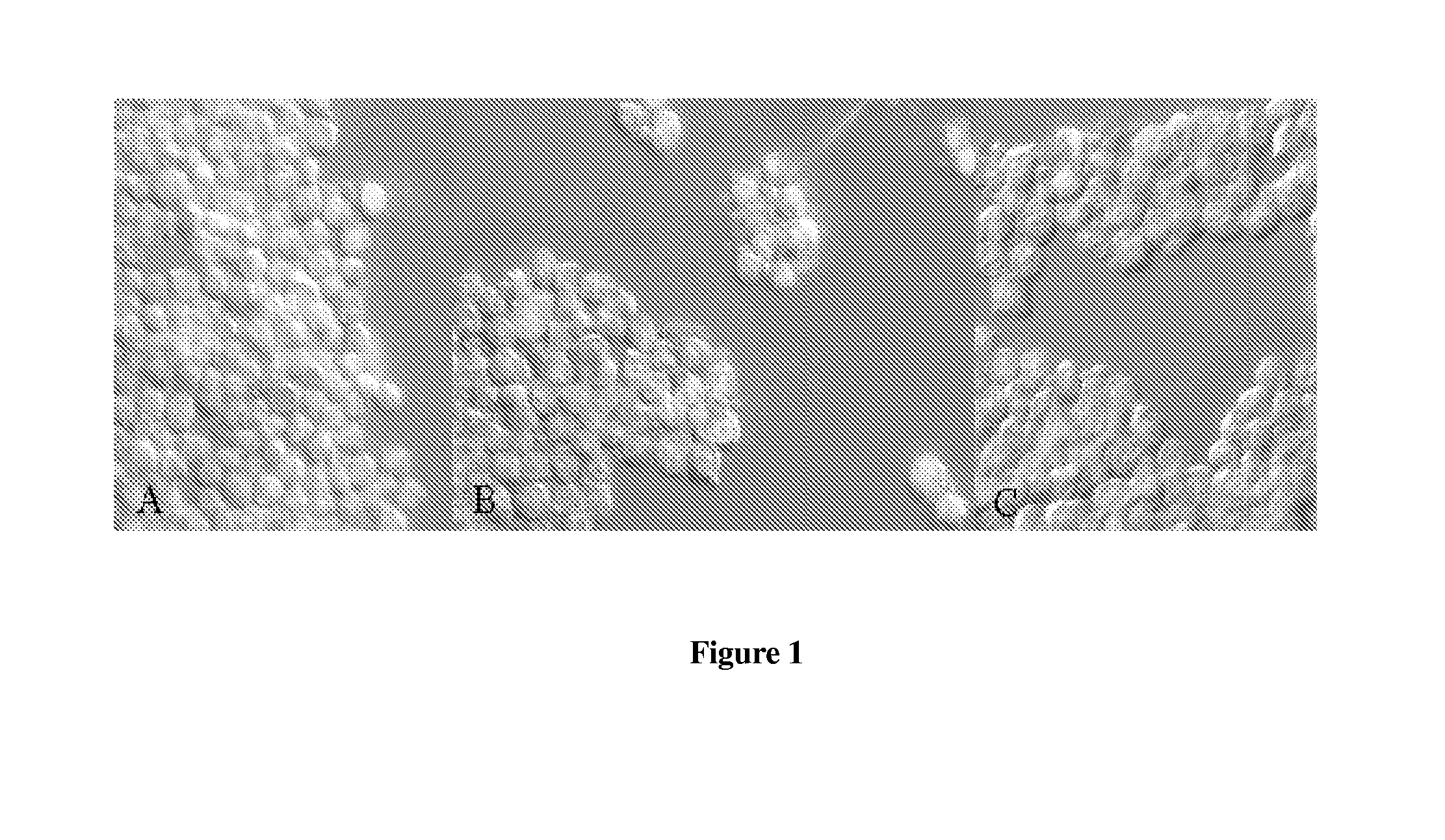
[0038] In FIG. 1, the anti-human EpCAM antibody was labeled with NIR fluorescent dye was used to incubate human epithelial cancer cell lines, breast cancers SKBR3 (A) and MDA-MB-231 (B) and human, non-epithelial melanoma cancer cell line, M21 (C). Fluorescence microscopy showed that the imaging conjugate (red color) binds to the epithelial cells and minimally to the non-epithelial cancer cells as determined by the red labeling of the cells in FIG. 1 parts A, and B. The cell nuclei were stained Sytox green for reference.

[0039] The results showed that the imaging agent binds to epithelial cells in culture, but not to non-epithelial cell lines. It is important to note that 90% of all human cancers are epithelial and therefore can be targeted with the imaging agent.

Demonstration of NIR-Dye Labeled Anti-EpCAM (Mouse) Targeting to Murine 4T1 Mammary Carcinoma Cells in Axillary Lymph Nodes Associat...


PUM


Abstract

Methods and imaging agents for imaging epithelial cancer cells in the lymphatic system are disclosed herein. In an embodiment, an imaging agent for imaging cancer cells in a lymphatic system comprises a fluorescent dye conjugated to one or more antibodies. The antibodies are capable of specific binding to an epithelial cell adhesion molecule (Ep-CAM). In addition, embodiments of the imaging agent may be administered to the lymphatic system where the disclosed imaging agents may bind to an epithelial cell adhesion molecule. The bound imaging agents may be excited with excitation light to image cancer cells in the lymphatic system.

Description

CROSS-REFERENCE TO RELATED APPLICATIONS [0001] The present application claims the benefit of U.S. Provisional Application Ser. No. 60 / 823,476 filed Aug. 24, 2006, herein incorporated by reference in its entirety for all purposes.STATEMENT REGARDING FEDERALLY SPONSORED RESEARCH OR DEVELOPMENT [0002] This work was supported by National Institutes of Health (R01 CA112679) and America Cancer Society (RSG-06-213-01-LR).BACKGROUND [0003] 1. Field of the Invention [0004] This invention relates generally to the field of cancer diagnostics. More specifically, the invention relates to a method of imaging and identifying cancer cells in the lymphatic system through near infrared fluorescence labeling. [0005] 2. Background of the Invention [0006] The lymph plexus consists of loosely connected epithelium without the structural integrity of smooth muscle cells for efficient collection of fluid and foreign particles. The plexus is located beneath the epidermis and provides the route of entry into ...

Claims


Application Information

Patent Timeline
IPC IPC(8): A61B5/00
CPCA61B5/0059A61B5/418A61B5/415A61B5/0073
Inventor ADAMS, KRISTENSEVICK-MURACA, EVA
Owner BAYLOR COLLEGE OF MEDICINE
Learn More