Virtual fly over of complex tubular anatomical structures

a tubular anatomical and virtual technology, applied in the field of medical imaging, can solve the problems of insufficient examination, time-consuming, and lower surface visibility coverag

Inactive Publication Date: 2008-03-20
UNIV OF LOUISVILLE RES FOUND INC
View PDF8 Cites 47 Cited by
  • Summary
  • Abstract
  • Description
  • Claims
  • Application Information

AI Technical Summary

Benefits of technology

[0008] An embodiment of the invention is method, which can be implemented in software, firmware, hardware, etc., for virtual fly over inspection of complex anatomical tubular structures. In a preferred embodiment, the method is implemented in software, and the software reconstructs the tubular anatomical structure from a binary imaging data that is originally acquired from computer aided tomography scan or comparable biological imaging system. The software of the invention splits the entire tubular anatomy into exactly two halves. The software assigns a virtual camera to each half to perform fly-over navigation. Through controlling the elevation of the virtual camera, there is no restriction on its field of view (FOV) angle, which can be greater than 90 degrees, for example. The camera viewing volume is perpendicular to each half of the tubular anatomical structure, so potential structures of interest, e.g., polyps hidden behind haustral folds in a colon are easily found. The orientation of the splitting surface is controllable, the navigation can be repeated at another or a plurality of another split orientations. This avoids the possibility that a structure of interest, e.g., a polyp that is divided between the two halves of the anatomical structure in a first fly over is missed during a virtual inspection. Preferred embodiment software conducts virtual colonoscopy fly over. Experimental virtual fly over colonoscopy software of the invention that performed virtual fly over 15 clinical datasets demonstrated average surface visibility coverage is 99.59+0.2%

Problems solved by technology

Although optical colonoscopy detects more than 90% of colorectal cancers, it is invasive, uncomfortable, inconvenient, and sometimes can not reach the colon caecum, resulting in an incomplete exam.
In general, fly-through based-methods suffer from the following limitations: (1) the camera's field of view is limited, and hence results in lower surface visibility coverage; and (2) the navigation must be done in both antegrade and retrograde directions to maximize visualized surface areas, and hence it is very time consuming.
Because flattening methods are based on geometric mapping, geometric distortion is inevitable.
One of the main drawbacks of this method is that a polyp may split among several of the rendered views.
In addition, the layout of several views, such as an unfolded cube is hard to follow, since a medical professional must observe views simultaneously that are sometimes oriented in different directions (horizontal and vertical).

Method used


Image

  • Virtual fly over of complex tubular anatomical structures
  • Virtual fly over of complex tubular anatomical structures
  • Virtual fly over of complex tubular anatomical structures

Examples


Embodiment Construction

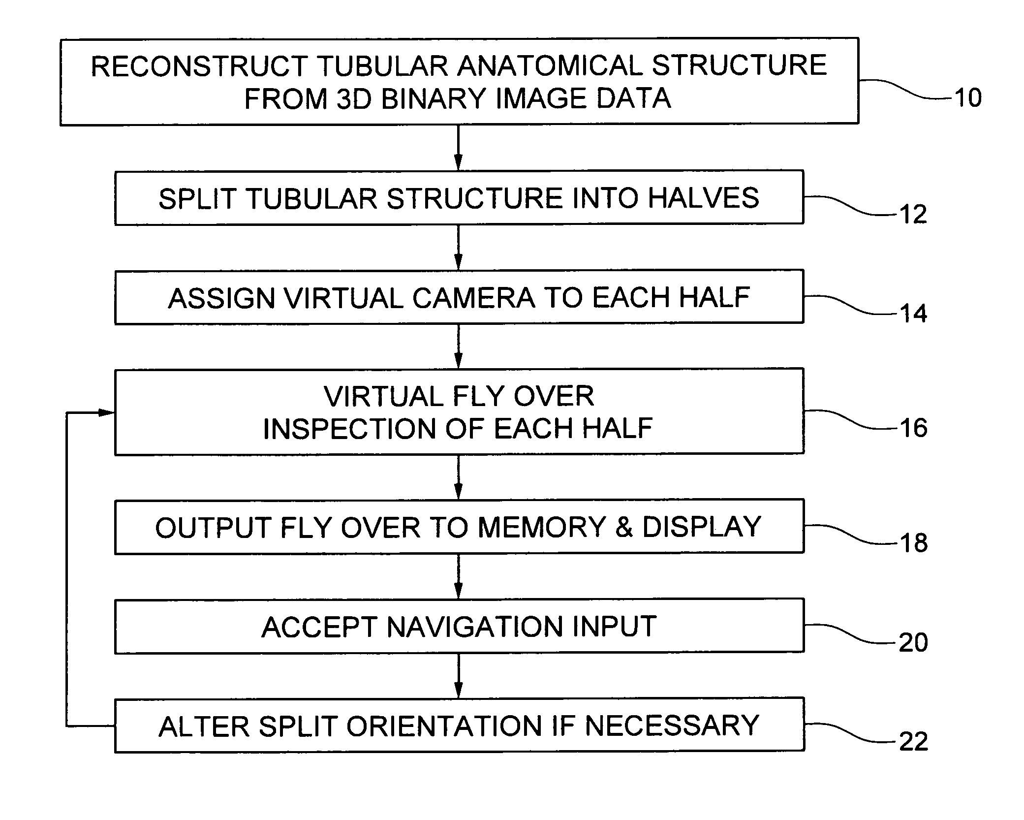
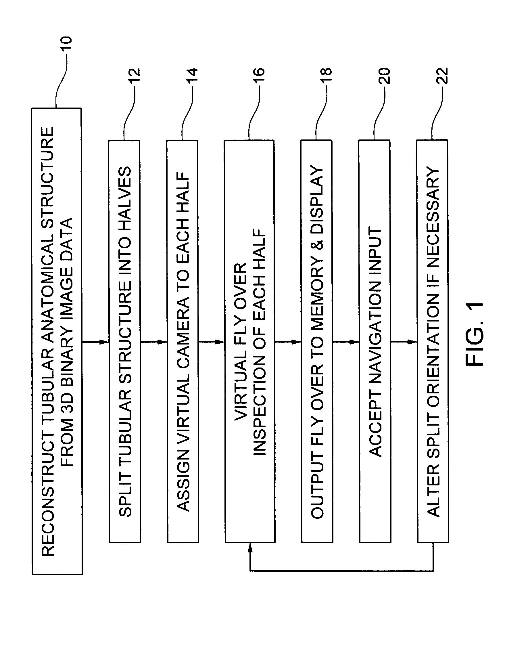
[0013] An embodiment of the invention is method, which can be implemented in software, firmware, hardware, etc., for virtual fly over inspection of complex anatomical tubular structures. In a preferred embodiment, the method is implemented in software, and the software reconstructs the tubular anatomical structure from a binary imaging data that is originally acquired from computer aided tomography scan or comparable biological imaging system. The software of the invention splits the entire tubular anatomy into exactly two halves. The software assigns a virtual camera to each half to perform fly-over navigation. Through controlling the elevation of the virtual camera, there is no restriction on its field of view (FOV) angle, which can be greater than 90 degrees, for example. The camera viewing volume is perpendicular to each half of the tubular anatomical structure, so potential structures of interest, e.g., polyps hidden behind haustral folds in a colon are easily found. The orient...


PUM


Abstract

An embodiment of the invention is method, which can be implemented in software, firmware, hardware, etc., for virtual fly over inspection of complex anatomical tubular structures. In a preferred embodiment, the method is implemented in software, and the software reconstructs the tubular anatomical structure from a binary imaging data that is originally acquired from computer aided tomography scan or comparable biological imaging system. The software of the invention splits the entire tubular anatomy into exactly two halves. The software assigns a virtual camera to each half to perform fly-over navigation. Through controlling the elevation of the virtual camera, there is no restriction on its field of view (FOV) angle, which can be greater than 90 degrees, for example. The camera viewing volume is perpendicular to each half of the tubular anatomical structure, so potential structures of interest, e.g., polyps hidden behind haustral folds in a colon are easily found. The orientation of the splitting surface is controllable, the navigation can be repeated at another or a plurality of another split orientations. This avoids the possibility that a structure of interest, e.g., a polyp that is divided between the two halves of the anatomical structure in a first fly over is missed during a virtual inspection. Preferred embodiment software conducts virtual colonoscopy fly over. Experimental virtual fly over colonoscopy software of the invention that performed virtual fly over on 15 clinical datasets demonstrated average surface visibility coverage is 99.59+ / −0.2%.

Description

PRIORITY CLAIM AND REFERENCE TO RELATED APPLICATION [0001] This application claims priority pursuant to 35 U.S.C. §120 from prior co-pending provisional application Ser. No. 60 / 842,850, filed Sep. 7, 2006.FIELD [0002] A field of the invention is medical imaging. Example applications of the invention include virtual colonoscopy. In general, the invention can be used to virtually inspect complex tubular anatomical structures, e.g., the colon & arteries. BACKGROUND [0003] Techniques for the examination of tubular anatomical structures, such as the trachea, arteries, colon, digestive tract, etc., are important procedures for detecting abnormalities. Such abnormalities can include polyps, the latter of which are indicators of developing cancer. For example, optical colonoscopies are an important procedure to detect polyps that are early stage indicators of cancer. [0004] Colorectal colon cancer is the third most common form of cancer and the second leading cause of death among cancers in...

Claims


Application Information

Patent Timeline
Patent Type & Authority Applications(United States)
IPC IPC(8): G06Q50/00
CPCG06F19/3437G06T2210/41G06T19/003G16H50/50
Inventor FARAG, ALY A.HASSOUNA, M. SABRY
Owner UNIV OF LOUISVILLE RES FOUND INC
Why Eureka
Try Eureka