Eureka AIR delivers breakthrough ideas for toughest innovation challenges, trusted by R&D personnel around the world.

Isolation of inner cell mass for the establishment of human embryonic stem cell (hESC) lines

a technology of embryonic stem cells and inner cells, which is applied in the field of isolate the inner cell mass for the establishment of human embryonic stem cells (hesc) lines, can solve the problems of low percentage of blastocysts hatching spontaneously in humans, the chance of isolating trophectoderm cells, and the inability to isolate the complete inner cell mass

Inactive Publication Date: 2007-11-13
RELIANCE LIFE SCI PVT
View PDF5 Cites 59 Cited by
  • Summary
  • Abstract
  • Description
  • Claims
  • Application Information

AI Technical Summary

Benefits of technology

[0055]2. It is another object of the present invention to isolate ICM using laser ablation technique without using any animal generated antibodies and sera, thereby preventing the possibility of transmission of animal organism to human and thus can safely be used on commercial scale.
[0057]4. It is another object of the present invention to isolate inner cell mass (ICM) by simple, shorter and easily feasible way without affecting / destroying the inner cell mass (ICM).
[0058]5. It is still another object of the present invention to ensure the purity of inner cell mass (ICM) by rupturing completely trophectoderm thereby minimising the contamination of inner cell mass (ICM).
[0061]The present invention relates to isolation of inner cell mass, using laser ablation of zona pellucida (ZP) and trophectoderm (TE) and aspiration of inner cell mass for establishing embryonic stem cell lines. In the present invention, the non contact diode laser used is highly accurate and reliable tool for cellular microsurgery. The system incorporates the latest in fiber optic technology to provide the most compact laser system currently available. The 1.48 μm diode non-contact Saturn Laser System is mounted / implanted via the epifluorescence port to inverted microscope fitted with micromanipulators. A pilot laser is used to target the main ablation laser and a series of LEDs inform the user when the laser is primed and is ready to fire. A two-second-operation window is used to reduce the possibility of accidentally firing the laser. The spot diameter of the laser can be varied according to the hole size required.
[0069]A pilot laser was used to target the main ablation laser and a series of LED's inform the user when the laser is primed and ready to discharge the laser beam. A two-second-operation window was used to reduce the possibility of accidentally firing the laser. The spot diameter of the laser can be varied according to the ablation size required.c) Laser Ablation and Isolation of Inner Cell Mass.
[0080]Although the isolation of inner cell mass using both the methods did not show any significant difference, however, of isolation of inner cell mass by laser ablation has distinct advantage. This method will eliminate the use of antibodies and sera of animal origin. Isolation of inner cell mass by laser ablation method can be further cultured in the presence or absence of feeder layer. However, culturing of inner cell mass in a feeder free condition will further eliminate the possibilities of contamination of ES cell lines with animal viruses or bacteria and can be commercially utilized for human transplantation studies. In the current experiments, efforts were made to establish ES cell line in the absence of feeder cells.

Problems solved by technology

Secondly, while removing the outgrowth of the inner cell mass mechanically, there is a chance of isolating trophectoderm cells.
Thirdly, the percentage of blastocysts hatching spontaneously in humans is very low.
This procedure too is disadvantageous as the possibility to isolate the complete inner cell mass is low and many a time cells get disintegrated.
It is a very tedious procedure and may cause severe damage to the embryo.
However, this method has several disadvantages.
Firstly, the embryo is exposed for a long time to acid tyrode or pronase causing deleterious effects on embryo, thereby reducing the viability of embryos proper.
Secondly, it is time consuming procedure as it takes about 1.5 to 2.0 hours.
Thirdly, the yield of inner cell mass (ICM) per blastocyst is low.
Fourthly, critical storage conditions are required for antibody and complement used in the process.
Lastly, it involves the risk of transmission of virus and bacteria of animal origin to humans, as animal derived antibodies and complement are used in the process.
Other possible disadvantages of the existing cell lines are as follows:1. Use of feeder cells for culturing the human embryonic stem cell (hESC ) lines produces mixed cell population that require the Embryonic stem cells (ESC) to be separated from feeder cell components and this impairs scale up.2. Embryonic stem cells (ESC) get contaminated by transcripts from feeder cells and cannot be used on a commercial scale.
However, the main disadvantage of this procedure is that the inner cell mass (ICM) is isolated by immunosurgery method, wherein the initial derivation of Embryonic Stem Cells is carried out using feeder layer containing xenogenic components.
This raises the issue of possible contamination with animal origin viruses and bacteria.
Using a laser for thinning the ZP, embryologists are able to achieve accurate reduction of the ZP by 50%, which is very difficult with acidic Tyrode's solution.
Presence of Acid Tyrode's solution near the embryo may also be detrimental.

Method used


Image

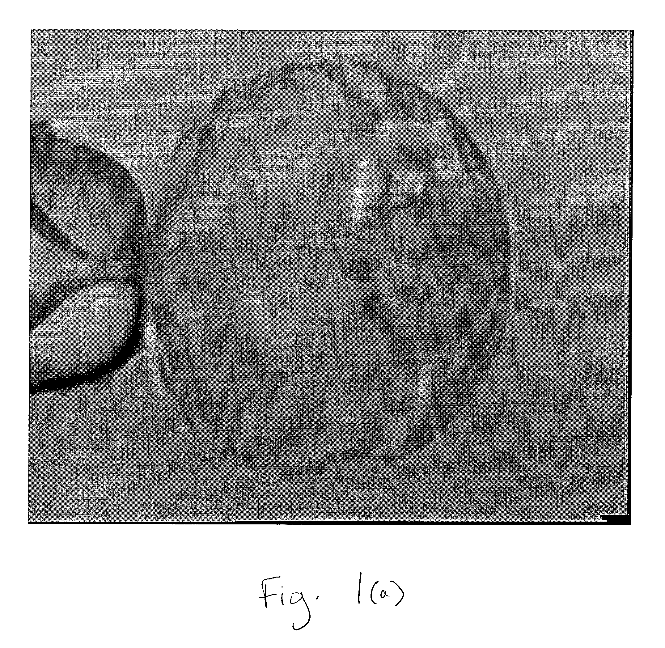
  • Isolation of inner cell mass for the establishment of human embryonic stem cell (hESC) lines
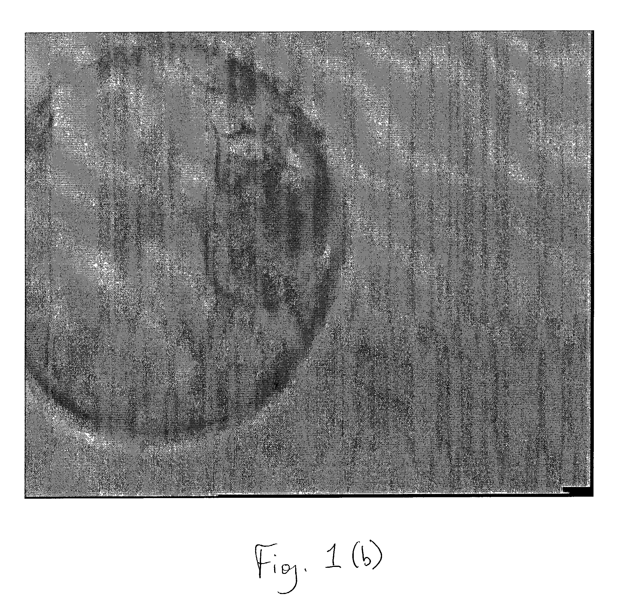
  • Isolation of inner cell mass for the establishment of human embryonic stem cell (hESC) lines
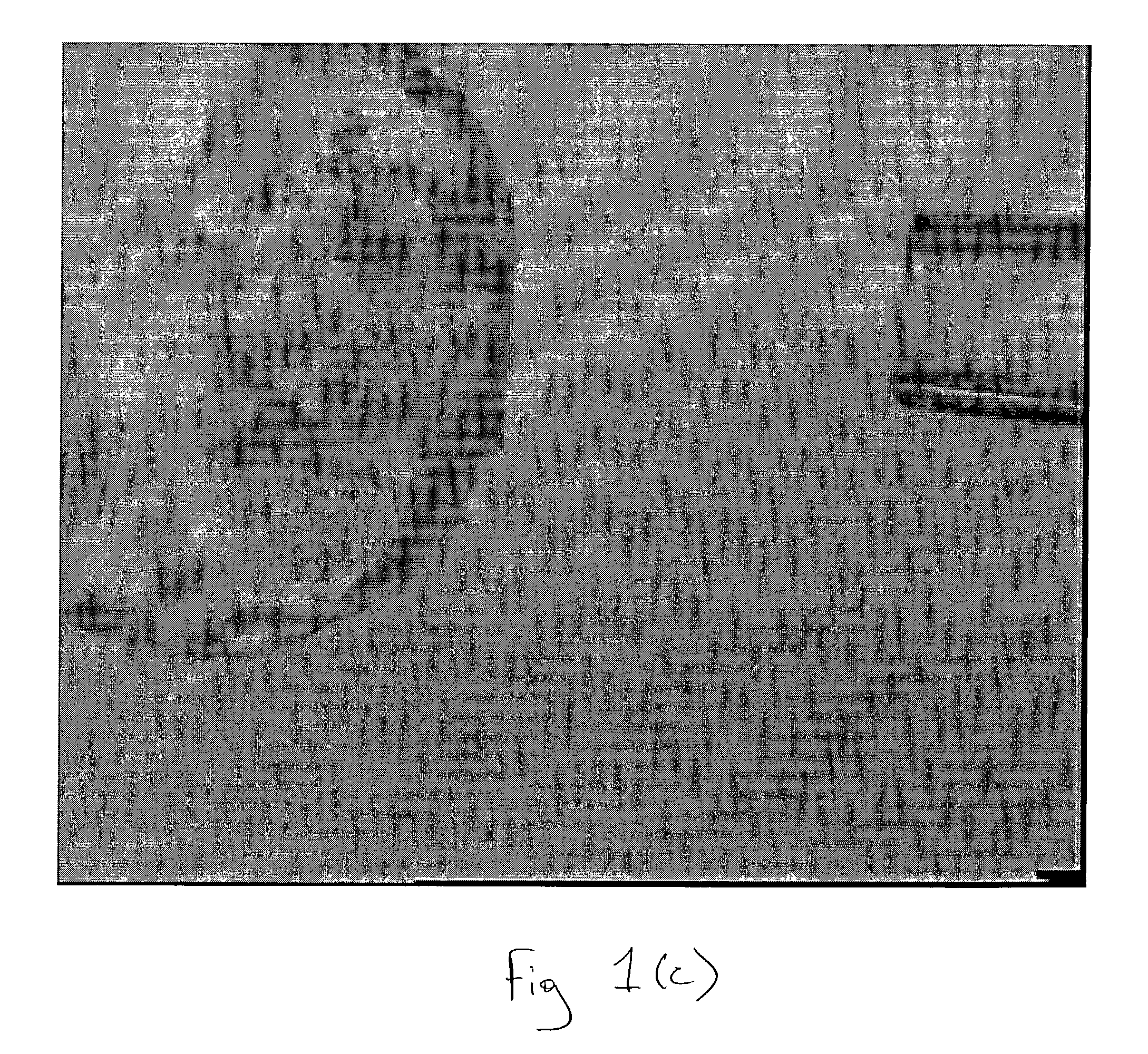
  • Isolation of inner cell mass for the establishment of human embryonic stem cell (hESC) lines

Examples


example 1

[0074]Total of 24 blastocyst stage human embryos were used for the isolation of inner cell mass. Embryos were washed several times in blastocyst culture medium (ISM-2 medium, Medicult, Denmark). Individual blastocyst was then placed in the 50 μl drop of Ca++ / Mg++ free embryo biopsy medium(EB 10 medium, Scadinavian). Micro drops were covered with mineral oil. Micromanipulator was set up. A glass holding pipette with outer diameter 75 μm and inner diameter 15 μm was used to secure the embryo. Biopsy pipette with an outer diameter of roughly 49 μm and inner diameter 35 μm was used for aspiration of inner cell mass. A pilot laser was used to target the main ablation laser. Embryo was immobilized on to the holding pipette in such a way that inner cell mass remained at 3 O'clock position and the zona pellucida and trophectoderm close to inner cell mass positioned to the aiming spot. The hole was induced by a local thermo-dissolution of the zona. Trophectoderm cells were ablated by giving ...

example 2

[0077]The objective was to determine efficiency of isolation of inner cell mass with conventional method I.e. immunosurgery and compared with newly invented laser ablated method.

[0078]21 blastocyst stage human embryo were used for isolation of inner cell mass. Embryos were washed several times with blastocyst culture medium (ISM-2 medium) and followed by ES medium. Individual blastocyst stage embryo was then placed in 50 μl micodrops of 1:50 anti-human antibody (Sigma) for 30 minutes at 37° C. and 5% CO2 in air. Blastocyst stage embryos were then washed four times after incubation with ES medium. Blastocysts were then again placed in 50 μl of microdrops of guinea pig complement at the concentration of 1:10 for 10 minutes at 37° C. and 5% CO2 in air. After incubation blastocyst stage embryos were washed several times in ES medium using fine bore glass pipette in order to remove trophectoderm. Isolated inner cell mass was then washed with ES medium and cultured in 96 well plate in the...


PUM


Abstract

A method for isolating an inner cell mass comprising the steps of immobilizing a blastocyst stage embryo having a zona pellucida, trophectoderm, and inner cell mass, creating an aperture in the blastocyst stage embryo by laser ablation, and removing the inner cell mass from the blastocyst stage embryo through the aperture. The aperture is through the zona pellucida and the trophectoderm. The laser ablation is acheived using a non-contact diode laser. The inner cell mass removed from the blastocyst stage embryo is used to establish human Embryonic Stem Cell lines.

Description

RELATED APPLICATION[0001]This application claims priority to the provisional application Ser. No. 60 / 314,323 filed on Aug. 23, 2001.FIELD OF THE INVENTION[0002]The present invention relates to a method of isolation of inner cell mass (ICM) derived from blastocyst stage mammalian embryo for establishing human embryonic stem cell (hESC) lines, using a non-contact diode laser technique.BACKGROUND OF THE INVENTION[0003]The isolation of human stem cells offers the promise of a remarkable array of novel therapeutics. Biologic therapies derived from such cells through tissue regeneration and repairs as well as through targeted delivery of genetic material are expected to be effective in the treatment of a wide range of medical conditions. Efforts to analyze and assess the safety of using human stem cells in the clinical setting are vitally important to this endeavor.[0004]Embryonic stem (ES) cells are the special kind of cells that can both duplicate themselves (self renew) and produce dif...

Claims


Application Information

Patent Timeline
Patent Type & Authority Patents(United States)
IPC IPC(8): C12N5/00A01N1/00C12N15/00C12N5/02C12N5/08C12N5/071A61B17/435A61B18/20A61K35/54C12N5/07C12N5/0735C12N13/00
CPCA61B17/435C12N5/0606A61B18/20
Inventor PARIKH, FIRUZA RAJESHTOTEY, SATISH MAHADOERAOSAXENA, SHAILAJA ANUPAM
Owner RELIANCE LIFE SCI PVT
Why Eureka
Eureka Blog
Learn More