

Compensating for truncated CT images for use as attenuation maps in emission tomography
a technology of emission tomography and compensation, which is applied in the field of medical diagnostic imaging and to correction of medical images, can solve the problems of insufficient computational power, inability to use iterative techniques, and inability to achieve the effect of sufficient computational power
- Summary
- Abstract
- Description
- Claims
- Application Information
AI Technical Summary
Benefits of technology
Problems solved by technology
Method used
Image

Examples
Embodiment Construction
[0034]As required, disclosures herein provide detailed embodiments of the present invention; however, the disclosed embodiments are merely exemplary of the invention that may be embodied in various and alternative forms. Therefore, there is no intent that specific structural and functional details should be limiting, but rather the intention is that they provide a basis for the claims and as a representative basis for teaching one skilled in the art to variously employ the present invention.
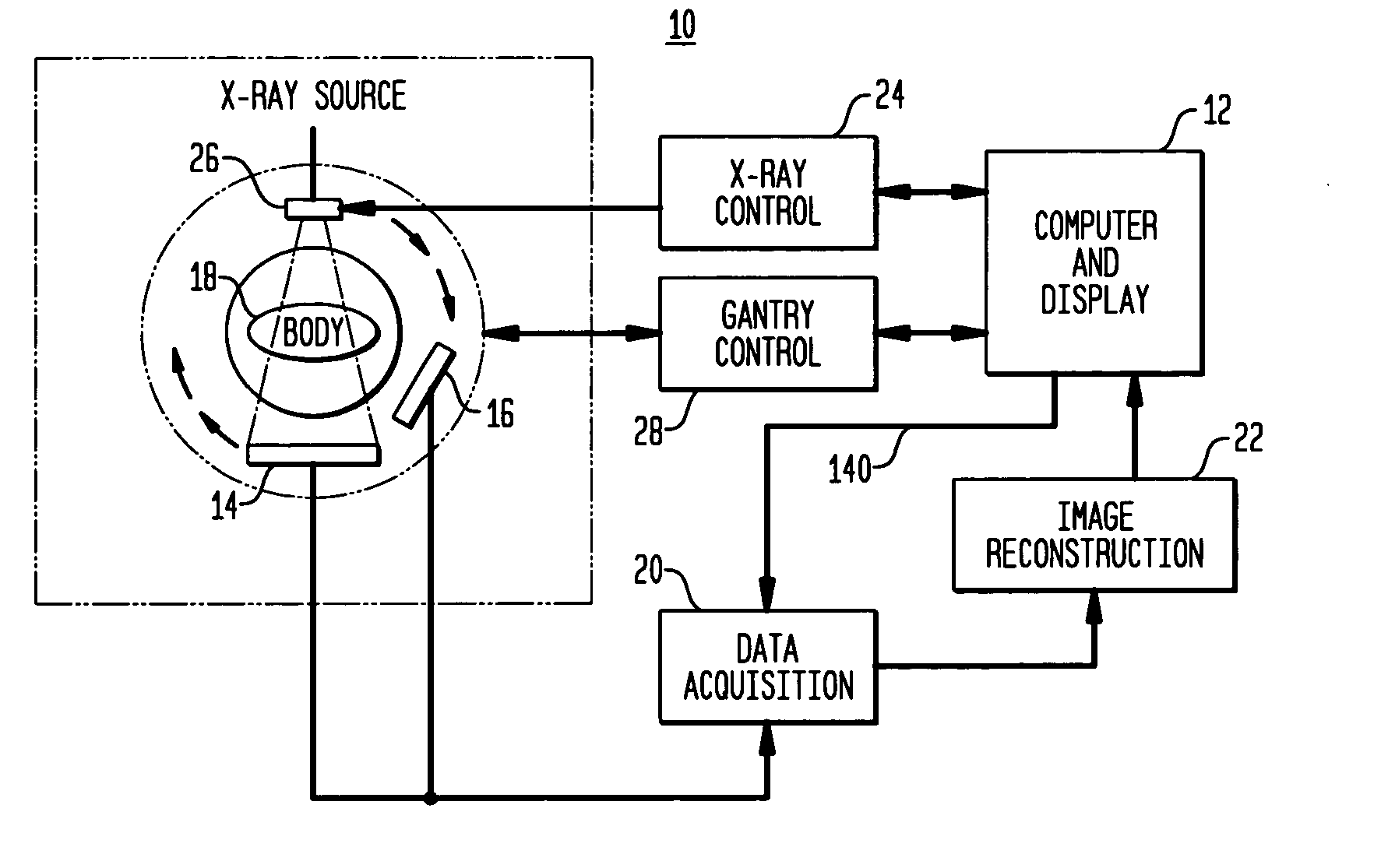
[0035]A process according to the present invention uses an imaging system as a means to reveal the presence of tumors or other defects in the organs or tissues of a patient who exhibits symptoms of an undesirable condition. The imaging system first requires that the patient adopt a position for collection of data from the organ or area of tissue under study, also referred to herein as the imaged object. Data collection proceeds using a dual modality technique that may include either simultaneous ...
PUM
 Abstract
Description
Claims
Application Information