
Fixing device for endotracheal tube

Inactive Publication Date: 2008-04-24
KANG SIN BUM
View PDF19 Cites 13 Cited by
  • Summary
  • Abstract
  • Description
  • Claims
  • Application Information

AI Technical Summary

Benefits of technology

[0029]To solve the foregoing problems, it is an object of the present invention to provide a device for fixing an endotracheal tube that can fix the endotracheal tube faster and more firmly when intubation of the endotracheal tube is required in an emergency or for putting a patient under anesthesia.
[0030]It is another object of the present invention to provide a device for fixing an endotracheal tube that enables a single person to fix the endotracheal tube easily and exactly.
[0031]It is another object of the present invention to provide an endotracheal tube fixing device comprising a multipurpose hole, which provides tighter fixation as well as simpler and easier suction when such suction is necessary.

Problems solved by technology

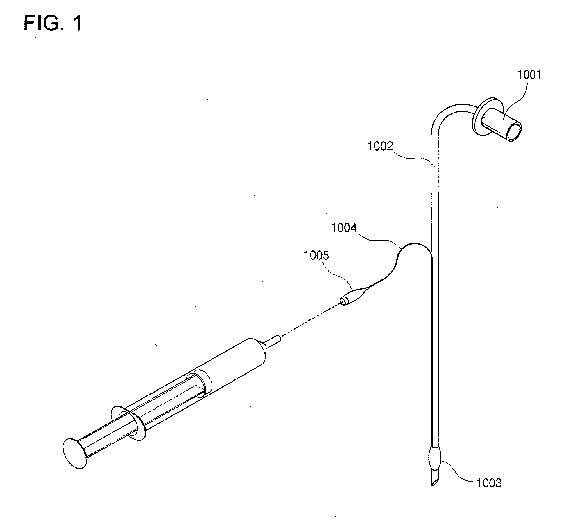
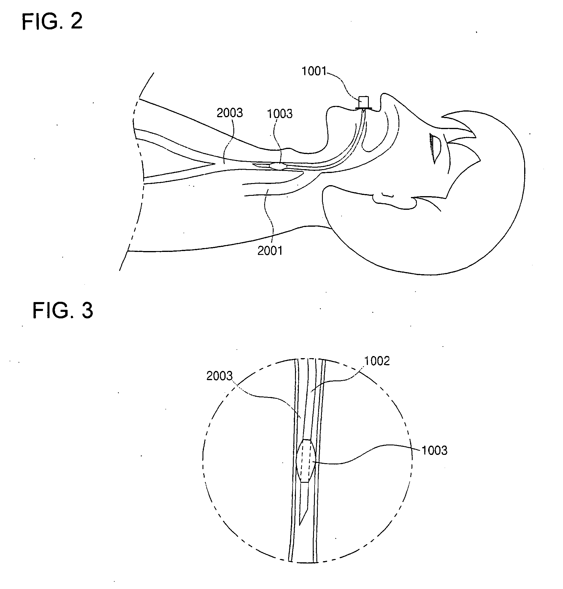
However, it is difficult to fix the endotracheal tube firmly at an exact position by means only of the cuff 1003.
Unless a suitable ventilating device or a fixing device thereof is provided, brain damage or even death may result.
While the endotracheal tube has many merits, it can cause a complication after the surgery has been completed, such as a sore throat and hoarseness.
Even though the sore throat is a temporary complication from which the patient can recover naturally within 72 hours, it can bring about serious discomfort to the patient.
However, recent research shows that lidocaine is not so effective.
However, in conventional devices, the fixing of the cuff has been achieved only by the cuff itself, which inevitably has brought about an overexpansion of the cuff, that is, an expansion beyond the minimum range of expansion needed to prevent the oxygen or anesthetic from flowing backward.
In practice, it is very hard to select an appropriate endotracheal tube and to intubate and fix it.
Even an experienced doctor frequently makes a mistake, which may resulting from any of many factors, such as the physical diversity of patients and the diversity of situations where the airway should be fixed.
In addition, over-intubation may force the endotracheal tube to be intubated into a blood vessel that is connected to only a single lung of the patient, so that it provides air to one lung rather than to both lungs.
In such a case, the result may be fatal.
As a result, it is true that a variety of fatal medical accidents can occur when the endotracheal tube is not fixed firmly enough.
Further, even when the endotracheal tube is intubated correctly, a number of different difficulties may follow in fixing the intubated tube.
However, such a method does not fix the intubated endotracheal tube strongly enough as reliably to prevent it from moving.
Especially, an endotracheal tube intubated into the airway of a young child or an infant may deviate from the correct position in the airway even with a small movement of the endotracheal tube.
Further, the adhesive bandage may bring about a contact dermatitis to a patient with sensitive skin.
Moreover, generally two persons are required to fix the endotracheal tube by using the adhesive bandage, wasting manpower.
These problems are most serious in a young child or an infant.
However, even with this technique, it is still very difficult to fix the endotracheal tube perfectly in the exact position.
In such a case the doctor should remove the band, relocate the endotracheal tube and bind the band again, which is burdensome in a medical emergency.
However, in the conventional way of fixing the endotracheal tube by using a band or the like, such suction cannot be performed efficiently because of the difficulty in securing enough space for performing suction or the difficulty in moving the suction device.
Further, when performing a surgical operation in the interior of the mouth or on a patient in a prone position, the conventional method of binding a band is not sufficient for performing the surgery because it makes movement of the fixed endotracheal tube more probable.
Thus, the conventional method has provided medical services that are neither fast nor proper.
The problem increases in the case of surgery for a patient in a prone position, where it is impossible to fix the intubated endotracheal tube completely by the conventional band binding method.
Thus, the possibility still exists that permanent damage including death could occur.
Moreover, the cuff is more likely to be overinflated in the conventional method, resulting in many complications after the intubation of the endotracheal tube, such as a sore throat.
Considering that the endotracheal tube is usually required in a very urgent emergency, the conventional method of fixing the endotracheal tube by binding a band is very problematic both in the speed and the reliability of the fixation.
When a dentist or a plastic surgeon performs surgery on the interior of the mouth, or an otorhinolaryngologist performs surgery on the tonsils, the procedure for moving the fixed endotracheal tube in the conventional method is very burdensome and difficult, as aforementioned.

Method used


Image

  • Fixing device for endotracheal tube
  • Fixing device for endotracheal tube
  • Fixing device for endotracheal tube

Examples


Embodiment Construction

[0104]Reference now will be made in detail to embodiments of the present invention which are illustrated in the accompanying drawings.

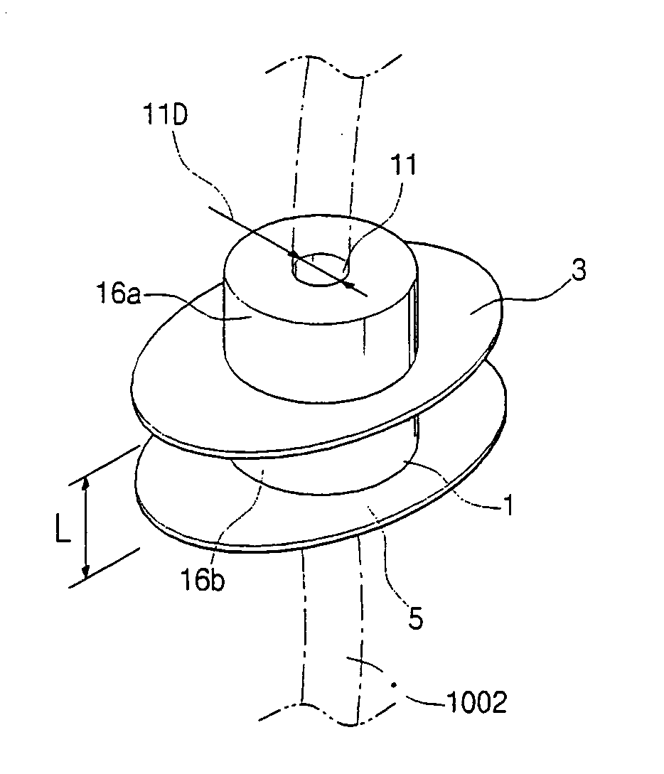
[0105]Referring to FIG. 5, an endotracheal tube fixing device according to an embodiment of the present invention comprises a body 1, and an upper wing 3 and a lower wing 5 that are generally disc-shaped and surround the body 1.

[0106]The body 1 is hollowed to define an insertion hole 11 through which the endotracheal tube is inserted, and, preferably, has shape of a hollow cylinder. The diameter 11D of the insertion hole 11 should be determined according to the diameter d of the endotracheal tube which is inserted. The diameter 11D will differ among various embodiments of the present invention as will be described in detail below.

[0107]The body also has a first portion 16a that protrudes from the upper wing 3 and a second portion 16b between the upper wing 3 and the lower wing 5. The first portion 16a, protruding from the upper wing 3, protrudes from ...


PUM


Abstract

A device for fixing an endotracheal tube. The device includes a body having an insertion hole, an upper wing provided on a periphery of the body and a lower wing provided on the periphery of the body that is spaced from the upper wing such that a patient's teeth are accommodated between the lower wing and the upper wing. The device also includes a cap for accommodating a portion of the body. The device fixes the endotracheal tube firmly so that it secures the endotracheal tube when surgery is being performed on a patient in a prone position. The device is movable along the teeth so that it allows surgery on the interior of the mouth or the tonsils to be performed more efficiently.

Description

BACKGROUND OF THE INVENTION[0001]1. Field of the Invention[0002]The present invention relates to a device for fixing an endotracheal tube. More specifically, the present invention is a fixing device for an endotracheal tube having a body that has a through hole, an upper wing and a lower wing spaced apart from the upper wing so as to accommodate a patient's teeth therebetween. The present invention may further comprise a cap that can be connected to the body so as to fix an intubated endotracheal tube more firmly, to fix the endotracheal tube sufficiently during a surgical operation when the patient's position makes it difficult to fix the endotracheal tube, such as a prone position, and to offer more convenience when a dentist or a plastic surgeon performs surgery on the interior of the mouth of the patient or an otorhinolaryngologist performs surgery on the patient's tonsils, by making it possible easily to move, disconnect, or reconnect the endotracheal tube.[0003]2. Description ...

Claims


Application Information

Patent Timeline
IPC IPC(8): A61M16/04
CPCA61M16/0463A61M16/0488A61M25/02A61M16/0493A61M2210/0625A61M2230/005A61M16/0495
Inventor KANG, SIN-BUM
Owner KANG SIN BUM
Learn More