Apparatus for supporting a patient in a prone position during diagnostic imaging

a technology for diagnosing imaging and prone positions, which is applied in the field of prone positions for positioning patients during medical imaging procedures, can solve the problems of degrading image quality or the necessity to repeat data acquisition, creating image artifacts, neck strain and patient motion,

Inactive Publication Date: 2009-10-20
GENERAL ELECTRIC CO
View PDF38 Cites 34 Cited by
  • Summary
  • Abstract
  • Description
  • Claims
  • Application Information

AI Technical Summary

Problems solved by technology

In other cases, such as during nuclear imaging, patient motion may create image artifact, degraded image quality or the necessity to repeat the data acquisition.
These positions cause neck strain and patient motion, as well as potentially limiting the breathing space and contributing to feelings of claustrophobia in some patients.
Thus, the prone position is not ergonomic and can be difficult, uncomfortable, and / or painful to hold for the length of time necessary to acquire the scan, and patient movement may cause artifacts and blurring of the image.
In NM, this causes image quality degradation as image quality decreases with increased distance between the imaged organ and the detector.
Additionally, the arms may cause artifacts due to the absorption of photons in NM, PET and CT.

Method used


Image

  • Apparatus for supporting a patient in a prone position during diagnostic imaging
  • Apparatus for supporting a patient in a prone position during diagnostic imaging
  • Apparatus for supporting a patient in a prone position during diagnostic imaging

Examples


Embodiment Construction

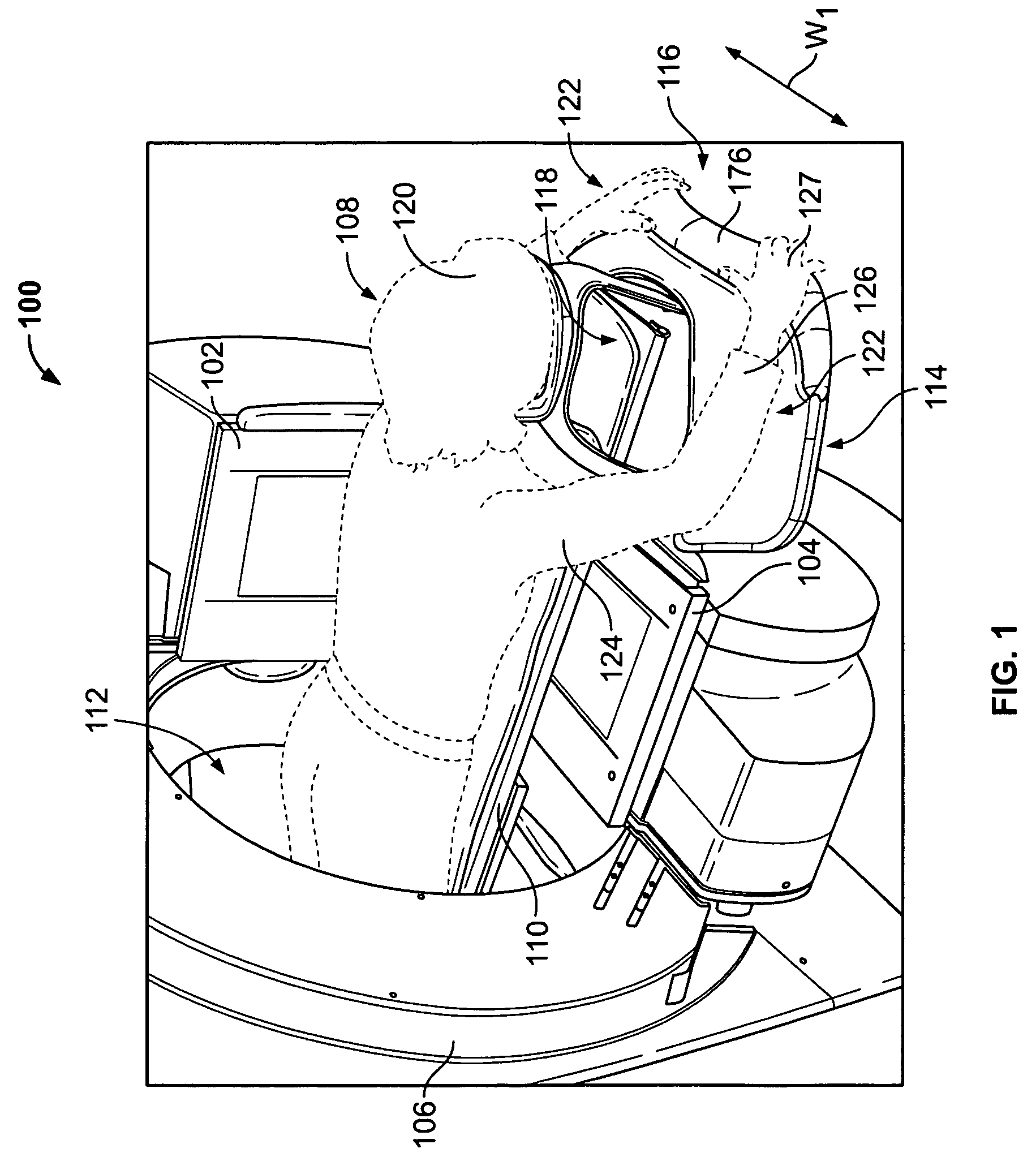
[0016]FIG. 1 illustrates an imaging system 100 with a patient 108 in a prone position on a patient table 110. Positioning apparatus 114 supports and holds the patient's head 120 and arms 122 while in the prone position. The patient 108 is facing a table top 118 of the patient table 110 with their arms 122 positioned away from their torso. In the exemplary illustration, the imaging system 100 is an NM system having first and second imaging detectors 102 and 104 mounted on a gantry 106. It should be understood that other diagnostic imaging modalities may be used, such as CT, MR and multi-modality systems. The patient table 110 extends through an opening 112 in the gantry 106 and is narrow in width W1 to allow the first and second detectors 102 and 104 to be as close to the patient 108 as possible when acquiring diagnostic data.

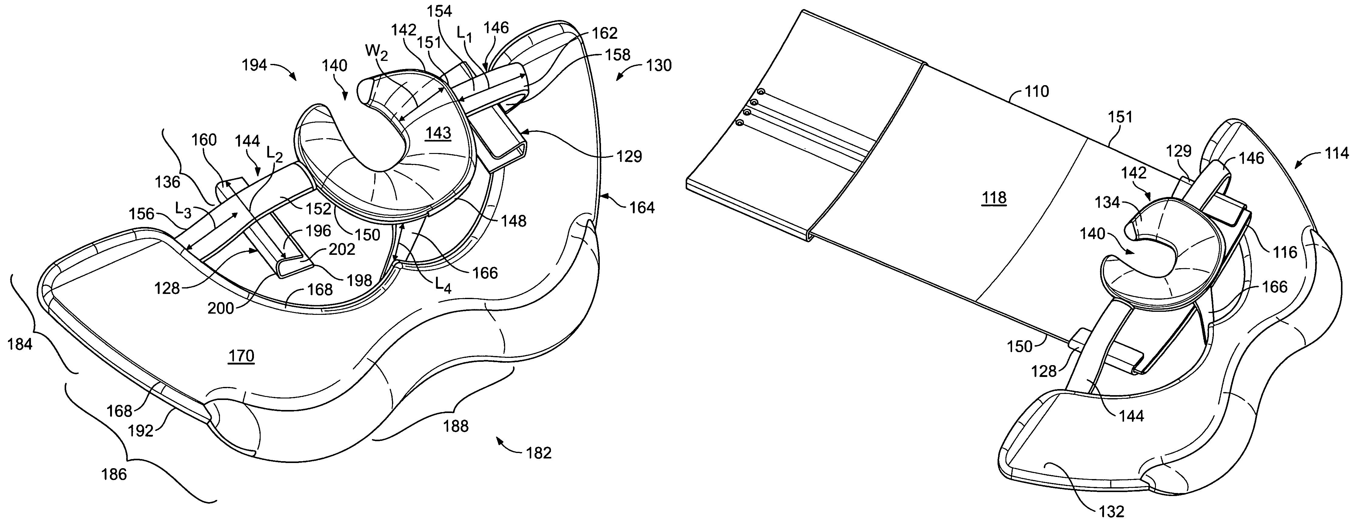
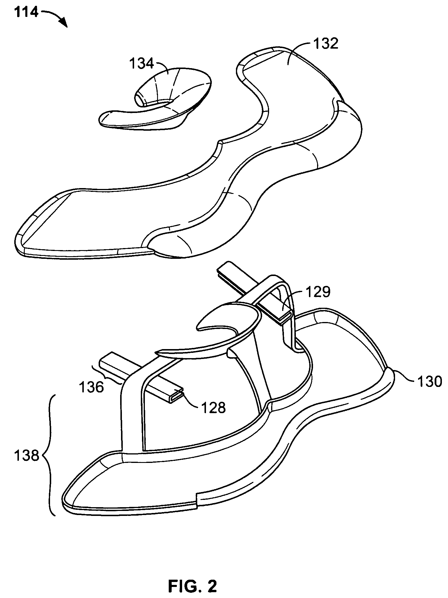
[0017]The prone patient positioning apparatus 114 is removably mounted to a first end 116 of the patient table 110. The positioning apparatus 114 elevates the p...


PUM


Abstract

A patient positioning apparatus to support a patient in a prone position on a patient table during a diagnostic imaging scan comprises a head support and an arm support. The head support is configured to extend above the patient table and supports a patient's head above a table top of the patient table while the patient lies on the patient table in a prone position. The arm support is joined to the head support and is configured to extend below the patient table to support a patient's forearms below the patient table.

Description

BACKGROUND OF THE INVENTION[0001]This invention relates generally to apparatus for positioning a patient during a medical imaging procedure, and more particularly, to apparatus for positioning a patient in a prone position during a medical imaging procedure.[0002]Diagnostic images of a patient are acquired using one or more modalities, such as Nuclear Medicine (NM), Computed Tomography (CT), Magnetic Resonance (MR), Positron Emission Tomography (PET), X-ray and Ultrasound. Some procedures for imaging anatomy, particularly within the torso, require the patient to lie prone on a patient table for a length of time without moving. In some cases, for example during nuclear imaging, an examination may last for an extended duration and thus patient comfort is advantageous. In other cases, such as during nuclear imaging, patient motion may create image artifact, degraded image quality or the necessity to repeat the data acquisition.[0003]The patient table is often narrow to allow the imagin...

Claims


Application Information

Patent Timeline
Patent Type & Authority Patents(United States)
IPC IPC(8): A61G13/12A47C16/00A47C20/00A61B6/04
CPCA47C20/00
Inventor ZELNIK, DEBORAH RUTH
Owner GENERAL ELECTRIC CO
Why Eureka
Try Eureka