Method and device for percutaneous left ventricular reconstruction

a left ventricular reconstruction and percutaneous technology, applied in the direction of prosthesis, catheter, therapy, etc., can solve the problems of back-up of pressure in the vascular system behind the ventricle, heart failure, and impaired pumping ability of the heart, so as to reduce the size and minimize the volume of the left ventricle

Active Publication Date: 2008-04-24
BIOVENTRIX A CHF TECH
View PDF59 Cites 56 Cited by
  • Summary
  • Abstract
  • Description
  • Claims
  • Application Information

AI Technical Summary

Benefits of technology

[0021] 2. reducing left ventricle volume while preserving the apex by realigning the ventricular apex to treat patients with congestive heart failure.
[0023] This invention relates to a device and method for treatment of congestive heart failure patients, and particularly to sizing device and method for reducing ventricular volume in patients suffering from congestive heart failure. The device and method may be performed thoracoscopically off-pump which is less expensive and less traumatic to the patient than an open-chest and open-heart surgical technique.
[0025] In yet another embodiment, this invention describes a method for performing minimally invasive left ventricular volume reduction in a congestive heart failure heart using a catheter by (a) accessing septum of the heart through the right ventricle; (b) identifying infracted tissue in the septum; (c) advancing the catheter through the infracted tissue of the septum; (d) identifying infracted tissue on the left ventricle wall; (e) bringing together the infracted tissue of the left ventricle and the septum, thereby minimizing the volume of the left ventricle; and (f) recreating an apex of the heart such that the procedure results in the desired geometric configuration and ejection fraction.
[0026] Once the boundaries of the scar have been defined using a sensing technique, achieving the clinical goal is dependent on realignment of the apical spatial relationships, and a decrease in the size of the left ventricle to improve ejection fraction and stroke volume. In the process, this invention will diminish wall tension, and therefore oxygen consumption, as well as solving potential alignment disparities by repositioning the apex to an appropriate anatomical configuration.
[0027] Another embodiment of this invention comprises a catheter-based, minimally invasive procedure of introducing a catheter into the right ventricle of the heart, via the septum, and into the left ventricle to reduce the volume of the left ventricle while preserving the apex of the left ventricle. This method comprises performing minimally invasive left ventricular volume reduction in a congestive heart failure heart using a catheter by (a) accessing septum of the heart through the right ventricle; (b) identifying tissue in the septum; (c) advancing the catheter through the infarcted tissue of the septum; (d) identifying infracted tissue on the left ventricular wall; and (e) bringing the infracted tissue of the left ventricle and the septum closer to each other and thereby minimizing the volume of the left ventricle.
[0028] Another aspect of the present invention pertains to suturing device, and related suturing methods, for thoracoscopy device placement on the heart. The sutures of the present inventions may be placed thoracoscopically. The inventive techniques and devices are thus minimally invasive and less risky to patients.

Problems solved by technology

Heart failure occurs when the pumping ability of the heart becomes impaired.
In certain pathological conditions, the ventricles of the heart become ineffective in pumping the blood, causing a back-up of pressure in the vascular system behind the ventricle.
The reduced effectiveness of the heart is usually due to an enlargement of the heart.
Acute myocardial infarction (AMI) due to obstruction of a coronary artery is a common initiating event that can lead ultimately to heart failure.
A myocardial ischemia may, for example, cause a portion of a myocardium of the heart to lose its ability to contract.
Once this tissue dies, it no longer as a muscle and cannot contribute to the pumping action of the heart.
As this situation worsens, the local area of compromised myocardium may in fact bulge out as the heart contracts, further decreasing the hearts ability to move blood forward.
One problem with a large dilated left ventricle is that there is a significant increase in wall tension and / or stress both during diastolic filling and during systolic contraction.
However, in a failing heart, the ongoing dilation is greater than the hypertrophy and the result is a rising wall tension requirement for systolic contraction.
This is felt to be an ongoing insult to the muscle myocyte resulting in further muscle damage.
This vicious cycle of cardiac failure results in the symptoms of congestive heart failure such as shortness of breath on exertion, edema in the periphery, nocturnal dypsnia (a characteristic shortness of breath that occurs at night after going to bed), weight gain, and fatigue, to name a few.
The stress increase requires a larger amount of oxygen supply, which can result in exhaustion of the myocardium leading to a reduced cardiac output of the heart.
Thus, it is not surprising that the cost of providing advanced medical care for the millions of patients suffering from HF is extraordinarily high—now estimated at more than $38 billion annually.
In certain pathological conditions, the ventricles of the heart become ineffective in pumping the blood, causing a back-up of pressure in the vascular system behind the ventricle.
The reduced effectiveness of the heart is usually due to an enlargement of the heart.
Acute myocardial infarction (AMI) due to obstruction of a coronary artery is a common initiating event that can lead ultimately to heart failure.
A myocardial ischemia may, for example, cause a portion of a myocardium of the heart to lose its ability to contract.
Once this tissue dies, it no longer functions as a muscle and cannot contribute to the pumping action of the heart.
As this situation worsens, the local area of compromised myocardium may in fact bulge out as the heart contracts, further decreasing the hearts ability to move blood forward.
One problem with a large dilated left ventricle is that there is a significant increase in wall tension and / or stress both during diastolic filling and during systolic contraction.
However, in a failing heart, the ongoing dilatation is greater than the hypertrophy and the result is a rising wall tension requirement for systolic contraction.
This is felt to be an ongoing insult to the muscle myocyte resulting in further muscle damage.
This vicious cycle of cardiac failure results in the symptoms of congestive heart failure such as shortness of breath on exertion, edema in the periphery, nocturnal dypsnia (a characteristic shortness of breath that occurs at night after going to bed), weight gain, and fatigue, to name a few.
The stress increase requires a larger amount of oxygen supply, which can result in exhaustion of the myocardium leading to a reduced cardiac output of the heart.
During a closed chest procedure where the heart is accessed using minimally invasive techniques, as contemplated here, it is not possible to visually differentiate the infracted tissue from viable or reversibly-ischemic myocardium.
While MRI and nuclear-uptake studies can identify the presence of scar, these are currently not feasible intraoperatively.

Method used


Image

  • Method and device for percutaneous left ventricular reconstruction
  • Method and device for percutaneous left ventricular reconstruction
  • Method and device for percutaneous left ventricular reconstruction

Examples


Embodiment Construction

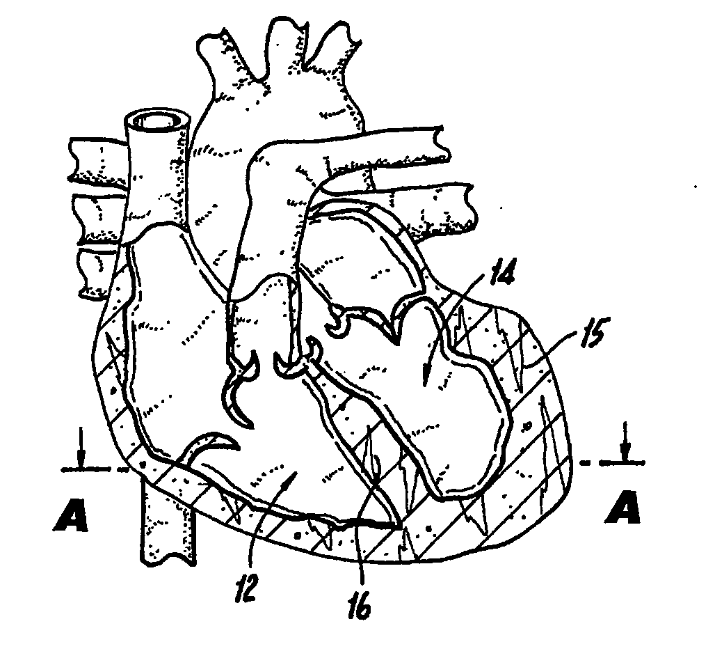
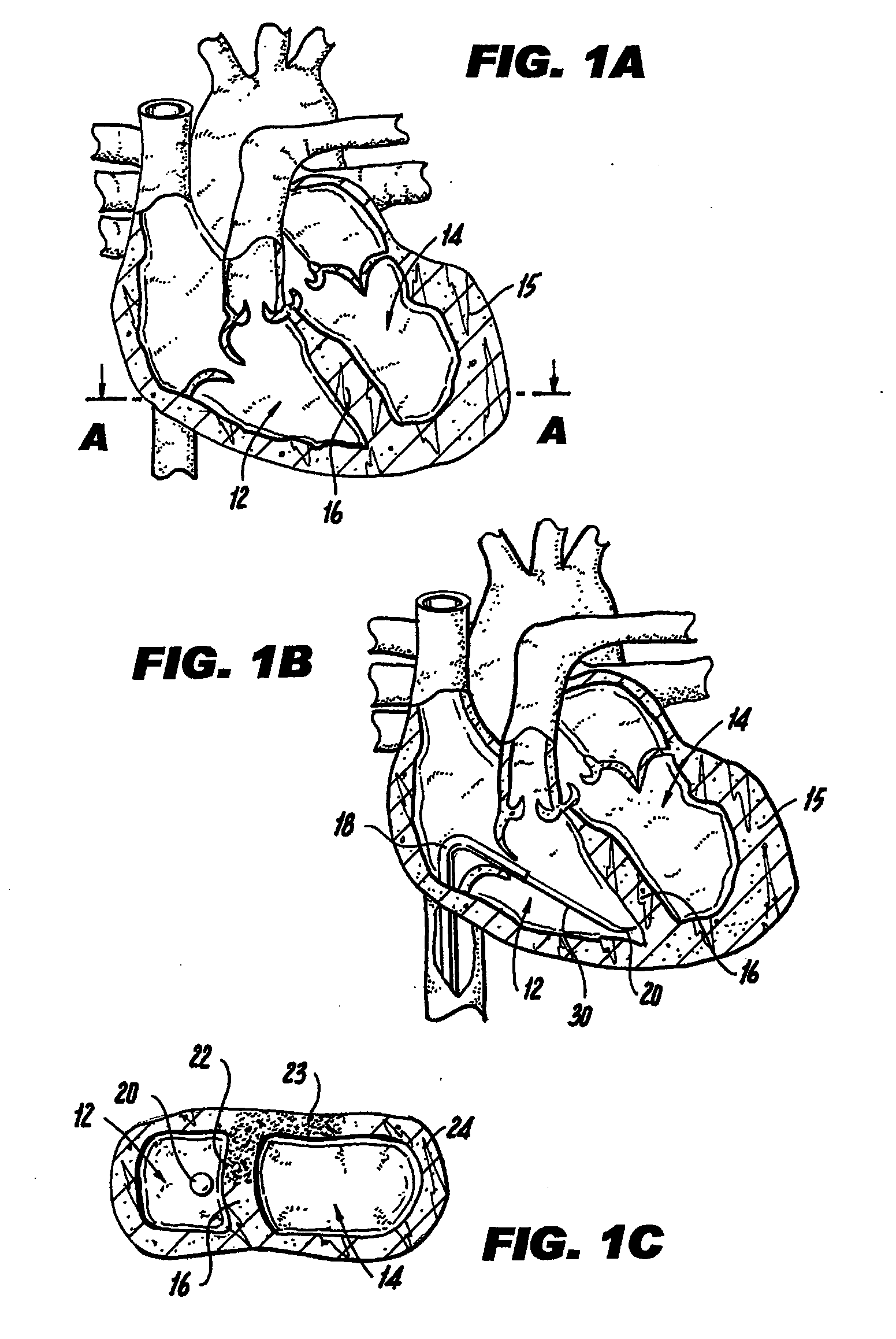
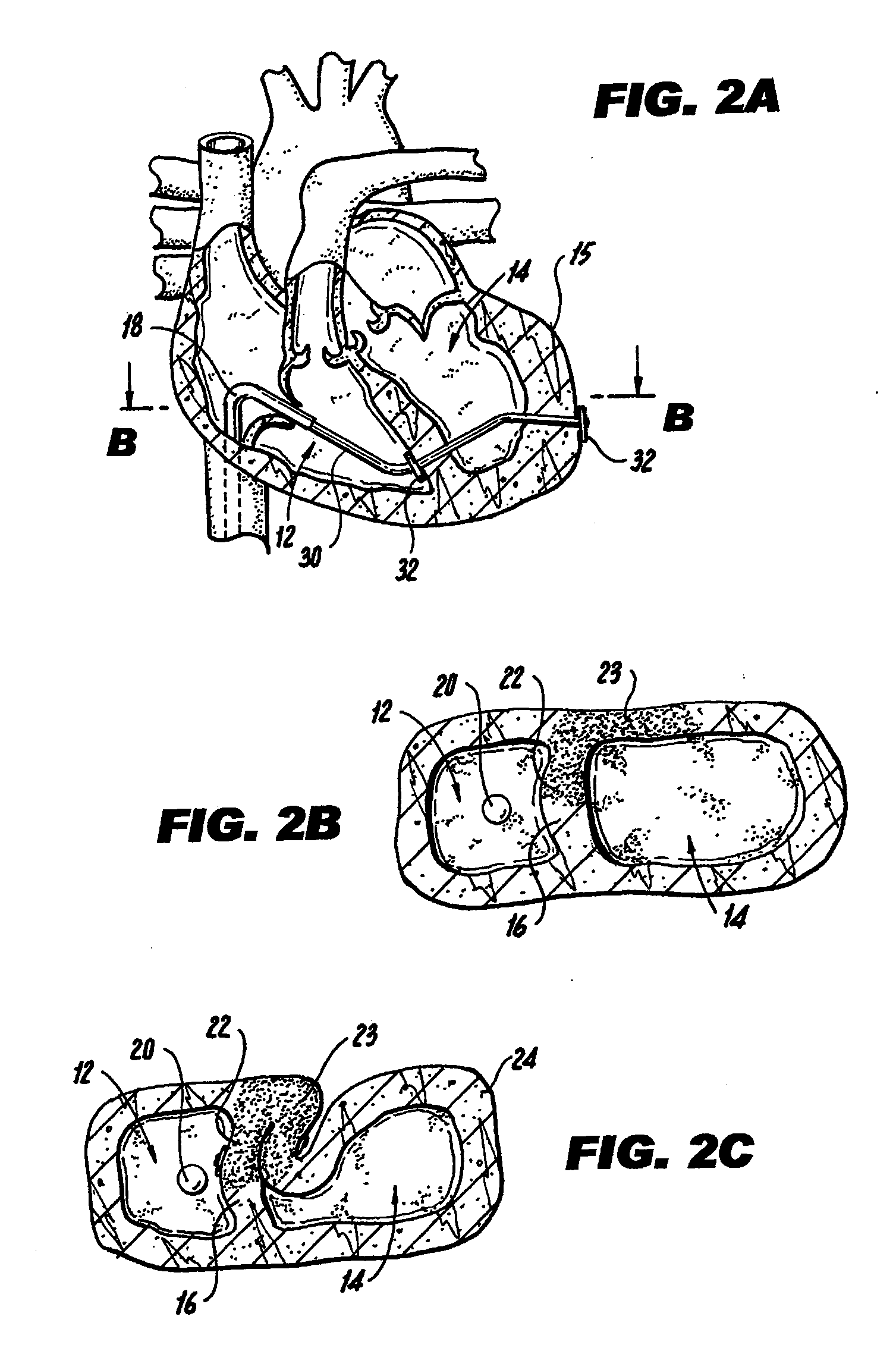
[0041]FIG. 1A illustrates a mammalian heart 10 and identifies the right ventricle 12, left ventricle 14, left ventricle wall 15 and septum 16. Right ventricle 12 may also be referred to interchangeably herein and in the figures as “RV”. Left ventricle 14 may also be referred to interchangeably herein and in the figures as “LV”. Additionally, left ventricle 14 is also referred to herein as “left ventricle chamber.”

[0042]FIGS. 1A-1C and 2A-2C illustrate a method of percutaneously accomplishing left ventricular restoration (“LVR”). In accordance with this method, a catheter 18 with a sensing element 20 is threaded through the femoral vein (not shown) into the right ventricle 12 of heart 10. It is to be understood that the invention is not limited to insertion of catheter 18 via the femoral vein and catheter 18 may be inserted via other arteries or veins. The sensing element 20 locates the infarcted tissue 22 of the interventricular septum 16. A commercially available device (EP Technol...


PUM


Abstract

A method for reducing left ventricular volume, which comprises identifying infarcted tissue during open chest surgery; reducing left ventricle volume while preserving the ventricular apex; and realigning the ventricular apex, such that the realigning step comprises closing the lower or apical portion of said ventricle to achieve appropriate functional contractile geometry of said ventricle in a dyskinetic ventricle of a heart.

Description

CROSS-REFERENCES TO RELATED APPLICATIONS [0001] This application is a continuation of U.S. patent application Ser. No. 11 / 248,521 filed on Oct. 12, 2005, which claims the benefit of under 35 U.S.C. §109(e) of U.S. Provisional Patent Application No. 60 / 618,835, filed on Oct. 13, 2004, the full disclosures of which are incorporated herein by reference.STATEMENT AS TO RIGHTS TO INVENTIONS MADE UNDER FEDERALLY SPONSORED RESEARCH OR DEVELOPMENT [0002] NOT APPLICABLE REFERENCE TO A “SEQUENCE LISTING,” A TABLE, OR A COMPUTER PROGRAM LISTING APPENDIX SUBMITTED ON A COMPACT DISK [0003] NOT APPLICABLE BACKGROUND OF THE INVENTION [0004] 1. Field of the Invention [0005] This invention relates to methods and devices for reconstructing the ventricle and for sensing infarcted septum and left ventricle wall during percutaneous left ventricle reconstruction, particularly where there is congestive heart failure. [0006] This invention also relates generally to devices for sizing a ventricle and relate...

Claims


Application Information

Patent Timeline
Patent Type & Authority Applications(United States)
IPC IPC(8): A61F2/00
CPCA61B5/042A61F2/2487A61B17/0401A61B2017/00022A61B2017/00243A61B2017/00247A61B2017/00575A61B2017/00579A61B2017/00606A61B2017/00632A61B2017/0412A61B2017/0419A61B2017/0427A61B2017/0464A61B2018/00392A61B8/12A61B5/283A61F2/2481A61F2002/2484A61B2090/376A61B2090/3782A61B1/3137A61B17/0469A61B2017/00044A61B2017/00106A61B2017/306A61M25/09
Inventor CHIN, SING-FATTBERTOLERO, ARTHURANNEST, LON
Owner BIOVENTRIX A CHF TECH
Why Eureka
Try Eureka