Radiotherapeutic Apparatus

Inactive Publication Date: 2008-06-05
ELEKTA AB
View PDF10 Cites 6 Cited by
  • Summary
  • Abstract
  • Description
  • Claims
  • Application Information

AI Technical Summary

Benefits of technology

[0009]As noted above, the apparatus can also comprise a further scanner for obtaining three-dimensional volume data regarding the volume. This will ideally use investigative means other than x-rays in order to minimise the dosage delivered to the patient. A magnetic resonance imaging scanner is ideal for the purpose. There will then preferably be a means for registration of the three-dimensional outputs of the reconstruction means and the further scanner, thereby to produce tissue type data and three-dimensional structure data in a region of interest within the volume.
[0010]The tissue type data can be passed to a dosage calculation means for determination of radiation dosage. The greater accuracy of the three-dimensional electron density data that can then be used given that the internal variation of tissue type is known will mean that the treatment can be modelled more accurately and, therefore, a more optimal treatment can be determined.

Problems solved by technology

These approaches are cumbersome, especially in the context of stereotactic radiosurgery, which normally does not require the acquisition of computerized tomograms for dose calculation.
While CT scanning is nearly ideal in terms of the accuracy of tissue density classification, the acquisition of computerized tomograms for the purposes of treatment planning is time consuming, results in the delivery of extraneous radiation dose to the patient, and is costly.
Approaches based on an assumption of tissue homogeneity are, in general, vulnerable to inhomogeneities in the volume of interest such as air cavities, and thus are unreliable and inaccurate in the vicinity of (for example) the patient's skull and lungs.
Some attempts have been made based on magnetic resonance imaging, but these are limited due to the erratic nature of automatic segmentation methods and the amount of manual labor required to segment the volume of interest manually.

Method used


Image

  • Radiotherapeutic Apparatus
  • Radiotherapeutic Apparatus

Examples


Embodiment Construction

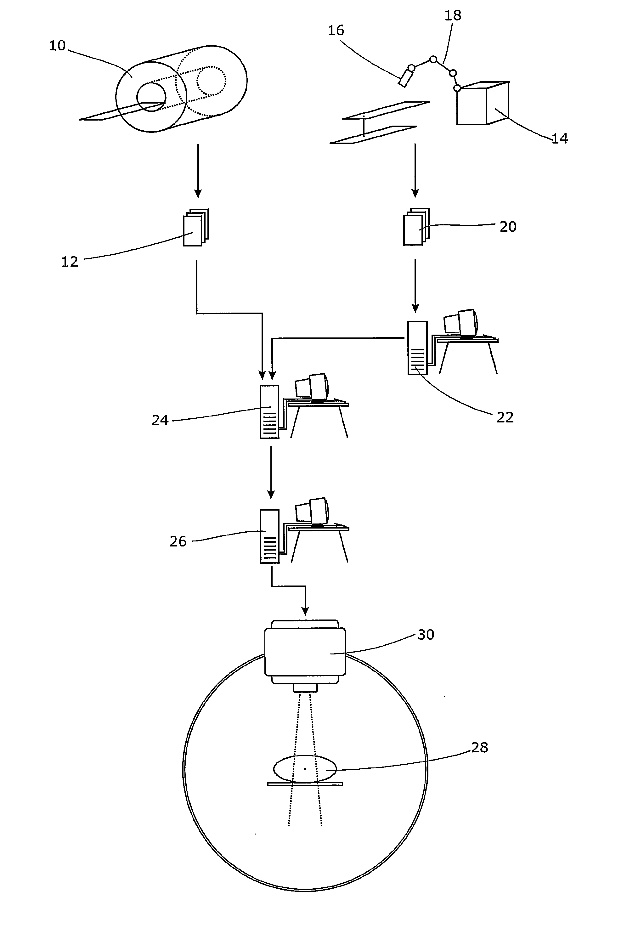
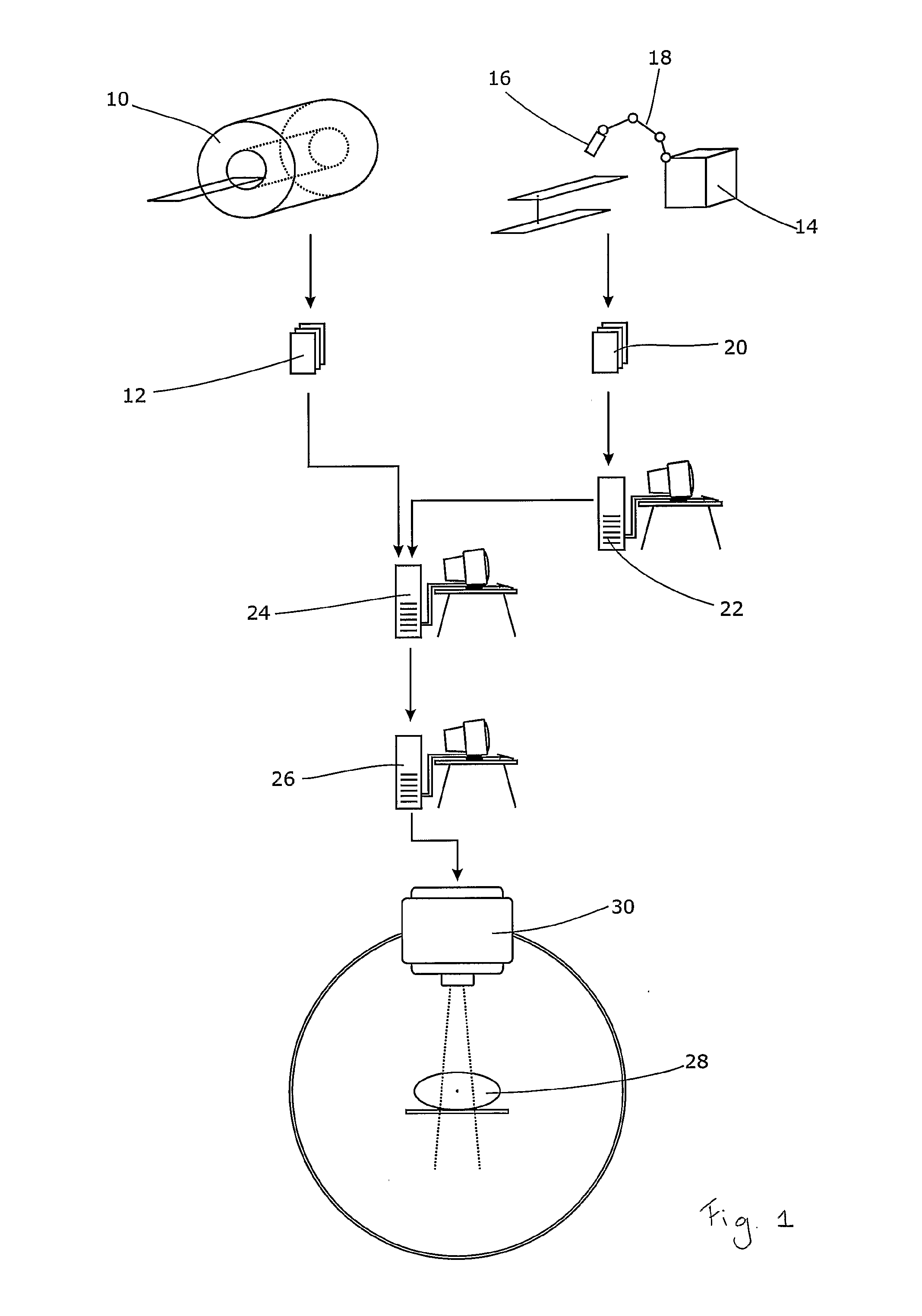
[0013]In technical terms, the solution could comprise (a) a three-dimensional, real-time motion tracking system; (b) an ultrasound device; (c) software for reconstruction of a three-dimensional ultrasound acquisition; (d) software for tissue classification; and (e) software for co-registering the classified three-dimensional sonographic acquisition with magnetic resonance images. The real-time motion tracking system provides the positions and orientations of the relevant part of the subject's body and of the ultrasound probe. The ultrasound device is used to interactively scan the volume of interest, the result of which is a volumetric reconstruction of the sonography. The volumetric reconstruction is subsequently classified such that density values comparable to those available from computerized tomography result. The classified volumetric reconstruction is finally co-registered with a magnetic resonance image set and used in place of a computerized tomogram as the basis of the dos...


PUM


Abstract

The co-registration of a volumetric ultrasound acquisition of the volume of interest with a magnetic resonance image allows the former to providing the basis for calculation of the dose distribution and the latter to provide the basis for target delineation and isocenter placement. The present invention therefore provides an apparatus for planning the radiotherapeutic treatment of a volume of tissue, comprising a sonographic apparatus for acquiring acoustic data relating to the volume, a means for reconstruction of an internal structure of the volume on the basis of the acoustic data, and a means for classification of the material type within that internal structure to one or more tissue types. A three-dimensional motion-tracking device for the sonographic device will assist. The sonographic device will typically be an ultrasound probe. The tissue type data can be passed to a dosage calculation means for determination of radiation dosage. The greater accuracy of the three-dimensional electron density data that can then be used given that the internal variation of tissue type is known will mean that the treatment can be modelled more accurately and, therefore, a more optimal treatment can be determined using, for instance, magnetic resonance imaging (MRI) alone.

Description

FIELD OF THE INVENTION[0001]The present invention relates to a radiotherapeutic apparatus. It seeks to address issues relating to the planning of radiotherapeutic treatment.BACKGROUND ART[0002]Most current dose calculation algorithms for stereotactic radiosurgery or radiation therapy either make use of electron-density values derived from computerized tomography (CT) investigation, or they assume that the body consists of homogenous material, such as water. This is then used as the basis for estimating attenuation of the radiation that will be used to treat the lesion. This may be x-radiation or other biologically effective radiation.[0003]These approaches are cumbersome, especially in the context of stereotactic radiosurgery, which normally does not require the acquisition of computerized tomograms for dose calculation. While CT scanning is nearly ideal in terms of the accuracy of tissue density classification, the acquisition of computerized tomograms for the purposes of treatment...

Claims


Application Information

Patent Timeline
IPC IPC(8): A61B8/00A61N5/00A61N5/10G01S7/52G01S15/89
CPCA61N5/103G01S15/8936G01S7/52036
Inventor PUTAALA, MIIKKA
Owner ELEKTA AB
Why Eureka
Try Eureka