System, method, and kit for processing a magnified image of biological material to identify components of a biological object

a biological object and magnified image technology, applied in image enhancement, image analysis, instruments, etc., can solve the problems of inability to identify cells in an image the inability to visualize cell structures by the automated image process, and the inability to distinguish cells whose membranes the automated image process has. to achieve the effect of improving the imag

Active Publication Date: 2008-06-19
VALA SCI
View PDF17 Cites 42 Cited by
  • Summary
  • Abstract
  • Description
  • Claims
  • Application Information

AI Technical Summary

Benefits of technology

[0010]A system and method for processing an original image of biological material to identify certain components of a biological object locates the biological object in the image, enhances the image by sharpening components of interest in the object, and applies a contour-finding function to the enh

Problems solved by technology

One difficult problem in the visualization of cell structures by automated image processes arises when the structures touch or overlap.
However, in cases where the cellular material is densely packed and/or agglomerated, an automated image process may have difficulty distinguishing cells whose membranes overlap and/or touch.
As a result, cells in an image may not be identified by the automated image process.
The failure of the process to identify and/or count multiple cells with abutting boundaries may lead

Method used


Image

  • System, method, and kit for processing a magnified image of biological material to identify components of a biological object
  • System, method, and kit for processing a magnified image of biological material to identify components of a biological object
  • System, method, and kit for processing a magnified image of biological material to identify components of a biological object

Examples


Embodiment Construction

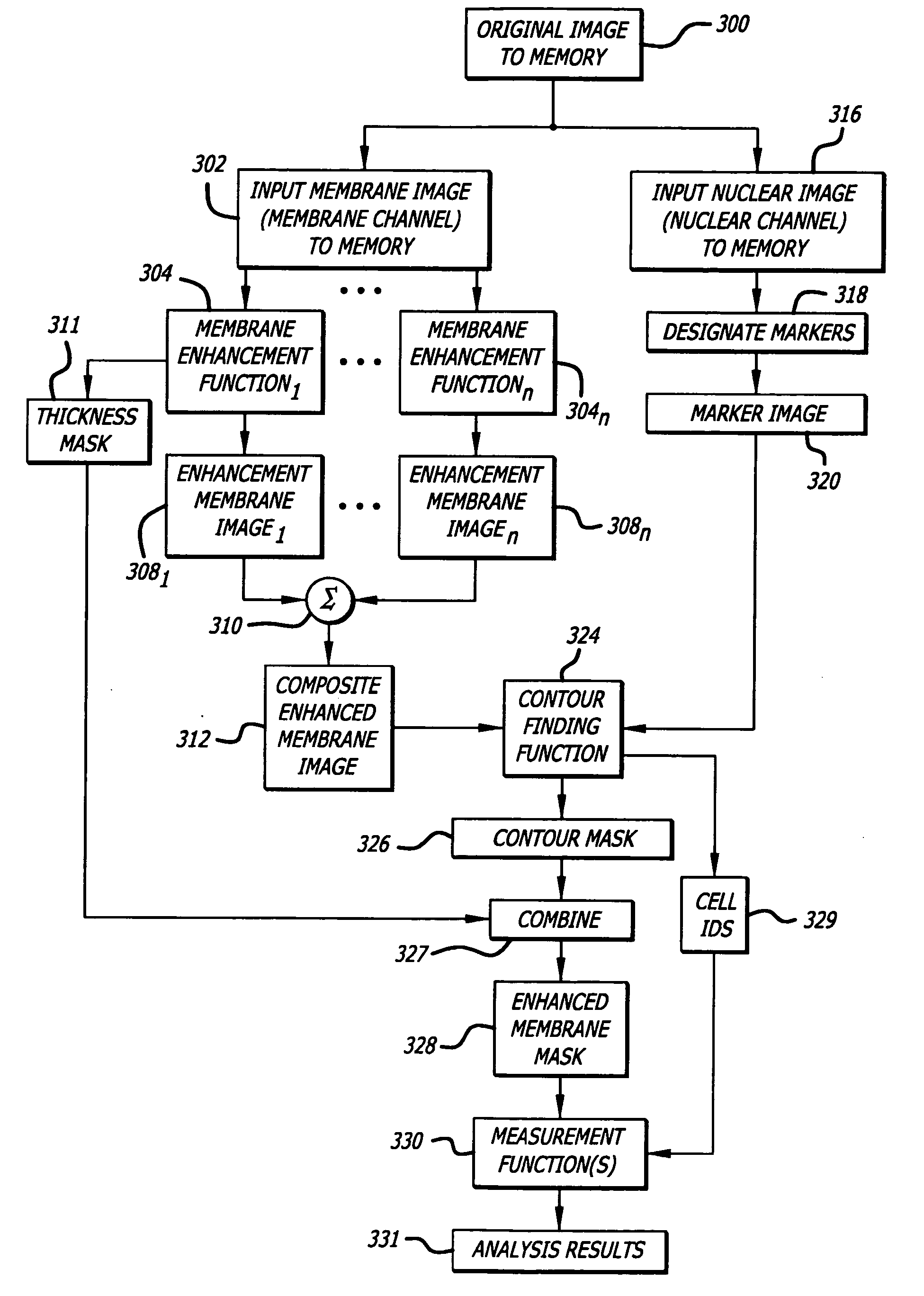
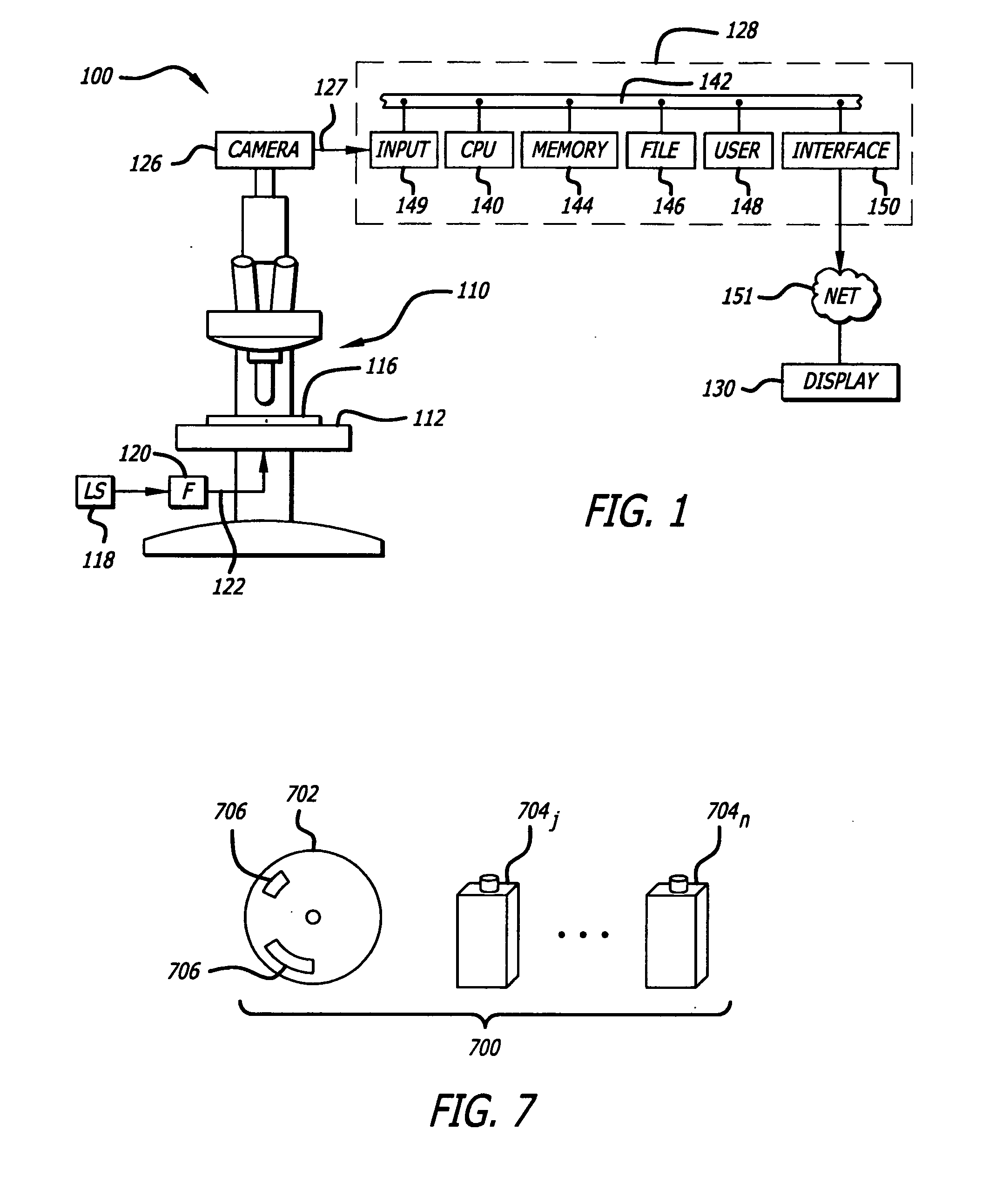
[0023]In this specification, the components to be identified are surface and / or structural features in magnified images of cells. The features are useful in enabling an automated image process to distinguish structures such as individual cells and / or components of cells in a digital image in order to obtain information useful in making measurements. An automated image process is a stream of activity conducted by a machine or capable of being conducted by a machine that processes information in a digital image. The components of interest are those features in an image that represent some shape, structure, form, or appearance of all or a part of a surface and / or a structure of cellular material. Examples of such components include, as examples and without limitation, boundaries between cells, outlines of cell membranes, outlines of intracellular membranes, and boundaries and / or outlines of intracellular objects. The automated image process may be executed by an automated high-throughp...


PUM


Abstract

A system, method and kit for processing an original image of biological material to identify certain components of a biological object by locating the biological object in the image, enhancing the image by sharpening components of interest in the object, and applying a contour-finding function to the enhanced image to create a contour mask. The contour mask may be processed to yield a segmented image divided by structural units of the biological material.

Description

BACKGROUND OF THE INVENTION[0001]The technical field concerns image processing. More particularly, the technical field covers processing a magnified image of biological material to identify one or more components of an object in the image. In addition, the technical field includes a combination of reagents to enhance the visualization of one or more components of an object in a magnified image of stimulated biological material and an automated process adapted to identify the components.[0002]Biological material may include cells disposed in or on plastic or glass culture dishes, cells disposed on plastic or glass slides, and / or tissue sections mounted to plastic or glass slides.[0003]Measurements of object features in magnified images of biological material are of increasing importance in the analysis of biological processes in automated high throughput screening (HTS) and in high content screening (HCS). For example, in HTS procedures developed for assaying cellular activity, each ...

Claims


Application Information

Patent Timeline
IPC IPC(8): G06K9/00G06K9/48
CPCG01N15/1475G06K9/00127G06T7/0083G06T7/0091G06T2207/10064G06T2207/20152G06T2207/30024G06T2207/10056G06T7/12G06T7/155G06V20/69
Inventor HUNTER, EDWARDMC DONOUGH, PATRICK M.MIKIC, IVANAPRICE, JEFFREY H.
Owner VALA SCI
Why Eureka
Try Eureka