
Method and system for processing an image of a biological specimen

a biological specimen and image processing technology, applied in image analysis, image enhancement, instruments, etc., can solve problems such as less accurate diagnosis

Active Publication Date: 2008-06-19
CYTYC CORP
View PDF13 Cites 6 Cited by
  • Summary
  • Abstract
  • Description
  • Claims
  • Application Information

AI Technical Summary

Benefits of technology

[0009]In one embodiment, a method of processing an image of a biological specimen having a plurality of cells includes identifying components of the biological specimen based on spectral characteristics of the components, selecting an identified component, and determining whether cells having the selected component have a likelihood of being cancerous or non-cancerous based on one or more physical characteristics of the identified component. By way of non-limiting examples, the identified components of the specimen may include one or more of background, cytoplasm and nuclear (e.g., nucleolus) components of the specimen. By way of further non-limiting examples, the components of the specimen may be identified using one or more light wavelengths in a range from about 400 nm to about 720 nm. The components may be identified by classifying a pixel of the image as a certain component of the biological specimen based on spectral characteristics of the pixel. The method may optionally further include determining whether cells corresponding to the selected components are cancer cells or normal repair cells. The determination of whether cells having the selected component have a likelihood of being cancerous or non-cancerous may be based on one or more of a shape, a texture or a size of the identified component. Additionally or alternatively, the determination of whether cells having the selected component have a likelihood of being cancerous or non-cancerous may be based on a transmittance or gray value of the identified component or of an image pixel of the identified component.
[0010]In another embodiment, a method of processing an image of a biological specimen having a plurality of cells includes identifying components of the biological specimen based on spectral characteristics of the components, the identified components including nucleus components. The method further includes selecting regions of the image having nucleus components having one or more nucleolus components, and determining whether cells in the selected regions are cancer cells or repair cells based on one or more physical characteristics of the nucleolus components. Again, the determination of whether cells in the selected regions are cance

Problems solved by technology

Thus, the cytotechnologist may not be presented with the most relevant fields of view, possibly resulting in a less accurate diagnosis.

Method used


Image

  • Method and system for processing an image of a biological specimen
  • Method and system for processing an image of a biological specimen
  • Method and system for processing an image of a biological specimen

Examples


Embodiment Construction

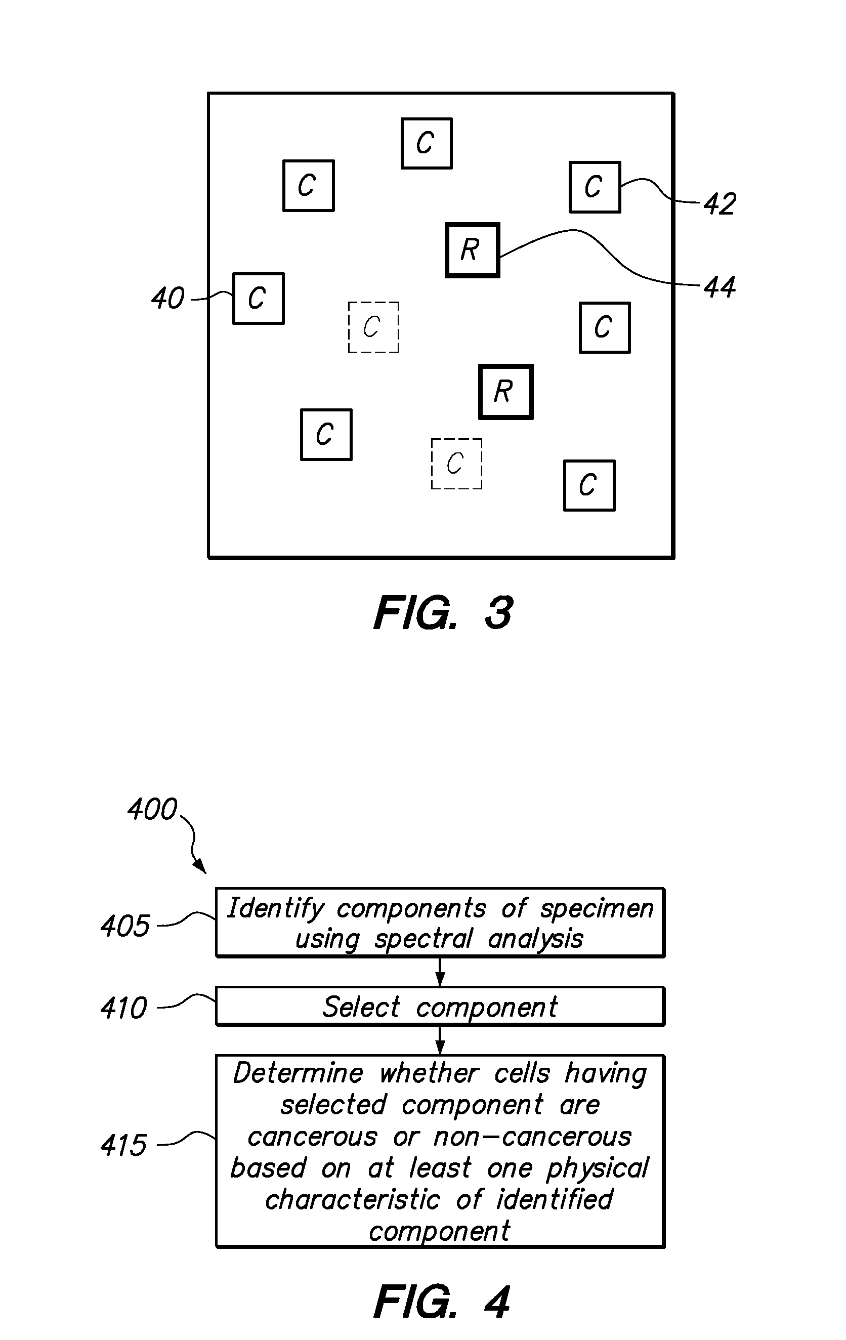
[0036]Referring to FIG. 4, according to one embodiment of the invention, a method 400 of processing an image of a biological specimen, e.g. a cytological specimen, includes identifying components of the specimen using spectral analysis in step 405. After components are identified, in step 410, one of the components is selected. For example, the selected component may be a nucleolus of a nucleus. Then, in step 415, a determination is made whether cells that include the selected component are cancerous or non-cancerous based on one or more physical characteristics of the component.

[0037]Referring to FIG. 5, a method 500 of processing an image of a biological specimen includes receiving a pixel that is to be imaged in step 505. In step 510, spectral analysis is performed to determine whether a pixel is background, cytoplasm, nucleus or nucleolus. For purposes of explanation and illustration, reference is made to nucleus and nucleolus components since background and cytoplasm components...


PUM


Abstract

Components, e.g., background, cytoplasm, nucleus and nucleolus, of a biological specimen are identified using multi-wavelength analysis. Specimen components, such as nucleoli, are selected, and a determination is made whether cells having nucleoli are cancer cells or regular repair cells based on one or more physical characteristics of the identified component. The physical characteristics can be one or more of a shape, size, texture and gray value.

Description

RELATED APPLICATION DATA[0001]The present application claims the benefit under 35 U.S.C. § 119 to U.S. provisional patent application Ser. No. 60 / 870,838, filed Dec. 19, 2006. The foregoing application is hereby incorporated by reference into the present application in its entirety.FIELD OF INVENTION[0002]The invention relates to imaging and analysis of biological specimens, and more particularly, to reducing the number of non-cancerous repair cells that are selected by an imaging system for subsequent review.BACKGROUND[0003]Medical professionals and cytotechnologists frequently review biological specimens affixed to a specimen carrier, such as a slide, to analyze whether a person from whom the specimen was obtained has or may have a particular medical condition. For example, it is well known to examine a cytological specimen in order to detect the presence of malignant or pre-malignant cells as part of a Papanicolaou (Pap) smear test. To facilitate this review process, automated sy...

Claims


Application Information

Patent Timeline
IPC IPC(8): G06K9/00
CPCG06K9/00147G06T7/0012G06T2207/30024G06T2207/10056G06T7/402G06T7/42G06V20/698
Inventor WONG, KAM LINZAHNISER, DAVIDMUI, KATHYLINDER, JIMSHEETS, ELLEN
Owner CYTYC CORP
Learn More